Categories
Health & Fitness

Healthy Knee is Friendly Indeed

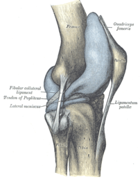
Capsule of right knee-joint (distended). Later...
Image via Wikipedia

[amazon_link asins=’B01A5Q746G,B01LX86E55,B00KYDCP7O,0979303605,1579548717,B01K3CXMB8,B01MRF7JRM,B01CTRKP2O,B071Y32649′ template=’ProductCarousel’ store=’finmeacur-20′ marketplace=’US’ link_id=’8f3c0421-79a1-11e7-9ce0-051296b748ee’]

Whether you are a ballerina, mountaineer, a weekend gladiator or just an office warrior, learn to protect your knees because the knee is a critical link in the kinetic chain that allows you to walk on two feet.
….....CLICK & SEE

Anit Ghosh, a former national footballer, suffered a career-threatening injury to his knee ligament five years ago. Regular and diligent post-injury rehabilitation work under the author’s guidance helped him gradually return to competitive football. Today, he turns out for Mohammedan Sporting and has learned to manage a problematic knee.

In  therapy practice, over half the ladies and about one in 10 men complain of knee pain. After back pain, knee pain is the most common cause of disability and time lost from work or training.

The knee is the largest joint in the human body and is formed by the articulation of three bones, the lower end of the thighbone (femur), the upper end of the shinbone (tibia) and the kneecap (patella). It may appear like a simple hinge, but besides the routine functions of bending and straightening, the knee joint performs a host of complex functions — it slides, glides, pivots, rolls and rotates — sometimes sequentially and at other times simultaneously. All these movements make the knee joint very vulnerable to shearing forces and dependant on good functional stability from the surrounding soft tissue network of ligaments, tendons and the two menisci, tough crescent-shaped cushions within the joint. In addition to the above, the knee joint also includes small, fluid-filled membranous sacs lying between the ligaments or skin, and the bones to provide smooth and frictionless gliding, like ball bearings in a machine. Furthermore, the entire articular surfaces, i.e. those that rub against one another, are covered with a tough, rubbery slippery tissue called cartilage.

Of these parts mentioned above can be a source of joint pain. Sometimes, knee pain can be caused by poor body mechanics and tight muscles elsewhere in the body and can easily be corrected by a slight alteration in gait and mechanics. For example, poor flexibility around the ankle and hip can transfer a lot of shearing forces onto the knee even though pathologically the knee is normal. The knee then is merely the “site” of the pain. The villain or “source” of pain may lie elsewhere.

The most common causes of knee pain  are described below..>..CLICK & SEE

*One of the most crippling forms of knee ailment is arthritis caused by the degeneration of the cartilage coating. The cartilage has very poor blood supply and consequently nutrient supply and therefore once traumatised, has hardly any chance of healing itself. The inherent nature of the cartilage is a huge limiting factor for arthritis rehabilitation.

*Chondromalacia is the softening or the wearing away of the articular cartilage under the kneecap. The articular cartilage on the inside aspect of the kneecap comes in constant contact with the articular surfaces of the femur during normal knee motion. The knee motion can sometimes become abnormal or faulty due to muscle imbalance or biomechanical misalignment and cause the patella to rub against the femoral surfaces. Repetitive ‘rubbing’ of the surfaces causes chronic inflammation sometimes popularly known as “jumpers knee”.

*One of the most common causes of pain inside the joint is a torn meniscus. The crescent-shaped spongy tissues act as shock absorbers within the joint and when torn, either by injury or degeneration, tends to get caught in the joint, causing pain and instability.

*When the articular cartilage begins fragmenting and eroding due to extreme softening, the underlying bone gets exposed. This is a condition called osteoarthritis.

*Often traumatic injuries or contact sports mishaps cause the ligaments within the knee joint to snap. This is a very painful condition and more often than not, needs surgical correction where the surgeon has to reconstruct the ligament necessitating a long healing period.

WHAT YOU CAN DO TO MANAGE, EVEN PREVENT KNEE PAIN?

*Stretch regularly. Regular stretching of the hip flexors, hip extensors and the iliotibial band (a sheath of muscle lying on the outside of your thigh extending from the hip to the lateral aspect of the knee) will ensure good gait and running mechanics and spare the knee of shearing forces.

*Train with weights. Loading the knee and hips early in life with weights will build density in the bones and prevent erosion in later life.

*Work the hamstrings. The average person has stronger quadriceps compared to the hamstrings. Increase hamstring strength for better muscle balance and correct alignment of the kneecap. This will avoid compression forces within the knee.

*Strengthen the vastus medialis muscle — the muscles lying in the inside aspect of your front thigh. This will help to realign and track the kneecap to its normal pathway.

*Exercise discretion while performing repetitive knee motions like running, skipping, jumping etc. If you must run, learn proper running technique. Let’s face it — nine out of 10 people who visit lifestyle and recreation gyms do not have good technique. Running on the treadmill for these people is sheer disaster!

*If you are an active sort of a person, check with your doctor whether you should supplement with Glucosamine sulphate and Chondroitin. They are known to have shown results in preventing degeneration of the knee joint.

*Avoid knee extensions. The leg extension exercise is treated as a panacea for all sorts of knee ailments by trainers and therapists alike. In reality, open-chain movements like the knee extension exercise is potentially more dangerous than closed-chain movements like the lunge and squat.

*The leg extension movement causes compression between the kneecap and the thighbone and I would recommend even healthy knee-owners to stay far away from it. Choose multi-joint exercises that make the quadriceps and hamstrings work together in unison.

The best exercises for the knee are:

*One-legged squats
*Glute ham raises
*Lunges
*Split squats

Sources: The Telegraph (Kolkata, India)

Reblog this post [with Zemanta]
Categories
Ailmemts & Remedies

Sprain & Strain

[amazon_link asins=’B01GY7AQME,B001M0A5QQ,B008VWW1YM,B00DQR62Q0,B06XBLNK3T,B001VF6TMQ,B004FJHGEA,B001S6RJ5O’ template=’ProductCarousel’ store=’finmeacur-20′ marketplace=’US’ link_id=’57e7b78b-651d-11e7-b896-17c9292a9941′]

Definition:
A sprain (from the French espraindre – to wring) is an injury which occurs to ligaments caused by being stretched beyond their normal capacity and possibly torn. Muscular tears caused in the same manner are referred to as a strain. In cases where either ligament or muscle tissue is torn, immobilization and surgical repair may be necessary.

CLICK & SEE THE PICTURES

Sprain. A sprain is a stretching or tearing of ligaments. Ligaments are tough bands of fibrous tissue that connect one bone to another. Common locations for sprains are your ankles and knees.

Strain. A strain is a stretching or tearing of muscle or tendon. People commonly call strains “pulled” muscles. Hamstring and back injuries are among the most common strains.


Degrees:

Although some signs and symptoms can be used to assess the severity of a sprain, the most definitive method is with the use of Magnetic Resonance Imaging (MRI). Sprains are graded in four degrees.

*The first degree is only a minor tear or stretch of a ligament.

*The second degree is a tear of a ligament, which is usually followed by pain or swelling.

*The third degree is a complete rupture.

*The fourth degree is the most severe and actually breaks the ligament, along with some small bones if severe enough, and requires surgery to repair.

Causes:
Sprains and strains occur commonly, and most result in minor injuries.

Sprains. A sprain occurs when you overextend or tear a ligament while severely stressing a joint. Ligaments are tough bands of fibrous tissue that connect one bone to another. They help to stabilize joints, preventing excessive movement. You may sprain your knee or ankle when walking or exercising on an uneven surface. A sprain also may occur when you land awkwardly, either at the end of a jump or while pivoting during an athletic activity.

Strains. A muscle becomes strained or pulled — or may even tear — when it stretches unusually far or abruptly. This type of injury — an acute strain — often occurs when muscles suddenly and powerfully contract. A muscle strain may occur when you slip on ice, run, jump, throw, lift a heavy object or lift in an awkward position. A chronic strain results from prolonged, repetitive movement of a muscle.

Signs & Symptoms:
The typical signs and symptoms associated with a sprain are the cardinal signs of a sprain.

*Inflammation

*Localized pain

*Swelling

*Loss of function

*Loss of normal limb function

*Elasticity of ligament decrease

Joints involved:
Although any joint can experience a sprain, some of the more common include:

*The ankle. It is the most common, and has been said that sprains such as serious ankle sprains are more painful and take longer to heal than actually breaking the bones in that area. See ->sprained ankle for more details.

.

*The knee. Perhaps one of the more talked about sprains is that to the anterior cruciate ligament (ACL) of the knee. This is a disabling sprain common to athletes, especially in basketball, football, and judo. See Anterior cruciate ligament injury.

*The fingers.

*The wrist.

*The toes.

Risk factors:
Factors contributing to sprains and strains include:

*Poor conditioning. Lack of conditioning can leave your muscles weak and more likely to sustain injury.

*Poor technique. The way you land from a jump — for example, when skiing or practicing martial arts — may affect your risk of injury to a ligament in your knee called the anterior cruciate ligament (ACL). Past research has shown that landing with an inward rotation at the knee (“knock-kneed” position) can predispose you to an ACL sprain.

*Fatigue. Tired muscles are less likely to provide good support for your joints. When you’re tired, you’re also more likely to succumb to forces that could stress a joint or overextend a muscle.

*Improper warm-up. Properly warming up before vigorous physical activity loosens your muscles and increases joint range of motion, making the muscles less tight and less prone to trauma and tears.

Treatment:
The first modality for a sprain can be remembered using the acronym R.I.C.E.

*Rest: The sprain should be rested. No additional force should be applied on site of the sprain. If, for example, the sprain were an ankle sprain, then walking should be kept to a minimum.

*Ice: Ice should be applied immediately to the sprain to minimize swelling and ease pain. It can be applied for 20-30 minutes at a time, 3-4 times a day. Ice can be combined with a wrapping to minimize swelling and provide support.

*Compression: Dressings, bandages, or ace-wraps should be used to immobilize the sprain and provide support.

*Elevation: Keeping the sprained joint elevated above heart level will also help to minimize swelling.

*Ice and compression (cold compression therapy) will not completely stop swelling and pain, but will help to minimize them as the sprain begins to heal itself. Careful management of swelling is critical to the healing process as additional fluid may pool in the sprained area.

Click to see :
->Sprain: First aid

Prevention:
Sprains can best be prevented by proper use of safety equipment (wrist, ankle guards), warm-ups and cool-downs (including stretching), being aware of your surroundings and maintaining strength and flexibility. Physical conditioning is the best way to avoid or lessen the degree of sprains.

Lifestyle and home remedies:
For immediate self-care of a sprain or strain, try the P.R.I.C.E. approach — protection, rest, ice, compression, elevation. In most cases beyond a minor strain or sprain, you’ll want your doctor and physical therapist to help you with this process:

*Protection. Immobilize the area to protect it from further injury. Use an elastic wrap, splint or sling to immobilize the area. If your injury is severe, your doctor or therapist may place a cast or brace around the affected area to protect it and instruct you on how to use a cane or crutches to help you get around, if necessary.

*Rest. Avoid activities that cause pain, swelling or discomfort. But don’t avoid all physical activity. Instead, give yourself relative rest. For example, with an ankle sprain you can usually still exercise other muscles to prevent deconditioning. For example, you could use an exercise bicycle, working both your arms and the uninjured leg while resting the injured ankle on a footrest peg. That way you still exercise three limbs and keep up your cardiovascular conditioning.

*Ice. Even if you’re seeking medical help, ice the area immediately. Use an ice pack or slush bath of ice and water for 15 to 20 minutes each time and repeat every two to three hours while you’re awake for the first few days following the injury. Cold reduces pain, swelling and inflammation in injured muscles, joints and connective tissues. It also may slow bleeding if a tear has occurred. If the area turns white, stop treatment immediately. This could indicate frostbite. If you have vascular disease, diabetes or decreased sensation, talk with your doctor before applying ice.

*Compression. To help stop swelling, compress the area with an elastic bandage until the swelling stops. Don’t wrap it too tightly or you may hinder circulation. Begin wrapping at the end farthest from your heart. Loosen the wrap if the pain increases, the area becomes numb or swelling is occurring below the wrapped area.

*Elevation. To reduce swelling, elevate the injured area above the level of your heart, especially at night. Gravity helps reduce swelling by draining excess fluid.

*Continue with P.R.I.C.E. treatment for as long as it helps you recover. Over-the-counter pain medications such as ibuprofen (Advil, Motrin, others) and acetaminophen (Tylenol, others) also can be helpful. If you want to apply heat to the injured area, wait until most of the swelling has subsided.

After the first two days, gently begin to use the injured area. You should see a gradual, progressive improvement in the joint’s ability to support your weight or your ability to move without pain.

Mild and moderate sprains usually heal in three to six weeks. If pain, swelling or instability persists, see your doctor. A physical therapist can help you to maximize stability and strength of the injured joint or limb.

Disclaimer: This information is not meant to be a substitute for professional medical advise or help. It is always best to consult with a Physician about serious health concerns. This information is in no way intended to diagnose or prescribe remedies.This is purely for educational purpose

Resources:
http://en.wikipedia.org/wiki/Sprain
MayoClinic.com

Enhanced by Zemanta
Categories
Ailmemts & Remedies

Knee Injury

The knee has a simple purpose. It needs to flex (bend) or extend (straighten) to allow the body to perform many activities like running, walking, kicking, and sitting. Imagine standing up from a chair if your knees couldn’t bend.

Knee Anatomy
While there are four bones that come together at the knee, only the femur (thigh bone) and the tibia (shin bone) form the joint itself. The head of the fibula (strut bone on the outside of the leg) provides some stability, and the patella (kneecap) helps with joint function. Movement and weight-bearing occur where the ends of the femur called the femoral condyles match up with the top flat surfaces of the tibia (tibial plateaus).

There are two major muscle groups that are balanced and allow movement of the knee joint. Contracting the quadriceps muscles on the front of the thigh extends the knee, while the hamstring muscles on the back of the thigh flex the knee when they contract. The muscles cross the knee joint and are attached to the tibia by tendons. The quadriceps tendon is a little special, in that it contains the patella within it. The patella allows the quadriceps muscle/tendon unit to work more efficiently. This tendon is renamed the patellar tendon in the area below the kneecap to its attachment to the tibia.

The stability of the knee joint is maintained by four ligaments, thick bands of tissue that stabilize the joint. The medial collateral ligament (MCL) and lateral collateral ligament (LCL) are on the sides of the knee and prevent the joint from sliding sideways. The anterior cruciate ligament (ACL) and posterior cruciate ligament (PCL) form an “X” on the inside of the knee and prevent the knee from sliding back and forth. These limitations on knee movement allow the knee to concentrate the forces of the muscles on flexion and extension.

Inside the knee, there are two shock-absorbing pieces of cartilage called menisci (singular meniscus) that sit on the top surface of the tibia. The menisci allow the femoral condyle to move on the tibial surface without friction, preventing the bones from rubbing on each other.

Bursas surround the knee joint and are fluid filled sacs that cushion the knee during its range of motion. In the front of the knee, there is a bursa between the skin and the kneecap called the prepatellar bursa and another above the kneecap called the suprapatellar bursa (supra=above).

Each part of the anatomy needs to function properly for the knee to work. Acute injury and trauma as well as chronic overuse can both cause inflammation and its accompanying symptoms of pain, swelling, redness and warmth.

Causes:.....
While direct blows will occur, the knee is more susceptible to twisting or stretching injuries, taking the joint through a greater range of motion than it can tolerate.

If the knee is stressed from a specific direction, then the ligament trying to hold it in place against that force can tear. Ligament stretching or tears are called sprains. These sprains are graded as first, second, or third degree based upon how much damage has occurred. Grade one sprains stretch the ligament but don’t tear the fibers; grade two sprains partially tear the fibers, but the ligament remains intact; and grade three tears completely disrupt the ligament.

Twisting injuries to the knee put stress on the cartilage or meniscus and can pinch it between the tibial surface and the edges of the femoral condyle, causing tears.

Injuries of the muscles and tendons surrounding the knee are caused by acute hyperflexion or hyperextension of the knee or by overuse. These injuries are called strains.

There can be inflammation of the bursas (known as bursitis) of the knee that can occur because of direct blows or chronic use and abuse.

Acute knee injuries fall into two groups; those where there is almost immediate swelling in the joint associated with the inability to bend the knee and bear weight, and those where there is discomfort and perhaps localized pain to one side of the knee, but have minimal swelling and minimal effects on walking.

Symptoms:
Acute knee injuries can cause pain and swelling with difficulty bending the knee and weight-bearing.

Longer-term symptoms that point to knee problems will include pain and swelling in addition to other complaints. Inflammation in the joint may be caused by even minor activity. Swelling may be intermittent, brought on by activity, and may gradually resolve as the inflammation decreases.

Pain, too, may come and go and may not occur right away with activity but might be delayed as the inflammation develops. Pain can also be felt with specific activities. Pain while climbing stairs is a symptom of meniscus injury, where the cartilage is being pinched in the joint as it narrows with bending. Pain with walking down stairs suggests patellar pain, where the kneecap is being forced onto the femur.

Giving way, or a feeling of instability, of the knee is a symptom of ligament injury, while popping or grinding in the knee is more associated with cartilage or meniscus tears. “Locking” is the term used when the knee joint refuses to completely straighten, and this is always due to torn cartilage. In this situation, the torn piece of cartilage folds upon itself and doesn’t allow the knee to extend.


Emergent medical care should be sought if, due to injury,

1.there is almost immediate swelling in the knee,
2.if the bones appear deformed,
3.if there is inability to bear weight,
4.if the pain is intolerable,
5.if there is loss of sensation below the injury site
6.or if the foot and ankle turn cold and no pulses can be felt.

Diagnosis:
The initial evaluation by the physician or health care provider will begin with a medical history. Whether the evaluation is occurring immediately after the injury or weeks later, the physician will ask about the mechanism of injury to help isolate what structures in the knee might be damaged. Is the injury due to a direct blow that might suggest a fracture or contusion or bruise? Was it a twisting injury that causes a cartilage or meniscus tear? Was there an injury associated with a planted foot to place stress and potentially tear a ligament?

Further questions will address other symptoms. Was swelling present, and if so, did it occur right away or was it delayed by hours? Did the injury prevent weight-bearing or walking? Does going up or down steps cause pain? Is there associated hip or ankle pain?

Physical examination of the knee begins with inspection, in which the physician will look at the bones and make certain they are where they belong. With fractures of the kneecap or patellar tendon injuries, the kneecap can slide high out of position. Also, patellar dislocations, where the kneecap slides to the outside or lateral part of the knee, are easily evident on inspection. Looking at how the knee is held is also important. If the knee is held slightly flexed, it can be a clue that there is fluid in the joint space, since joint space is maximal at 15 degrees of flexion.

Palpation (feeling) is the next part of the exam, and knowing the anatomy, the physician can feel where any pain might exist and correlate that to the underlying structures like ligaments or muscle-insertion points. Palpation over the joint line, the space between the bones in the front part of the knee, can show fluid or tenderness associated with meniscus injury. This is also the part of the exam where the ligaments are stressed to make certain that they are intact. Sometimes, the physician will also stress the uninjured knee to see how loose or tight the normal ligaments are as a comparison. Finally, the blood supply and nerve supply to the leg and foot will be assessed.

Sometimes x-rays of the knee are required to make certain there are no broken bones, but often with stress or overuse injuries where no direct blow has occurred, plain x-rays are not needed and imaging of the knee may wait until a later date, where an MRI might be considered.

Treatment:
Almost all knee injuries will need more than one visit to the doctor. If no operation is indicated, then RICE (rest, ice, compression, and elevation) with some strengthening exercises and perhaps physical therapy will be needed. Sometimes the decision for surgery is delayed to see if the RICE and physical therapy will be effective. Each injury is unique, and treatment decisions depend on what the expectation for function will be. As an example, a torn ACL would usually require surgery in a young athlete, but the ACL may be allowed to heal with physical therapy in an 80-year-old who is not very mobile.

With the technology available, many knee injuries that require surgery can be treated surgically with an arthroscope, where a camera is used and small punctures are made in the knee to insert instruments. Patients usually begin their post-op rehabilitation within days of the surgery.

Sometimes surgery is delayed to allow the patient to strengthen the muscles around the knee joint. If there is no rush to operate, then opportunity exists to strengthen the quadriceps and hamstring muscles beforehand to allow for easier post-operative therapy.

Diagnosis and Treatment of Specific Types of Knee Injuries
Muscle Tendon Injuries:-
Almost all of these strains are treated with ice, elevation and rest. Sometimes compression with an ace wrap or knee sleeve is recommended, and crutches may be used for a short time to assist with walking. Ibuprofen can be used as an anti-inflammatory medication.

The mechanism of injury is either hyperextension, in which the hamstring muscles can be stretched or torn, or hyperflexion, in which the quadriceps muscle is injured. Rarely with a hyperflexion injury, the patellar or quadriceps tendon can be damaged and rupture. This injury is characterized by the inability to extend the knee and a defect that can be felt either above or below the patella. Surgery is required to repair this injury.

MCL and LCL Injuries
These ligaments can be stretched or torn when the foot is planted and a sideways force is directed to the knee. This can cause significant pain and difficulty walking as the body tries to protect the knee, but there is usually little swelling within the knee. The treatment for this injury may include a knee immobilizer, a removable Velcro splint that keeps the knee straight and keeps the knee stable. RICE (rest, ice, compression, and elevation) are the mainstays of treatment.

ACL Injuries
If the foot is planted and there is force applied from the front to back to the knee, then the cruciate ligaments can be damaged. Swelling in the knee occurs within minutes, and attempts at walking are difficult. The definitive diagnosis is difficult in the emergency department because the swelling and pain make it hard to test if the ligament is loose. Long-term treatment may require surgery and significant physical therapy to return good function of the knee joint.

Mensicus Tears
The cartilage of the knee can be acutely injured or can gradually tear. Acutely, the injury is of a twisting nature where the cartilage that is attached to and lays flat on the tibia is pinched between the femoral condyle and the tibial plateau. Pain and swelling occur gradually over many hours (as opposed to an ACL tear which swells much more quickly). Sometimes the injury seems trivial and no care is sought, but chronic pain occurs over time. There may be intermittent swelling, pain with walking uphill or climbing steps or giving way of the knee that results in near falls. History and physical examination often can make the diagnosis and MRI may be used to confirm it.

Fractures
Fractures of the bones of knee are relatively common. The patella, or kneecap, may fracture due to a fall directly onto it or in car accidents, when the knee is driven into the dashboard. If the bone is pulled apart, surgery will be required for repair, but if the bone is in good position, a knee immobilizer and watchful waiting may be all that is required.

The head of the fibula on the lateral side of the knee joint can be fractured either by a direct blow or as part of an injury to the shin or ankle. This bone usually heals with little intervention, but fractures of this bone can have a major complication. The peroneal nerve wraps around the bone and can be damaged by the fracture. This will cause a foot drop, so do not be surprised if the physician examines your foot when you complain of knee problems.

With jumping injuries, the surface of the tibia can be damaged, resulting in a fracture to the tibial plateau. Since this is where the femoral condyle sits to move the knee joint, it is important that it heals in the best position possible. For that reason, after plain x-rays reveal this fracture, a CT scan is done to make certain that there is no displacement of the bones. Occasionally this type of fracture requires surgery for repair.

Fractures of the femur require significant force, but in people with osteoporosis, less force is needed to cause a fracture of this large bone. In people with knee replacements who fall, there is a potential weakness at the site of the knee replacement above the femoral condyle, and this can be a site of fracture. The decision to operate or treat by immobilization with a cast will be made by the orthopedist.

Bursa Inflammation
Housemaid’s knee (prepatellar bursitis) is due to repetitive kneeling and crawling on the knees. The bursa or space between the skin and kneecap becomes inflamed and fills with fluid. It is a localized injury and does not involve the knee itself. Treatment includes padding the knee and using ibuprofen as an anti-inflammatory medication.

Patellar Injuries
The kneecap sits within the tendon of the quadriceps muscle, in front of the femur, just above the knee joint. It is held in place by the muscles of the knee.

The patella can dislocate laterally (toward the outside of the knee). This occurs more commonly in women because of anatomic differences in the angle aligning the femur and tibia. Fortunately, the dislocation is easily returned to the normal position by straightening out the knee, usually resulting in the kneecap popping into place. Unfortunately, physical therapy and muscle strengthening are needed to prevent recurrent dislocations.

Patello-
femoral syndrome occurs when the underside of the patella becomes inflamed if irritation develops as it rides its path with each flexion and extension of the knee, and it does not track smoothly. This inflammation can cause localized pain, especially with walking down stairs and with running. Treatment includes ice, anti-inflammatory medication, and exercises to balance the quadriceps muscle. More severe cases may require arthroscopic surgery to remove some of the inflamed cartilage and realign parts of the quadriceps muscle.

Click to see to learn more:->………..(1).(2)…..(3)……(4)....(5)…..(6)(7)

Prevention:
Accidents happen, and while many knee injuries occur during recreational activities or sports, more happen at work and at home.

Strong muscles stabilize joints. With the knee, having strong and flexible quadriceps and hamstring muscles can prevent minor stresses to the knee from causing significant injury.

Proper footwear can also minimize the risk for knee injury. Wearing shoes that are appropriate for the activity can lessen the risk of twisting and other forces that can stress the knee.

Disclaimer: This information is not meant to be a substitute for professional medical advise or help. It is always best to consult with a Physician about serious health concerns. This information is in no way intended to diagnose or prescribe remedies.This is purely for educational purpose
Reblog this post [with Zemanta]
Categories
Ailmemts & Remedies

Knee pain

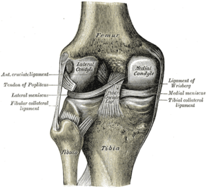
Left knee-joint from behind, showing interior ...
Image via Wikipedia

Common Causes

Knee pain usually results from overuse,but in several cases poor form during physical activity, not warming up or cooling down, or inadequate stretching. Simple causes of knee pain often clear up on their own with self care. Being overweight can put you at greater risk for knee problems.

Other Knee pain causes :-

  • Arthritis — including rheumatoid, osteoarthritis, and gout, or other connective tissue disorders like lupus.
  • Bursitis — inflammation from repeated pressure on the knee (like kneeling for long periods of time, overuse, or injury).
  • Tendinitis — a pain in the front of your knee that gets worse when going up and down stairs or inclines. Happens to runners, skiers, and cyclists.
  • Baker’s cyst — a fluid-filled swelling behind the knee that may accompany inflammation from other causes, like arthritis. If the cyst ruptures, pain in the back of your knee can travel down your calf.
  • Torn cartilage (a meniscus tear) — can cause pain on the inside or outside of the knee joint.
  • Torn ligament (ACL tear) — can cause pain and instability of the knee.
  • Strain or sprain — minor injuries to the ligaments caused by sudden or unnatural twisting.
  • Dislocation of the kneecap.
  • Infection in the joint.
  • Knee injuries — can cause bleeding into your knee, which worsens the pain.
  • Hip disorders — may cause pain that is felt in the knee. For example, iliotibial band syndrome is injury to the thick band that runs from your hip to the outside of your knee.

Less common conditions that can lead to knee pain include the following:

Home Care

Many causes of knee pain, especially those related to overuse or physical activity, respond well to self-care:

  • Rest and avoid activities that aggravate the pain, especially weight bearing activities.
  • Apply ice. First, apply it every hour for up to 15 minutes. After the first day, apply it at least 4 times per day.
  • Keep your knee elevated as much as possible to bring any swelling down.
  • Gently compress the knee by wearing an ace bandage or elastic sleeve. Either can be purchased at most pharmacies. This may reduce swelling and provide support.
  • Take acetaminophen for pain or ibuprofen for pain and swelling.
  • Sleep with a pillow underneath or between your knees

YOGA EXERCISE

Several kind of knee pain problem can be cured through yoga exercise under some Yoga Expart.

css.php