Categories
Ailmemts & Remedies Pediatric

Plagiocephaly

Definition:
The skull isn’t perfectly smooth – it’s covered with lumps, dips and some flatter areas. But sometimes a large area of flattening distorts the skull, making it look parallelogram-shaped. This is known as plagiocephaly.

The most common form is positional plagiocephaly. It occurs when a baby’s head develops a flat spot due to pressure on that area. Babies are vulnerable because their skull is soft and pliable when they’re born.

CLICK & SEE THE PICTURES

Positional plagiocephaly typically develops after birth when babies spend time in a position that puts pressure on one part of the skull. Because babies spend so much time lying on their back, for example, they may develop a flat spot where their head presses against the mattress.

Starting in the early 1990s, parents were told to put their babies to sleep on their back to reduce the risk of SIDS. While this advice has saved thousands of babies’ lives, experts have noticed a fivefold increase in misshapen heads since then.

More rarely, babies develop positional plagiocephaly when movement in the uterus is constricted for some reason – because their mother is carrying more than one baby, for example. It can also happen to breech babies who get wedged under their mother’s ribs.

Another type of plagiocephaly is craniosynostosis, a birth defect in which the joints between the bones of the skull close early. Babies born with craniosynostosis need surgery to allow their brain to grow properly.

Symptoms
Plagiocephaly may become apparent at different ages, depending on the cause. Some babies are born with a flat head (this may be a temporary deformity due to the baby’s passage down the birth canal), while others develop it later as the bones of the skull fuse. The abnormal shape can best be seen if you look down on the baby’s head from above.

Signs of plagiocephaly include:
CLICK & SEE THE PICTURES
•Parallelogram-shaped skull when viewed from above
•Flattening on one side at the back of the head, with a compensatory protrusion or bulge in the forehead on the same side
•Eyes appearing to have unequal positioning
•A bald spot on flattened side (may be asymmetrical)

To learn more you may click to see :Plagiocephaly, Brachycephaly, Brachycephaly with Plagiocephaly and Scaphocephaly

Causes & Risk Factors:
A baby’s skull is very soft and can be forced to grow in different directions fairly easily. When the skull is kept in one particular position for long periods – because the baby is sleeping in a set position (such as on his back) or because muscles attached to the skull go into spasm (known as torticollis) – areas of the skull may be squashed or pulled flat. This is known as positional or deformation plagiocephaly. It generally gets better by itself over time.

Other factors that increase the risk of plagiocephaly include a multiple birth pregnancy (as the babies ‘squash together’ in the womb), prematurity, poor muscle tone and a condition known as oligohydramnios, where there’s insufficient fluid in the womb to cushion the baby.

In the US at least, plagiocephaly has become more common in recent years. Statistics show that while one in 300 healthy infants was affected in 1992, by 1999 one in 60 had the condition.

This increase is thought to be due to the Back to Sleep campaign, designed to reduce the number of sudden infant deaths (cot deaths).

It’s possible to prevent positional plagiocephaly by changing your baby’s resting position frequently. Your baby still needs to be laid on their back to sleep, but try to alternate the position of the head and encourage them to spend time on their tummy while they’re awake and supervised.

Switch between putting them in a sloping chair, car seat or sling, or on a flat surface, so there’s no constant pressure on one area of the skull.Prolonge keeping the baby in the carseat is  very dengerous for the babies.

Plagiocephaly may also be caused by the bones of the skull joining together abnormally early. These bones normally grow together slowly so the skull expands in all directions. But if some fuse too soon (craniosynostosis), that part of the skull can’t grow in the way it should, pulling the head out of shape. This may occur in isolation, or as part of a genetic syndrome such as Apert syndrome or Crouzon syndrome.

Many vaginally delivered babies are born with an oddly shaped head caused by the pressure of passing through the birth canal. This usually corrects itself within about six weeks. But if your baby’s head hasn’t rounded out by age 6 weeks – or if you first notice that your baby has a flat spot on her skull after 6 weeks of age – it’s probably a case of positional plagiocephaly.

Plagiocephaly shows up most often in babies who are reported to be “good sleepers,” babies with unusually large heads, and babies who are born prematurely and have weak muscle tone.

Babies with torticollis can also develop a flat spot on their skull because they often sleep with their head turned to one side. Torticollis occurs when a tight or shortened muscle on one side of the neck causes the chin to tilt to the other side. Premature babies are especially prone to torticollis

Diagnosis:
Most often, your child’s doctor can make the diagnosis of positional plagiocephaly simply by examining your child’s head, without having to order lab tests or X-rays. The doctor will also note whether regular repositioning of your child’s head during sleep successfully reshapes the child’s growing skull over time, whereas craniosynostosis, on the other hand, typically worsens over time.

If there’s still some doubt, X-rays or a CT scan of the head will show your child’s doctor if the skull bones are normally separated or if they fused together too soon. If the bones aren’t fused, the doctor will probably rule out craniosynostosis and confirm that the child has positional plagiocephaly.

It’s important the type and cause of plagiocephaly are determined, as each requires different treatment. X-rays, CT scans and other tests may be needed to confirm diagnosis.

Treatment:
The condition will sometimes improve as the baby grows, but in many cases, treatment can significantly improve the shape of a baby’s head. Initially, treatment usually takes the form of reducing the pressure on the affected area through repositioning of the baby onto his or her tummy for extended periods of time throughout the day. Other treatments include repositioning the child’s head throughout the day so that the rounded side of the head is placed dependent against the mattress, repositioning cribs and other areas that infants spend time in so that they will have to look in a different direction to see their parents, or others in the room, repositioning mobiles and other toys for similar reasons, and avoiding extended time sleeping in car-seats (when not in a vehicle), bouncy seats, or other supine seating which is thought to exacerbate the problem. If the child appears to have discomfort or cries when they are repositioned, they may have a problem with the neck.  If this is unsuccessful, treatment using a cranial remoulding orthosis (baby helmet) can help to correct abnormal head shapes. These helmets are used to treat deformational plagiocephaly, brachycephaly, scaphocephaly and other head shape deformities in infants 3-18 months of age. For years, infants have been successfully treated with cranial remolding orthoses. A cranial remolding orthoses (helmet) provides painless total contact over the prominent areas of the skull and leaves voids over the flattened areas to provide a pathway for more symmetrical skull growth. Treatment generally takes 3-4 months, but varies depending on the infant’s age and severity of the cranial asymmetry.

Prognosis:
There are some beginning studies that indicate that babies with plagiocephaly tend to have learning difficulties later on in school, however these studies are still early, and do not yet represent a scientific consensus. Other more complete studies suggest that there is no evidence to suggest that plagiocephaly is harmful to brain development, vision, or hearing

Prevention:

To successfully prevent Plagiocephaly, prenatal education on skull deformation   is crucial.
Being aware of preventative measures can help reduce the chance your child will develop positional plagiocephaly. Different repositioning techniques and adequate Tummy Time are keys to prevention and also help your baby meet developmental milestones.

Disclaimer: This information is not meant to be a substitute for professional medical advise or help. It is always best to consult with a Physician about serious health concerns. This information is in no way intended to diagnose or prescribe remedies.This is purely for educational purpose

Resources:
http://www.bbc.co.uk/health/physical_health/conditions/plagiocephaly2.shtml
http://en.wikipedia.org/wiki/Plagiocephaly
http://www.babycenter.com/0_plagiocephaly-flat-head-syndrome_1187981.bc
http://www.cranialtech.com/index.php?option=com_content&view=article&id=73&Itemid=76
http://www.cranialtech.com/
http://www.monroeoandp.com/diagnosis_of_plagiocephaly.html

Enhanced by Zemanta
Categories
Ailmemts & Remedies Pediatric

Craniosynostosis

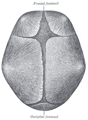
{{Gray's Anatomy plate|The skull at birth, sho...
Image via Wikipedia

[amazon_link asins=’B00SZAM9CE,019511843X,B00ED0SOG0,B01D55D63Y,B0177656AS,B00VQYG526,B01FHAM6L8,B0140HMV0K,B01EG9YT8E’ template=’ProductCarousel’ store=’finmeacur-20′ marketplace=’US’ link_id=’9de2275a-0733-11e7-b98f-e12794808e24′]

Deffinition:
Craniosynostosis is a defect in which one or more of the flexible and fibrous joints (cranial sutures) between the skull bones closes too soon; it occurs before birth or within a few months after birth.  It is a condition in which one or more of the fibrous sutures in an infant skull prematurely fuses. This results in restricted skull and brain growth. Because the brain cannot expand in the direction of the fused suture, it is forced to grow in the direction of the open sutures, often resulting in an abnormal head shape and facial features. Some cases of craniosynostosis may result in increased pressure on the brain and developmental delays.  It can be the result of an inherited syndrome or sporadic. In sporadic cases, the cause is unknown.Craniosynostosis can occur alone or as part of a syndrome of craniofacial defects.
CLICK & SEE THE PICTURES
Craniosynostosis is a very rare occurrence. The sagittal form of the disorder, in which the sagittal suture closes prematurely, is the most common form of craniosynostosis, occurring in three to five of every 1,000 babies, typically males. The frequencies of the various types of craniosynostosis are 50–60% sagittal, 20–30% coronal, 4–10% metopic, and 2–4% lambdoid.It is estimated that craniosynostosis affects 1 in 2,000 live births.

Description:
The skull of a newborn is composed of five bones that are held together by the fibrous sutures positioned at the front, top, sides, and back of the skull. By remaining open, the sutures allow the skull to normally expand in all directions as the brain is growing.

The premature closing of one or more of these cranial sutures stops the normal capacity of the skull to expand in early childhood. As not all of the cranial sutures will close, the skull expands in the areas that are still flexible. This results in an abnormally shaped skull or face. The forehead may be very pronounced and inclined forward. Viewed from above, the skull may be more rectangular in shape rather than oval.

Other forms of craniosynostosis include coronal craniosynostosis (affecting the coronal suture that crosses the top of the skull from temple to temple), metopic craniosynostosis (affecting the metopic suture of the forehead), sagittal craniosynostosis (affecting the sagittal suture that unites the two parietal bones), and lambdoidal craniosynostosis (affecting the lambdoid suture between the occipital and parietal bones of the skull).

Causes:
The cause of craniosynostosis is unknown.However, there’s a hereditary component to craniosynostosis when it occurs with certain genetic syndromes such as Apert’s syndrome and Crouzon syndrome.It is usually caused by a genetic mutation. Mutations in several genes (designated TWIST, FGFR-1, FGFR-2, and FGFR-3) have been linked with craniosynostosis. In particular, the protein encoded for by TWIST is critical in the initiation and maintenance of the cranial suture process. As of 2004, the favored hypothesis is that the protein that normally functions to ensure that the formation of the cranial sutures occurs at the right time in development somehow goes awry and causes premature fusion of the bones of the brain.

Symptoms:
Infant’s skull has seven bones. Normally, these bones don’t fuse until around age 2, giving your baby’s brain time to grow. Joints called cranial sutures, made of strong, fibrous tissue, hold these bones together. In the front of your baby’s skull, the sutures intersect in the large soft spot (fontanel) on the top of your baby’s head. Normally, the sutures remain flexible until the bones fuse.
Joints called cranial sutures, made of strong, fibrous tissue, hold the bones of your baby’s skull together until the bones fuse, normally around age 2. Until then, the sutures intersect at the fontanels, the soft spots on your baby’s head. The largest of the four fontanels is at the front of the skull (anterior); the next largest is at the back (posterior). Each side of the skull has a tiny fontanel.
Craniosynostosis signs in general
Signs of craniosynostosis include:

#A misshapen skull, with the shape depending on which of the cranial sutures are affected
#Abnormal feeling “soft spot” (fontanel) on your baby’s skull
#Early disappearance of the fontanel
#Slow or no growth of head as your baby grows
#Development of a raised, hard ridge along affected sutures
#Increased pressure within the skull (intracranial pressure)
The signs of craniosynostosis may not be noticeable at birth, but they will become apparent during the first few months of your baby’s life.

Main categories and characteristics :-
There are two categories of craniosynostosis:

1.Primary craniosynostosis. If your baby has primary craniosynostosis, usually one, but sometimes more, of the cranial sutures become rigid, fusing the connecting bones and inhibiting the brain’s ability to grow normally.
2.Secondary craniosynostosis. With secondary craniosynostosis, which occurs more frequently than the primary type, your infant’s brain stops growing, usually due to an underlying hereditary syndrome, allowing the sutures to fuse prematurely. Secondary craniosynostosis is often associated with facial deformities and developmental delays.

Common types and characteristics :-
The most common types of craniosynostosis are:

#Sagittal synostosis (scaphocephaly). Premature fusion of the suture at the top of the head (sagittal suture) forces the head to grow long and narrow, rather than wide. Scaphocephaly is the most common type of craniosynostosis, and it is more common in boys.
#Coronal synostosis (anterior plagiocephaly). Premature fusion of one of the sutures that run from each ear to the sagittal suture on top of the head may force your baby’s forehead to flatten on the affected side. It also may raise the eye socket and cause a deviated nose and slanted skull. This second most common type of craniosynostosis is more common in girls. Untreated, it may lead to vision loss on the affected side (amblyopia).
#Bicoronal synostosis (brachycephaly).
When both of the coronal sutures fuse prematurely, your baby may have a flat, elevated forehead and brow.


Rare types and characteristics :-
Two less common types of synostosis are:


#Metopic synostosis (trigonocephaly).
The metopic suture runs from the baby’s nose to the sagittal suture. Premature fusion gives the scalp a triangular appearance.
#Lambdoid synostosis (posterior plagiocephaly)
. This rare form of craniosynostosis involves the lambdoid suture, which runs across the skull near the back of the head. It may cause flattening of the head on the affected side.

Misshapen head may not mean craniosynostosis
:-
A misshapen head doesn’t always indicate craniosynostosis. For example, if the back of your baby’s head appears flattened, it could be the result of your baby’s sleeping on his or her back.

The “Back to Sleep” campaign, co-sponsored by the National Institute of Child Health & Human Development, encourages parents to put healthy babies to sleep on their backs to reduce the risk of sudden infant death syndrome (SIDS). The campaign has resulted in more cases of misshapen heads. In this case, the flattening is a result of positional molding, not craniosynostosis.

Risk Factors:
Babies with craniosynostosis, particularly those with an underlying syndrome, may develop increased intracranial pressure. Their skulls don’t expand enough to make room for their growing brain.

If untreated, increased intracranial pressure can cause these conditions:

#Blindness
#Seizures
#Brain damage
#Death, in very rare instances
In addition, facial deformities that affect the middle of your child’s face may cause:

#Upper airway obstructions, compromising your baby’s ability to breathe
#Permanent head deformity

Diagnosis:
Your doctor will feel your baby’s head for abnormalities such as suture ridges, perform a physical exam and look for facial deformities. In addition, your doctor may order other tests, including:

#Imaging studies. X-rays or a computerized tomography (CT) scan of your baby’s skull will show whether any sutures have fused. Fused sutures are identifiable by their absence, because they’re invisible once fused, and by the ridging of the suture line.

#Genetic testing. If your doctor suspects your baby’s misshapen skull is caused by an underlying hereditary syndrome, genetic testing may help identify the syndrome. Genetic tests usually require a blood sample.
Depending on what type of abnormality your doctor is looking for, your baby may be required to give a hair, skin or other tissue sample, such as cells from the inside of the cheek. The sample is then sent to a lab for analysis.

Treatments:-

Surgery is the common treatment for craniosynostosis:
But for Mild cases of craniosynostosis — those that involve only one suture and no underlying syndrome — may require no treatment. Skull abnormalities may become less obvious as your infant grows and develops hair.

Surgery
:-
For other infants, surgery, usually during infancy, is the primary treatment for craniosynostosis. However, the type and timing of surgery depend on the type of synostosis and whether there’s an underlying syndrome.

The purpose of surgery is to relieve pressure on the brain, create room for the brain to grow normally and improve your child’s appearance. A team that includes a specialist in surgery of the head and face (craniofacial surgeon) and a specialist in brain surgery (neurosurgeon) often performs the surgery.

#Traditional surgery. The surgeon makes an incision in your infant’s scalp and cranial bones, then reshapes the affected portion of the skull. Sometimes plates and screws, often made of material that is absorbed over time, are used to hold the bones in place. Surgery, which is performed during general anesthesia, may take up to seven hours.

After surgery, your infant remains in the hospital for at least three days. Some temporary facial swelling is common after surgery. Complications are rare. Some children may require a second surgery later because the synostosis recurs. Also, children with facial deformities often require future surgeries to reshape their faces.

#Endoscopic surgery
. This less invasive form of surgery isn’t an option for everyone. But in certain cases, the surgeon may use a lighted tube (endoscope) inserted through one or two small scalp incisions over the affected suture. The surgeon then opens the suture to enable your baby’s brain to grow normally. Endoscopic surgery usually takes about an hour, causes less swelling and blood loss, and shortens the hospital stay, often to one day after surgery.

If your baby has an underlying syndrome, your doctor may recommend regular follow-up visits after surgery to monitor head growth and check for increased intracranial pressure.

Recovery and rehabilitation:-
Regardless of the type of surgery performed to correct the defects associated with craniosynostosis, the child will be restricted from vigorous activity or rough play while healing. The protective helmet is required for children after endoscopic strip craniectomy, while permanent plates inserted during other corrective surgeries eliminate the need for the helmet. Children who have had surgery to repair craniosynostosis will continue to need periodic examination by the surgeon until approximately age 18, when the skull has grown to its adult size and shape.

Prognosis:-
The outlook for a complete recovery for a child with craniosynostosis depends on whether just one suture is involved or whether multiple sutures have closed. Also, the presence of other abnormalities can lessen the confidence of a satisfactory outcome. Without surgical intervention, craniosynostosis can lead to increased brain pressure, delayed mental development, mental retardation, seizures, or blindness. After surgery is accomplished, the prognosis is excellent.

Disclaimer: This information is not meant to be a substitute for professional medical advise or help. It is always best to consult with a Physician about serious health concerns. This information is in no way intended to diagnose or prescribe remedies.This is purely for educational purpose.

Resources:
http://en.wikipedia.org/wiki/Craniosynostosis
http://www.mayoclinic.com/health/craniosynostosis/
http://www.answers.com/topic/craniosynostosis

Categories
Ailmemts & Remedies Pediatric

Craniosynostosis

Alternative Names:Premature closure of sutures

Definition:
To allow for rapid brain growth, your baby’s skull cap is made up of five bones, held together by a fibrous-like material called “sutures”. Normally, they remain open as long as the brain grows, which gives the brain room needed to grow in all directions.

If any of the sutures close or fuse before the brain has finished growing, the condition known as craniosynostosis occurs. Other terms used to describe this problem include: synostosis, or cranial stenosis. Premature closure can take place before birth or at any time until the brain stops growing.

CLICK & SEE THE PICTURES

Because the brain is growing normally, it will take the path of least resistance which will result in a misshapen head or face.
The skull of an infant is made up of free-floating bones separated by fibers called sutures. This arrangement allows the infant’s head to pass through the birth canal and also enables the skull to grow with the brain in early infancy. Craniosynostosis is a birth defect of the brain characterized by the premature closure of one or more of the fibrous joints between the bones of the skull (called the cranial sutures) before brain growth is complete.Closure of a single suture is most common.
Skull of a newborn

The abnormally shaped skull that results is due to the brain not being able to grow in its natural shape because of the closure. Instead it compensates with growth in areas of the skull where the cranial sutures have not yet closed. The condition can be gene-linked, or caused by metabolic diseases, such as rickets or an overactive thyroid. Some cases are associated with other disorders such as microcephaly (abnormally small head) and hydrocephalus (excessive accumulation of cerebrospinal fluid in the brain).

Causes :
The cause of craniosynostosis is unknown. Which suture is involved determines the abnormal shape of the head.

A person’s genes may play a role in craniosynostosis. The hereditary form often occurs with other defects that can cause seizures, diminished intellectual capacity, and blindness. Genetic disorders commonly associated with craniosynostosis include Crouzon, Apert, Carpenter, Chotzen, and Pfeiffer syndromes.

However, most cases of craniosynostosis occur in a family with no history of the condition and children with craniosynostosis are otherwise healthy and have normal intelligence.

There are different types of craniosynostosis. Sagittal synostosis (scaphocephaly) is the most common type. It affects the main (sagittal) suture on the very top of the head. The early closing forces the head to grow long and narrow, instead of wide. Babies with this type of craniosynostosis tend to have a broad forehead. It is more common in boys than girls.

Frontal plagiocephaly is the next most common form. It is the closure of one side of the suture that runs from ear to ear on the top of the head. It is more common in girls.

Metopic synostosis is a rare form of craniosynostosis that affects the suture close to the forehead. The child’s head shape may be described as trigonocephaly, and the deformity may range from mild to severe.

Symptoms :

The first sign of craniosynostosis is changes in the shape of the head and face . Other features can include :-

*a full or bulging fontanelle (soft spot located on the top of the head)

*sleepiness (or less alert than usual)

*scalp veins may be very noticeable

*increased irritability

*high-pitched cry

*poor feeding

*projectile vomiting

*increasing head circumference

*seizures

*bulging eyes and an inability of the child to look upward with the head facing forward

*developmental delays

Diagnosis:
Craniosynostosis may be congenital (present at birth) or may be observed later, during a physical examination. The diagnosis is made after a thorough physical examination and after diagnostic testing. During the examination, a measurement of the circumference of your child’s head is taken and plotted on a graph to identify normal and abnormal ranges. In addition, the following tests may be ordered:

x-rays of the head – a diagnostic test that uses invisible electromagnetic energy beams to produce images of internal tissues and bones of the head onto film.
computerized tomography scan (Also called a CT or CAT scan.) of the head – a diagnostic imaging procedure that uses a combination of x-rays and computer technology to produce cross-sectional images (often called slices), both horizontally and vertically, of the head.

Treatment:

Treatment for craniosynostosis generally consists of surgery to relieve pressure on the brain and the cranial nerves. The goal of treatment is to reduce the pressure in the head and correct the deformities of the face and skull bones. The optimal time to perform surgery is before the child is 1 year of age since the bones are still very soft and easy to work with. Surgery may be necessary at a much earlier age depending upon the severity of the condition. For some children with less severe problems, cranial molds can reshape the skull to accommodate brain growth and improve the appearance of the head.

Possible Complications :

Craniosynostosis results in head deformity that can be severe and permanent if it is not corrected. Increased intracranial pressure, seizures, and developmental delay can occur.

Prognosis:
The prognosis for craniosynostosis varies depending on whether single or multiple cranial sutures are involved or other abnormalities are present. The prognosis is better for those with single suture involvement and no associated abnormalities. The key to treating craniosynostosis is early detection and treatment. Some forms of craniosynostosis can affect the brain and development of a child. The degree of the problems is dependent on the severity of the craniosynostosis, the number of sutures that are fused, and the presence of brain or other organ system problems that could affect the child.

Prevention:
Be sure to bring your child to well-child visits, so your pediatrician can routinely chart the growth of your infant’s head over time. This will help identify the problem early if it occurs.Persons with hereditary craniosynostosis might consider genetic counseling.

When to Contact a Medical Professional:
Call your health care provider if you think your child’s head has an unusual shape. A referral to a pediatric neurologist or neurosurgeon should follow.
Click to see also:->
Abnormal Head Shapes in Infants
My Baby’s Head is Flat!

Disclaimer: This information is not meant to be a substitute for professional medical advise or help. It is always best to consult with a Physician about serious health concerns. This information is in no way intended to diagnose or prescribe remedies.This is purely for educational purpose

Resources:
KidsGrowth.com
http://www.nlm.nih.gov/medlineplus/ency/article/001590.htm
http://www.craniosynostosis.net/what-is-craniosynostosis.shtml

Enhanced by Zemanta
css.php