Categories
Ailmemts & Remedies

Knee Problems and Injuries

ntroduction:
Most people have had a minor knee problem at one time or another. Most of the time our body movements do not cause problems, but it’s not surprising that symptoms develop from everyday wear and tear, overuse, or injury. Knee problems and injuries most often occur during sports or recreational activities, work-related tasks, or home projects..
The knee is the largest joint in the body. The upper and lower bones of the knee are separated by two discs (menisci). The upper leg bone (femur) and the lower leg bones (tibia and fibula) are connected by ligaments, tendons, and muscles. The surface of the bones inside the knee joint is covered by articular cartilage, which absorbs shock and provides a smooth, gliding surface for joint movement. See an illustration of the structures of the knee .

Although a knee problem is often caused by an injury to one or more of these structures, it may have another cause. Some people are more likely to develop knee problems than others. Many jobs, sports and recreation activities, getting older, or having a disease such as osteoporosis or arthritis increase your chances of having problems with your knees.

Sudden (acute) injuries

Injuries are the most common cause of knee problems. Sudden (acute) injuries may be caused by a direct blow to the knee or from abnormal twisting, bending the knee, or falling on the knee. Pain, bruising, or swelling may be severe and develop within minutes of the injury. Nerves or blood vessels may be pinched or damaged during the injury. The knee or lower leg may feel numb, weak, or cold; tingle; or look pale or blue. Acute injuries include:

•Sprains, strains, or other injuries to the ligaments and tendons that connect and support the kneecap.
•A tear in the rubbery cushions of the knee joint (meniscus).
•Ligament tears. The medial collateral ligament (MCL) is the most commonly injured ligament of the knee.
•Breaks (fracture) of the kneecap, lower portion of the femur, or upper part of the tibia or fibula. Knee fractures are most commonly caused by abnormal force, such as a falling on the knee, a severe twisting motion, severe force that bends the knee, or when the knee forcefully hits an object.
•Kneecap dislocation. This type of dislocation occurs more frequently in 13- to 18-year-old girls. Pieces of bone or tissue (loose bodies) from a fracture or dislocation may get caught in the joint and interfere with movement.
•Knee joint dislocation. This is a rare injury that requires great force. It is a serious injury and requires immediate medical care.

CLICK & SEE
Overuse injuries

Overuse injuries..Overuse injuries
Overuse injuries occur with repetitive activities or repeated or prolonged pressure on the knee. Activities such as stair climbing, bicycle riding, jogging, or jumping stress joints and other tissues and can lead to irritation and inflammation. Overuse injuries include:

•Inflammation of the small sacs of fluid that cushion and lubricate the knee (bursitis).
•Inflammation of the tendons (tendinitis) or small tears in the tendons (tendinosis).
•Thickening or folding of the knee ligaments (Plica syndrome).
•Pain in the front of the knee from overuse, injury, excess weight, or problems in the kneecap (patellofemoral pain syndrome).
•Irritation and inflammation of the band of fibrous tissue that runs down the outside of the thigh (iliotibial band syndrome).
Conditions that may cause knee problems
Problems not directly related to an injury or overuse may occur in or around the knee.

•Osteoarthritis (degenerative joint disease) may cause knee pain that is worse in the morning and improves during the day. It often develops at the site of a previous injury. Other types of arthritis, such as rheumatoid arthritis, gout, and lupus, also can cause knee pain, swelling, and stiffness.
•Osgood-Schlatter disease causes pain, swelling, and tenderness in the front of the knee below the kneecap. It is especially common in boys ages 11 to 15.
•A popliteal (or Baker’s) cyst causes swelling in the back of the knee.
•Infection in the skin (cellulitis), joint (infectious arthritis), bone (osteomyelitis), or bursa (septic bursitis) can cause pain and decreased knee movement.
•A problem elsewhere in the body, such as a pinched nerve or a problem in the hip, can sometimes cause knee pain.
•Osteochondritis dissecans causes pain and decreased movement when a piece of bone or cartilage or both inside the knee joint loses blood supply and dies.


.Treatment

Treatment for a knee problem or injury may include first aid measures, rest, bracing, physical therapy, medicine, and in some cases surgery. Treatment depends on the location, type, and severity of the injury as well as your age, health condition, and activity level (such as work, sports, or hobbies).

YOU MAY CLICK TO CHECK SYMPTOMS OF EMERGENCY & TAKE ACTION ACCORDINGLY:

Home Treatment
Home treatment may help relieve pain, swelling, and stiffness.
home treatment
•Rest and protect an injured or sore area. Stop, change, or take a break from any activity that may be causing your pain or soreness. When resting, place a small pillow under your knee.
•Ice will reduce pain and swelling. Apply ice or cold packs immediately to prevent or minimize swelling. Apply the ice or cold pack for 10 to 20 minutes, 3 or more times a day.
*For the first 48 hours after an injury, avoid things that might increase swelling, such as hot showers, hot tubs, hot packs, or alcoholic beverages.
*After 48 to 72 hours, if swelling is gone, apply heat and begin gentle exercise with the aid of moist heat to help restore and maintain flexibility. Some experts recommend alternating between heat and cold treatments.
•Compression, or wrapping the injured or sore area with an elastic bandage (such as an Ace wrap), will help decrease swelling.
*Don’t wrap it too tightly, since this can cause more swelling below the affected area. Loosen the bandage if it gets too tight. Signs that the bandage is too tight include numbness, tingling, increased pain, coolness, or swelling in the area below the bandage.
*Don’t expect the bandage to protect or stabilize a knee injury.
*Talk to your doctor if you think you need to use a wrap for longer than 48 to 72 hours; a more serious problem may be present.
•Elevate the injured or sore area on pillows while applying ice and anytime you are sitting or lying down. Try to keep the area at or above the level of your heart to help minimize swelling.

.
•Reduce stress on your sore knee (until you can get advice from your doctor):
*Use a cane or crutch in the hand opposite your painful knee.
*Use two crutches, keeping weight off the leg with the sore knee. You can get canes or crutches from most pharmacies. Crutches are recommended if a cane causes you to walk with a limp.
•Gently massage or rub the area to relieve pain and encourage blood flow. Do not massage the injured area if it causes pain.

.
•Try the following exercises to maintain flexibility:
*Hamstring stretch *Knee-to-chest exercise •Avoid high-impact exercise, such as running, skiing, snowboarding, or playing tennis, until your knee is no longer painful or swollen.
Do not smoke.
Smoking slows healing because it decreases blood supply and delays tissue repair. For more information, see the topic Quitting Tobacco Use.

Medicine you can buy without a prescription Try a nonprescription medicine to help treat your fever or pain: –

.•Acetaminophen, such as Tylenol or Panadol
Note: Do not use a nonsteroidal anti-inflammatory medicine, such as ibuprofen or aspirin, for the first 24 hours after an injury. Using these medicines may increase the time it takes your blood to clot and cause more severe bruising from bleeding under the skin.

.
•Nonsteroidal anti-inflammatory drugs (NSAIDs):
*Ibuprofen, such as Advil or Motrin
*Naproxen, such as Aleve or Naprosyn
•Aspirin (also a nonsteroidal anti-inflammatory drug), such as Bayer or Bufferin
Talk to your child’s doctor before switching back and forth between doses of acetaminophen and ibuprofen. When you switch between two medicines, there is a chance your child will get too much medicine.

Safety tips Be sure to follow these safety tips when you use a nonprescription medicine:
•Carefully read and follow all directions on the medicine bottle and box.
•Do not take more than the recommended dose.
•Do not take a medicine if you have had an allergic reaction to it in the past.
•If you have been told to avoid a medicine, call your doctor before you take it.
•If you are or could be pregnant, do not take any medicine other than acetaminophen unless your doctor has told you to.
•Do not give aspirin to anyone younger than age 20 unless your doctor tells you to.

.

Symptoms to Watch For During Home Treatment
Use the Check Your Symptoms section to evaluate your symptoms if any of the following occur during home treatment:

•Signs of infection develop.
•Numbness, tingling, or weakness develops.
•Your knee, lower leg, or foot becomes pale or cool or looks blue.
•Symptoms do not improve with home treatment.
•Symptoms become more severe or frequent.

Prevention
The following tips may prevent knee problems.


General prevention tips

•Wear your seat belt in a motor vehicle.
•Don’t carry objects that are too heavy. Use a step stool. Do not stand on chairs or other unsteady objects.
•Wear knee guards during sports or recreational activities, such as roller-skating or soccer.
•Stretch before and after physical exercise, sports, or recreational activities to warm up your muscles.
•Use the correct techniques or positions during activities so that you do not strain your muscles.
•Use equipment appropriate to your size, strength, and ability. Avoid repeated movements that can cause injury. In daily routines or hobbies, look at activities in which you make repeated knee movements.
•Consider taking lessons to learn the proper technique for sports. Have a trainer or person who is familiar with sports equipment check your equipment to see if it is well suited for your level of ability, body size, and body strength.
•If you feel that certain activities at your workplace are causing pain or soreness from overuse, call your human resources department for information on other ways of doing your job or to talk about using different equipment.

.
Tips specific to the knee
•Keep your knees and the muscles that support them strong and flexible. Warm up before activities. Try the following stretches:
?Hamstring stretch ?Knee-to-chest exercise ?Calf stretch ?Straight-leg raises •Avoid activities that stress your knees, such as deep knee bends or downhill running.
•Wear shoes with good arch supports.
•Do not wear high-heeled shoes.
•When playing contact sports, wear the right shoes that are made for the surface you are playing or running on, such as a track or tennis court.
•Replace running shoes every 300 to 500 miles (480 to 800 kilometers).


Tips specific to female athletes

Sports trainers recommend training programs that help women learn to run, jump, and pivot with knees bent to avoid knee injuries. In sports such as soccer, basketball, and volleyball, women who bend their knees and play low to the ground have fewer knee injuries than women who run and pivot with stiff legs.

Knee brace use
Some people use knee braces to prevent knee injuries or after a knee injury. There are many types of knee braces, from soft fabric sleeves to rigid, metal hinged braces, that support and protect the knee. If your doctor has recommended the use of a knee brace, follow his or her instructions. If you are using a knee brace to help prevent problems, follow the manufacturer’s instructions for use.

Keep bones strong
•Eat a nutritious diet with enough calcium and vitamin D, which helps your body absorb calcium. Calcium is found in dairy products, such as milk, cheese, and yogurt; dark green, leafy vegetables, such as broccoli; and other foods. For more information, see the topic Healthy Eating.
•Exercise and stay active. It is best to do weight-bearing exercise, such as walking, jogging, stair climbing, dancing, or lifting weights, for 45 to 60 minutes at least 4 days a week. Weight-bearing exercises stimulate new bone growth by working the muscles and bones against gravity. Exercises that are not weight-bearing, such as swimming, are good for your general health but do not stimulate new bone growth. Talk to your doctor about an exercise program that is right for you. Begin slowly, especially if you have been inactive. For more information, see the topic Fitness.
•Avoid drinking more than one alcoholic drink per day. People who drink more than this may be at higher risk for weakening bones (osteoporosis). Alcohol use also increases your risk of falling and breaking a bone.
•Stop or do not begin smoking. Smoking puts you at a much higher risk for developing osteoporosis. It also interferes with blood supply and healing. For more information, see the topic Quitting Tobacco Use.
Possible abuse
Bruises are often the first sign of abuse. Seek help if:

•You suspect abuse. Call your local child or adult protective agency, police, or a doctor, nurse, or counselor.
•You or someone you know is a victim of violence.
•You have trouble controlling your anger with a child or other person in your care. Resources are available for help.

Source: Health.com

Reblog this post [with Zemanta]
Categories
Health & Fitness

Healthy Knee is Friendly Indeed

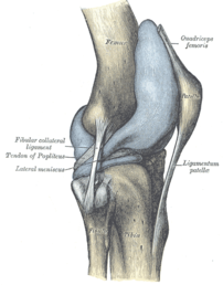
Capsule of right knee-joint (distended). Later...
Image via Wikipedia

[amazon_link asins=’B01A5Q746G,B01LX86E55,B00KYDCP7O,0979303605,1579548717,B01K3CXMB8,B01MRF7JRM,B01CTRKP2O,B071Y32649′ template=’ProductCarousel’ store=’finmeacur-20′ marketplace=’US’ link_id=’8f3c0421-79a1-11e7-9ce0-051296b748ee’]

Whether you are a ballerina, mountaineer, a weekend gladiator or just an office warrior, learn to protect your knees because the knee is a critical link in the kinetic chain that allows you to walk on two feet.
….....CLICK & SEE

Anit Ghosh, a former national footballer, suffered a career-threatening injury to his knee ligament five years ago. Regular and diligent post-injury rehabilitation work under the author’s guidance helped him gradually return to competitive football. Today, he turns out for Mohammedan Sporting and has learned to manage a problematic knee.

In  therapy practice, over half the ladies and about one in 10 men complain of knee pain. After back pain, knee pain is the most common cause of disability and time lost from work or training.

The knee is the largest joint in the human body and is formed by the articulation of three bones, the lower end of the thighbone (femur), the upper end of the shinbone (tibia) and the kneecap (patella). It may appear like a simple hinge, but besides the routine functions of bending and straightening, the knee joint performs a host of complex functions — it slides, glides, pivots, rolls and rotates — sometimes sequentially and at other times simultaneously. All these movements make the knee joint very vulnerable to shearing forces and dependant on good functional stability from the surrounding soft tissue network of ligaments, tendons and the two menisci, tough crescent-shaped cushions within the joint. In addition to the above, the knee joint also includes small, fluid-filled membranous sacs lying between the ligaments or skin, and the bones to provide smooth and frictionless gliding, like ball bearings in a machine. Furthermore, the entire articular surfaces, i.e. those that rub against one another, are covered with a tough, rubbery slippery tissue called cartilage.

Of these parts mentioned above can be a source of joint pain. Sometimes, knee pain can be caused by poor body mechanics and tight muscles elsewhere in the body and can easily be corrected by a slight alteration in gait and mechanics. For example, poor flexibility around the ankle and hip can transfer a lot of shearing forces onto the knee even though pathologically the knee is normal. The knee then is merely the “site” of the pain. The villain or “source” of pain may lie elsewhere.

The most common causes of knee pain  are described below..>..CLICK & SEE

*One of the most crippling forms of knee ailment is arthritis caused by the degeneration of the cartilage coating. The cartilage has very poor blood supply and consequently nutrient supply and therefore once traumatised, has hardly any chance of healing itself. The inherent nature of the cartilage is a huge limiting factor for arthritis rehabilitation.

*Chondromalacia is the softening or the wearing away of the articular cartilage under the kneecap. The articular cartilage on the inside aspect of the kneecap comes in constant contact with the articular surfaces of the femur during normal knee motion. The knee motion can sometimes become abnormal or faulty due to muscle imbalance or biomechanical misalignment and cause the patella to rub against the femoral surfaces. Repetitive ‘rubbing’ of the surfaces causes chronic inflammation sometimes popularly known as “jumpers knee”.

*One of the most common causes of pain inside the joint is a torn meniscus. The crescent-shaped spongy tissues act as shock absorbers within the joint and when torn, either by injury or degeneration, tends to get caught in the joint, causing pain and instability.

*When the articular cartilage begins fragmenting and eroding due to extreme softening, the underlying bone gets exposed. This is a condition called osteoarthritis.

*Often traumatic injuries or contact sports mishaps cause the ligaments within the knee joint to snap. This is a very painful condition and more often than not, needs surgical correction where the surgeon has to reconstruct the ligament necessitating a long healing period.

WHAT YOU CAN DO TO MANAGE, EVEN PREVENT KNEE PAIN?

*Stretch regularly. Regular stretching of the hip flexors, hip extensors and the iliotibial band (a sheath of muscle lying on the outside of your thigh extending from the hip to the lateral aspect of the knee) will ensure good gait and running mechanics and spare the knee of shearing forces.

*Train with weights. Loading the knee and hips early in life with weights will build density in the bones and prevent erosion in later life.

*Work the hamstrings. The average person has stronger quadriceps compared to the hamstrings. Increase hamstring strength for better muscle balance and correct alignment of the kneecap. This will avoid compression forces within the knee.

*Strengthen the vastus medialis muscle — the muscles lying in the inside aspect of your front thigh. This will help to realign and track the kneecap to its normal pathway.

*Exercise discretion while performing repetitive knee motions like running, skipping, jumping etc. If you must run, learn proper running technique. Let’s face it — nine out of 10 people who visit lifestyle and recreation gyms do not have good technique. Running on the treadmill for these people is sheer disaster!

*If you are an active sort of a person, check with your doctor whether you should supplement with Glucosamine sulphate and Chondroitin. They are known to have shown results in preventing degeneration of the knee joint.

*Avoid knee extensions. The leg extension exercise is treated as a panacea for all sorts of knee ailments by trainers and therapists alike. In reality, open-chain movements like the knee extension exercise is potentially more dangerous than closed-chain movements like the lunge and squat.

*The leg extension movement causes compression between the kneecap and the thighbone and I would recommend even healthy knee-owners to stay far away from it. Choose multi-joint exercises that make the quadriceps and hamstrings work together in unison.

The best exercises for the knee are:

*One-legged squats
*Glute ham raises
*Lunges
*Split squats

Sources: The Telegraph (Kolkata, India)

Reblog this post [with Zemanta]
Categories
Ailmemts & Remedies

Knee Injury

The knee has a simple purpose. It needs to flex (bend) or extend (straighten) to allow the body to perform many activities like running, walking, kicking, and sitting. Imagine standing up from a chair if your knees couldn’t bend.

Knee Anatomy
While there are four bones that come together at the knee, only the femur (thigh bone) and the tibia (shin bone) form the joint itself. The head of the fibula (strut bone on the outside of the leg) provides some stability, and the patella (kneecap) helps with joint function. Movement and weight-bearing occur where the ends of the femur called the femoral condyles match up with the top flat surfaces of the tibia (tibial plateaus).

There are two major muscle groups that are balanced and allow movement of the knee joint. Contracting the quadriceps muscles on the front of the thigh extends the knee, while the hamstring muscles on the back of the thigh flex the knee when they contract. The muscles cross the knee joint and are attached to the tibia by tendons. The quadriceps tendon is a little special, in that it contains the patella within it. The patella allows the quadriceps muscle/tendon unit to work more efficiently. This tendon is renamed the patellar tendon in the area below the kneecap to its attachment to the tibia.

The stability of the knee joint is maintained by four ligaments, thick bands of tissue that stabilize the joint. The medial collateral ligament (MCL) and lateral collateral ligament (LCL) are on the sides of the knee and prevent the joint from sliding sideways. The anterior cruciate ligament (ACL) and posterior cruciate ligament (PCL) form an “X” on the inside of the knee and prevent the knee from sliding back and forth. These limitations on knee movement allow the knee to concentrate the forces of the muscles on flexion and extension.

Inside the knee, there are two shock-absorbing pieces of cartilage called menisci (singular meniscus) that sit on the top surface of the tibia. The menisci allow the femoral condyle to move on the tibial surface without friction, preventing the bones from rubbing on each other.

Bursas surround the knee joint and are fluid filled sacs that cushion the knee during its range of motion. In the front of the knee, there is a bursa between the skin and the kneecap called the prepatellar bursa and another above the kneecap called the suprapatellar bursa (supra=above).

Each part of the anatomy needs to function properly for the knee to work. Acute injury and trauma as well as chronic overuse can both cause inflammation and its accompanying symptoms of pain, swelling, redness and warmth.

Causes:.....
While direct blows will occur, the knee is more susceptible to twisting or stretching injuries, taking the joint through a greater range of motion than it can tolerate.

If the knee is stressed from a specific direction, then the ligament trying to hold it in place against that force can tear. Ligament stretching or tears are called sprains. These sprains are graded as first, second, or third degree based upon how much damage has occurred. Grade one sprains stretch the ligament but don’t tear the fibers; grade two sprains partially tear the fibers, but the ligament remains intact; and grade three tears completely disrupt the ligament.

Twisting injuries to the knee put stress on the cartilage or meniscus and can pinch it between the tibial surface and the edges of the femoral condyle, causing tears.

Injuries of the muscles and tendons surrounding the knee are caused by acute hyperflexion or hyperextension of the knee or by overuse. These injuries are called strains.

There can be inflammation of the bursas (known as bursitis) of the knee that can occur because of direct blows or chronic use and abuse.

Acute knee injuries fall into two groups; those where there is almost immediate swelling in the joint associated with the inability to bend the knee and bear weight, and those where there is discomfort and perhaps localized pain to one side of the knee, but have minimal swelling and minimal effects on walking.

Symptoms:
Acute knee injuries can cause pain and swelling with difficulty bending the knee and weight-bearing.

Longer-term symptoms that point to knee problems will include pain and swelling in addition to other complaints. Inflammation in the joint may be caused by even minor activity. Swelling may be intermittent, brought on by activity, and may gradually resolve as the inflammation decreases.

Pain, too, may come and go and may not occur right away with activity but might be delayed as the inflammation develops. Pain can also be felt with specific activities. Pain while climbing stairs is a symptom of meniscus injury, where the cartilage is being pinched in the joint as it narrows with bending. Pain with walking down stairs suggests patellar pain, where the kneecap is being forced onto the femur.

Giving way, or a feeling of instability, of the knee is a symptom of ligament injury, while popping or grinding in the knee is more associated with cartilage or meniscus tears. “Locking” is the term used when the knee joint refuses to completely straighten, and this is always due to torn cartilage. In this situation, the torn piece of cartilage folds upon itself and doesn’t allow the knee to extend.


Emergent medical care should be sought if, due to injury,

1.there is almost immediate swelling in the knee,
2.if the bones appear deformed,
3.if there is inability to bear weight,
4.if the pain is intolerable,
5.if there is loss of sensation below the injury site
6.or if the foot and ankle turn cold and no pulses can be felt.

Diagnosis:
The initial evaluation by the physician or health care provider will begin with a medical history. Whether the evaluation is occurring immediately after the injury or weeks later, the physician will ask about the mechanism of injury to help isolate what structures in the knee might be damaged. Is the injury due to a direct blow that might suggest a fracture or contusion or bruise? Was it a twisting injury that causes a cartilage or meniscus tear? Was there an injury associated with a planted foot to place stress and potentially tear a ligament?

Further questions will address other symptoms. Was swelling present, and if so, did it occur right away or was it delayed by hours? Did the injury prevent weight-bearing or walking? Does going up or down steps cause pain? Is there associated hip or ankle pain?

Physical examination of the knee begins with inspection, in which the physician will look at the bones and make certain they are where they belong. With fractures of the kneecap or patellar tendon injuries, the kneecap can slide high out of position. Also, patellar dislocations, where the kneecap slides to the outside or lateral part of the knee, are easily evident on inspection. Looking at how the knee is held is also important. If the knee is held slightly flexed, it can be a clue that there is fluid in the joint space, since joint space is maximal at 15 degrees of flexion.

Palpation (feeling) is the next part of the exam, and knowing the anatomy, the physician can feel where any pain might exist and correlate that to the underlying structures like ligaments or muscle-insertion points. Palpation over the joint line, the space between the bones in the front part of the knee, can show fluid or tenderness associated with meniscus injury. This is also the part of the exam where the ligaments are stressed to make certain that they are intact. Sometimes, the physician will also stress the uninjured knee to see how loose or tight the normal ligaments are as a comparison. Finally, the blood supply and nerve supply to the leg and foot will be assessed.

Sometimes x-rays of the knee are required to make certain there are no broken bones, but often with stress or overuse injuries where no direct blow has occurred, plain x-rays are not needed and imaging of the knee may wait until a later date, where an MRI might be considered.

Treatment:
Almost all knee injuries will need more than one visit to the doctor. If no operation is indicated, then RICE (rest, ice, compression, and elevation) with some strengthening exercises and perhaps physical therapy will be needed. Sometimes the decision for surgery is delayed to see if the RICE and physical therapy will be effective. Each injury is unique, and treatment decisions depend on what the expectation for function will be. As an example, a torn ACL would usually require surgery in a young athlete, but the ACL may be allowed to heal with physical therapy in an 80-year-old who is not very mobile.

With the technology available, many knee injuries that require surgery can be treated surgically with an arthroscope, where a camera is used and small punctures are made in the knee to insert instruments. Patients usually begin their post-op rehabilitation within days of the surgery.

Sometimes surgery is delayed to allow the patient to strengthen the muscles around the knee joint. If there is no rush to operate, then opportunity exists to strengthen the quadriceps and hamstring muscles beforehand to allow for easier post-operative therapy.

Diagnosis and Treatment of Specific Types of Knee Injuries
Muscle Tendon Injuries:-
Almost all of these strains are treated with ice, elevation and rest. Sometimes compression with an ace wrap or knee sleeve is recommended, and crutches may be used for a short time to assist with walking. Ibuprofen can be used as an anti-inflammatory medication.

The mechanism of injury is either hyperextension, in which the hamstring muscles can be stretched or torn, or hyperflexion, in which the quadriceps muscle is injured. Rarely with a hyperflexion injury, the patellar or quadriceps tendon can be damaged and rupture. This injury is characterized by the inability to extend the knee and a defect that can be felt either above or below the patella. Surgery is required to repair this injury.

MCL and LCL Injuries
These ligaments can be stretched or torn when the foot is planted and a sideways force is directed to the knee. This can cause significant pain and difficulty walking as the body tries to protect the knee, but there is usually little swelling within the knee. The treatment for this injury may include a knee immobilizer, a removable Velcro splint that keeps the knee straight and keeps the knee stable. RICE (rest, ice, compression, and elevation) are the mainstays of treatment.

ACL Injuries
If the foot is planted and there is force applied from the front to back to the knee, then the cruciate ligaments can be damaged. Swelling in the knee occurs within minutes, and attempts at walking are difficult. The definitive diagnosis is difficult in the emergency department because the swelling and pain make it hard to test if the ligament is loose. Long-term treatment may require surgery and significant physical therapy to return good function of the knee joint.

Mensicus Tears
The cartilage of the knee can be acutely injured or can gradually tear. Acutely, the injury is of a twisting nature where the cartilage that is attached to and lays flat on the tibia is pinched between the femoral condyle and the tibial plateau. Pain and swelling occur gradually over many hours (as opposed to an ACL tear which swells much more quickly). Sometimes the injury seems trivial and no care is sought, but chronic pain occurs over time. There may be intermittent swelling, pain with walking uphill or climbing steps or giving way of the knee that results in near falls. History and physical examination often can make the diagnosis and MRI may be used to confirm it.

Fractures
Fractures of the bones of knee are relatively common. The patella, or kneecap, may fracture due to a fall directly onto it or in car accidents, when the knee is driven into the dashboard. If the bone is pulled apart, surgery will be required for repair, but if the bone is in good position, a knee immobilizer and watchful waiting may be all that is required.

The head of the fibula on the lateral side of the knee joint can be fractured either by a direct blow or as part of an injury to the shin or ankle. This bone usually heals with little intervention, but fractures of this bone can have a major complication. The peroneal nerve wraps around the bone and can be damaged by the fracture. This will cause a foot drop, so do not be surprised if the physician examines your foot when you complain of knee problems.

With jumping injuries, the surface of the tibia can be damaged, resulting in a fracture to the tibial plateau. Since this is where the femoral condyle sits to move the knee joint, it is important that it heals in the best position possible. For that reason, after plain x-rays reveal this fracture, a CT scan is done to make certain that there is no displacement of the bones. Occasionally this type of fracture requires surgery for repair.

Fractures of the femur require significant force, but in people with osteoporosis, less force is needed to cause a fracture of this large bone. In people with knee replacements who fall, there is a potential weakness at the site of the knee replacement above the femoral condyle, and this can be a site of fracture. The decision to operate or treat by immobilization with a cast will be made by the orthopedist.

Bursa Inflammation
Housemaid’s knee (prepatellar bursitis) is due to repetitive kneeling and crawling on the knees. The bursa or space between the skin and kneecap becomes inflamed and fills with fluid. It is a localized injury and does not involve the knee itself. Treatment includes padding the knee and using ibuprofen as an anti-inflammatory medication.

Patellar Injuries
The kneecap sits within the tendon of the quadriceps muscle, in front of the femur, just above the knee joint. It is held in place by the muscles of the knee.

The patella can dislocate laterally (toward the outside of the knee). This occurs more commonly in women because of anatomic differences in the angle aligning the femur and tibia. Fortunately, the dislocation is easily returned to the normal position by straightening out the knee, usually resulting in the kneecap popping into place. Unfortunately, physical therapy and muscle strengthening are needed to prevent recurrent dislocations.

Patello-
femoral syndrome occurs when the underside of the patella becomes inflamed if irritation develops as it rides its path with each flexion and extension of the knee, and it does not track smoothly. This inflammation can cause localized pain, especially with walking down stairs and with running. Treatment includes ice, anti-inflammatory medication, and exercises to balance the quadriceps muscle. More severe cases may require arthroscopic surgery to remove some of the inflamed cartilage and realign parts of the quadriceps muscle.

Click to see to learn more:->………..(1).(2)…..(3)……(4)....(5)…..(6)(7)

Prevention:
Accidents happen, and while many knee injuries occur during recreational activities or sports, more happen at work and at home.

Strong muscles stabilize joints. With the knee, having strong and flexible quadriceps and hamstring muscles can prevent minor stresses to the knee from causing significant injury.

Proper footwear can also minimize the risk for knee injury. Wearing shoes that are appropriate for the activity can lessen the risk of twisting and other forces that can stress the knee.

Disclaimer: This information is not meant to be a substitute for professional medical advise or help. It is always best to consult with a Physician about serious health concerns. This information is in no way intended to diagnose or prescribe remedies.This is purely for educational purpose

Reblog this post [with Zemanta]
Categories
Ailmemts & Remedies

Bone Fracture

DEFINITION:-
If more pressure is put on a bone than it can stand, it will split or break. A break of any size is called a fracture. If the broken bone punctures the skin, it is called an open fracture (compound fracture)….>…..click & see

A stress fracture .>....(click & see)...is a hairline crack ->. (click & see).….in the bone that develops because of repeated or prolonged forces against the bone.

A bone fracture (sometimes abbreviated FRX or Fx or Fx) is a medical condition in which a bone is cracked or broken. It is a break in the continuity of the bone. While many fractures are the result of high force impact or stress, bone fracture can also occur as a result of certain medical conditions that weaken the bones, such as osteoporosis, certain types of cancer or osteogenesis imperfecta. Although fractures are commonly referred to as bone breaks, the word break is not part of formal orthopaedic terminology.

Fractures, broken bones–you can call it what you wish, it means the same thing–are among the most common orthopedic problems, about 6.8 million come to medical attention each year in the United States. The average citizen in a developed country can expect to sustain two fractures over the course of their lifetime.

Fractures happen because an area of bone is not able to support the energy placed on it (quite obvious, but it becomes more complicated). Therefore, there are two critical factors in determining why a fracture occurs:

*the energy of the event

*the strength of the bone

The energy can being acute, high-energy (e.g. car crash), or chronic, low-energy (e.g. stress fracture). The bone strength can either be normal or decreased (e.g. osteoporosis). A very simple problem, the broken bone, just became a whole lot more complicated!

Different Types of Fractures:-
A doctor may be able to tell whether a bone is broken simply by looking at the injured area. But the doctor will order an X-ray to confirm the fracture and determine what type it is.

Reassure your child that, with a little patience and cooperation, getting an X-ray to look at the broken bone won’t take long. Then, he or she will be well on the way to getting a cool — maybe even colorful — cast that every friend can sign.

For little ones who may be scared about getting an X-ray, it might help to explain the process like this: “X-rays don’t hurt.

……....CLICK & SEE

Doctors use a special machine to take a picture to look at the inside of your body. When the picture comes out, it won’t look like the ones in your photo album, but doctors know how to look at these pictures to see things like broken bones.”However, a fracture through the growing part of a child’s bone (called the growth plate) may not show up on X-ray. If this type of fracture is suspected, the doctor will treat it even if the X-ray doesn’t show a break.

You may click to see the different pictures of broken bones

Children’s bones are more likely to bend than break completely because they’re softer. Fracture types that are more common in kids include:

*buckle or torus fracture: one side of the bone bends, raising a little buckle, without breaking the other side

*greenstick fracture: a partial fracture in which one side of the bone is broken and the other side bends (this fracture resembles what would happen if you tried to break a green stick)

Mature bones are more likely to break completely. A stronger force will also result in a complete fracture of younger bones.

A complete fracture may be a:

*closed fracture: a fracture that doesn’t break the skin

*open (or compound) fracture: a fracture in which the ends of the broken bone break through the skin (these have an increased risk of infection)

*non-displaced fracture: a fracture in which the pieces on either side of the break line up

*displaced fracture: a fracture in which the pieces on either side of the break are out of line (which might require surgery to make sure the bones are properly aligned before casting)

Other common fracture terms include:

*hairline fracture: a thin break in the bone
*single fracture: the bone is broken in one place
*segmental: the bone is broken in two or more places in the same bone
*comminuted fracture: the bone is broken into more than two pieces or crushed

CAUSES:-
The following are common causes of broken bones:

*Fall from a height

*Motor vehicle accidents

*Direct blow

*Child abuse

*Repetitive forces, such as those caused by running, can cause stress fractures of the foot, ankle, tibia, or hip

In children:-
In children, whose bones are still developing, there are risks of either a growth plate injury or a greenstick fracture.

*A greenstick fracture occurs because the bone is not as brittle as it would be in an adult, and thus does not completely fracture, but rather exhibits bowing without complete disruption of the bone’s cortex.

*Growth plate injuries, as in Salter-Harris fractures, require careful treatment and accurate reduction to make sure that the bone continues to grow normally.

*Plastic deformation of the bone, in which the bone permanently bends but does not break, is also possible in children. These injuries may require an osteotomy (bone cut) to realign the bone if it is fixed and cannot be realigned by closed methods.

SYMPTOMS:

*A visibly out-of-place or misshapen limb or joint

*Swelling, bruising, or bleeding

*Intense pain

*Numbness and tingling

*Broken skin with bone protruding

*Limited mobility or inability to move a limb

TREATMENT:-
FIRST AID :

*Check the person’s airway and breathing. If necessary, call 911 and begin rescue breathing, CPR, or bleeding control.Keep the person still and calm.

*Examine the person closely for other injuries.

*In most cases, if medical help responds quickly, allow the medical personnel to take further action.

*If the skin is broken, it should be treated immediately to prevent infection. Don’t breathe on the wound or probe it. If possible, lightly rinse the wound to remove visible dirt or other contamination, but do not vigorously scrub or flush the wound. Cover with sterile dressings.

*If needed, immobilize the broken bone with a splint or sling. Possible splints include a rolled up newspaper or strips of wood. Immobilize the area both above and below the injured bone.

*Apply ice packs to reduce pain and swelling.

*Take steps to prevent shock. Lay the person flat, elevate the feet about 12 inches above the head, and cover the person with a coat or blanket. However, DO NOT move the person if a head, neck, or back injury is suspected.

CHECK BLOOD CIRCULATION:-
Check the person’s blood circulation. Press firmly over the skin beyond the fracture site. (For example, if the fracture is in the leg, press on the foot). It should first blanch white and then “pink up” in about two seconds. Other signs that circulation is inadequate include pale or blue skin, numbness or tingling, and loss of pulse. If circulation is poor and trained personnel are NOT quickly available, try to realign the limb into a normal resting position. This will reduce swelling, pain, and damage to the tissues from lack of blood.

TREAT BLEEDING:-
*Place a dry, clean cloth over the wound to dress it.

*If the bleeding continues, apply direct pressure to the site of bleeding. DO NOT apply a tourniquet to the extremity to stop

the bleeding unless it is life-threatening.

DO NOT:-
*DO NOT move the person unless the broken bone is stable.

*DO NOT move a person with an injured hip, pelvis, or upper leg unless it is absolutely necessary. If you must move the

person, pull the person to safety by his clothes (such as by the shoulders of a shirt, a belt, or pant-legs).

*DO NOT move a person who has a possible spine injury.

*DO NOT attempt to straighten a bone or change its position unless blood circulation appears hampered.

*DO NOT try to reposition a suspected spine injury.

*DO NOT test a bone’s ability to move.

Call immediately for emergency medical assistance if:
Call 911 if:

*There is a suspected broken bone in the head, neck, or back.

*There is a suspected broken bone in the hip, pelvis, or upper leg.

*You cannot completely immobilize the injury at the scene by yourself.

*There is severe bleeding.

*An area below the injured joint is pale, cold, clammy, or blue.

*There is a bone projecting through the skin.

Even though other broken bones may not be medical emergencies, they still deserve medical attention. Call your health care  provider to find out where and when to be seen.

If a young child refuses to put weight on an arm or leg after an accident, won’t move the arm or leg, or you can clearly see a deformity, assume the child has a broken bone and get medical help.

First aid for fractures includes stabilizing the break with a splint in order to prevent movement of the injured part, which could sever blood vessels and cause further tissue damage. Waxed cardboard splints are inexpensive, lightweight, waterproof and strong. Compound fractures are treated as open wounds in addition to fractures.

At the hospital, closed fractures are diagnosed by taking an X-ray photograph of the injury.

Since bone healing is a natural process which will most often occur, fracture treatment aims to ensure the best possible function of the injured part after healing. Bone fractures are typically treated by restoring the fractured pieces of bone to their natural positions (if necessary), and maintaining those positions while the bone heals. To put them back into the natural positions, the doctor often “snaps” the bones back into place. This process is extremely painful without anesthesia, about as painful as breaking the bone itself. To this end, a fractured limb is usually immobilized with a plaster or fiberglass cast which holds the bones in position and immobilizes the joints above and below the fracture. If being treated with surgery, surgical nails, screws, plates and wires are used to hold the fractured bone together more directly. Alternatively, fractured bones may be treated by the Ilizarov method which is a form of external fixator.

Occasionally smaller bones, such as toes, may be treated without the cast, by buddy wrapping them, which serves a similar function to making a cast. By allowing only limited movement, fixation helps preserve anatomical alignment while enabling callus formation, towards the target of achieving union.

Surgical methods of treating fractures have their own risks and benefits, but usually surgery is done only if conservative treatment has failed or is very likely to fail. With some fractures such as hip fractures (usually caused by osteoporosis or Osteogenesis Imperfecta), surgery is offered routinely, because the complications of non-operative treatment include deep vein thrombosis (DVT) and pulmonary embolism, which are more dangerous than surgery. When a joint surface is damaged by a fracture, surgery is also commonly recommended to make an accurate anatomical reduction and restore the smoothness of the joint. Infection is especially dangerous in bones, due to their limited blood flow. Bone tissue is predominantly extracellular matrix, rather than living cells, and the few blood vessels needed to support this low metabolism are only able to bring a limited number of immune cells to an injury to fight infection. For this reason, open fractures and osteotomies call for very careful antiseptic procedures and prophylactic antibiotics.
Sometimes bones are reinforced with metal, but these fracture implants must be designed and installed with care. Stress shielding occurs when plates or screws carry too large of a portion of the bone’s load, causing atrophy. This problem is reduced, but not eliminated, by the use of low-modulus materials, including titanium and its alloys. The heat generated by the friction of installing hardware can easily accumulate and damage bone tissue, reducing the strength of the connections. If dissimilar metals are installed in contact with one another (i.e., a titanium plate with cobalt-chromium alloy or stainless steel screws), galvanic corrosion will result. The metal ions produced can damage the bone locally and may cause systemic effects as well.

Herbal Treatment For Bone Broken for quicker bone groth & healing:-

By eating garlic buds, frying it in ghee joins the broken bone and releives the fracture pain. Eat Agar Agar – sea weed boiled with water. Eat the powder of Vajiram – Pirandai.

Prevention:
*Wear protective gear while skiing, biking, roller blading, and participating in contact sports. This includes helmets, elbow pads, knee pads, and shin pads.

*Create a safe home for young children. Gate stairways and keep windows closed.

*Teach children how to be safe and look out for themselves.

*Supervise children carefully. There is no substitute for supervision, no matter how safe the environment or situation appears to be.

*Prevent falls by not standing on chairs, counter tops, or other unstable objects. Remove throw rugs and electrical cords from floor surfaces. Use handrails on staircases and non-skid mats in bathtubs. These steps are especially important for the elderly.

Disclaimer: This information is not meant to be a substitute for professional medical advise or help. It is always best to consult with a Physician about serious health concerns. This information is in no way intended to diagnose or prescribe remedies.This is purely for educational purpose.

Resources:
http://en.wikipedia.org/wiki/Bone_fracture
http://www.herbalking.in/diseases_b.htm#bonebroken
http://orthopedics.about.com/cs/otherfractures/a/fracture.htm
http://kidshealth.org/parent/general/aches/broken_bones.html

Reblog this post [with Zemanta]
Categories
Ailmemts & Remedies

Shin Pain

[amazon_link asins=’B00PZ5HTIG,B0186EMIJC,B01HORWZN6,B075WBCSKS,B06Y1TR11Q,B0101BXKHM,B06ZYHZ889,B072WQQLPV,B074H8XWCK’ template=’ProductCarousel’ store=’finmeacur-20′ marketplace=’US’ link_id=’09dcd0ce-ed12-11e7-9e14-37e9fbd710f0′]

Shin splints is a general term used to refer to a painful condition in the shins. It is often caused by running or jumping, and may be very slow to heal.

Shin pain is a very common lower leg complaint that usually manifests itself as shin splints. Shin splints are characterized by pain in the front or side of the lower leg, particularly near the shin and often involve small tears of the leg muscle near the shin bone. It is especially common among people involved in athletic pursuits such as running and walking, although non athletic activities such as standing all day at work can also lead to shin splints. Symptoms include tightness, tenderness on palpation of the edge of the shin bone (tibia) and throbbing of the shins during and after activity. The symptoms often disappear at rest then return as soon as activity is resumed…..click & see the pictures

Causes of Shin Pain:

Overused Muscle
One cause is overused muscle, either an acute injury or DOMS (delayed-onset muscle soreness). The muscle pain is caused by any activity that involves running, jumping, and sometimes even swimming. An individual not accustomed to running may experience pain in the shin muscles the next day even after a single, short bout of intense running. There may even be immediate discomfort in the muscles from a build-up of lactic acid.

Inflammation of Connective Tissue
Shin pain may also be the result of inflammation of connective tissue such as periosteum (periostitis). The pain may be caused by a stress fracture in the bone or some other problem like osteosarcoma. Pain in the lower leg may also be referred from a distant area of the body, such as pressure on the sciatic nerve (sciatica) near the foot joint.


Chronic Compartment Syndrome

A problem which can mimic anterior shin splints is chronic compartment syndrome (CCS). This is a serious problem which can lead to significant loss of function in the lower leg. CCS occurs when swelling within the indistensible anterior compartment of the leg reduces blood flow. This relative lack of blood, ischemia, can cause more swelling and generate a positive feedback loop. In severe cases the result can be acute compartment syndrome (ACS) which requires emergency surgery to prevent ischemic muscle necrosis, muscle death due to lack of blood.
The purpose of the muscles of the anterior shin (tibialis anterior) is to dorsiflex the foot (raise the toe). It may not be obvious why a muscle which raises the toe can be stressed or injured by running, given that it is not responsible for propulsion. The reason is that unskilled runners overstride, and land heavily on the heel with each footstrike. When this happens, the forefoot rapidly slaps down to the ground. Effectively, the foot, which is dorsiflexed prior to making contact with the ground, is forcefully extended. This forceful extension of the toe causes a corresponding rapid stretch in the attached muscles. A reflex in the muscles responds, causing a powerful contraction. It is this eccentric contraction which leads to muscle soreness and possible injury to the muscle, tendon or connective tissue.

In a similar way, improper pronation of the foot during the footstrike can also cause pain in the muscles which oppose pronation, on the inside or outside of the shin. In proper pronation the foot strikes the ground on the outside of the heel and then rolls toward the inside of the foot approximately 5%. The ideal degree of pronation varies slightly with the individual. It is determined by factors such as the height of the arch (a higher arch has more clearance for pronation than a low arch) and the flexibility of the arch. In over pronation, the foot rolls in too far. The result is that the foot pushes off almost entirely from the big toe, causing excessive strain on the big toe and the outside of the shin. In contrast, under pronation occurs when the foot does not roll enough. This causes the entire weight of the foot strike to concentrate on too small an area on the outside of the foot, which places a corresponding strain on the shin.

It is also commonly believed that a contributing cause of shin muscle pain in some cases is the relative weakness of the muscles on the anterior of the lower leg compared to those in the calf. In this case, exercises that preferentially strengthen the anterior muscles may help alleviate or avoid shin splints. The shin pain is attributed to a forced extension of the muscle, in this case by the opposing calf muscles which “overpower” the shin.

Bikers who have to use their feet to change gears or apply brakes can suffer from shin splints after long rides.

1.A sudden increase in activity is the most common cause either when first starting an exercise program or when increasing distances to quickly.

2.Exercising on hard surfaces.

3.Tight lower leg muscles.

4.Exercising in shoes with inadequate support and/or cushioning.

5.Biomechanical factors such as excessive pronation of the foot are considered to be significant contributing factors in the cause of shin pain. When the foot contacts the ground taking all our weight it begins to flatten (pronates) to absorb the shock of our body weight hitting the ground. One of the important muscle groups helping to control this motion are the muscles in our shins. When the foot flattens too far these muscles are overworked and become fatigued and painful. The muscle attached along the inside of the shin bone is the tibialis posterior muscle which is over worked when the tibia internally rotates as a result of this excessive pronating of the foot. The over repeated contraction of the muscle on the tibia bone will eventually cause irritation then pain.

Diagnosis:
Think of CCS when pain worsens steadily during exercise rather than improving as the ligaments and muscles warm. Tingling in the foot is a particular red flag; it indicates compression of the nerve.

If a bone problem is suspected to be causing inflammation of connective tissue, a bone scan can be useful in confirming the diagnosis.


Shin Pain -Modern Treatment & Prognosis
The aim of the treatment is to reduce the amount of the excessive pronation (flattening) of the foot which will in turn reduce the internal tibial (shin) rotation. This will reduce the over action of the tibialis posterior muscle which is one of the main causes of shin pain. An orthotic does this best by controlling the excessive pronation and flattening of the arch of the foot. Podlink orthotics offer excellent shock absorption due to the material used in the heel region and will restore the foot and its posture to the right biomechanical position.. The foot will adapt to this biomechanical posture reducing the symptoms quickly.
1.Reduce or stop exercising for a period of time to allow inflammation in the shin area to subside.
2.Icing the affected area.
CCS
If you suspect CCS seek medical attention before continuing to train. If you suspect ACS, seek medical attention immediately night or day.

Acute treatment
The immediate treatment for shin splints is rest. Running and other strenuous lower limb activities like soccer and other sports which include flexing the muscle, should be avoided until the pain subsides and is no longer elicited by activity. In conjunction with rest, anti-inflammatory treatments such as icing and drugs such as NSAIDs may be suggested by a doctor or trainer, though there is some controversy over their effectiveness. Some people will use acupuncture to treat shin splints though there has not been any conclusive or comprehensive study in the effects of acupuncture on shin splints.

Prevention :

Training
Like any muscle, the muscles of the anterior shin can be trained for greater static and dynamic flexibility through adaptation, which will diminish the contracting reflex, and allow the muscles to handle the rapid stretch. The key to this is to stretch the shins regularly. However, static stretching might not be enough. To adapt a muscle to rapid, eccentric contraction, it has to acquire greater dynamic flexibility as well. One way to work on the dynamic flexibility of the anterior shin is to subject it to exaggerated stress, in a controlled way. If the muscle is regularly subject to an even greater dynamic, eccentric contraction than during the intended exercise, it will become more capable of handling the ordinary amount of stress. Experienced long-distance runners practice controlled downhill running as a part of training, which places greater eccentric loads on the quadriceps as well as on the shins. A professional trainer or coach, or perhaps a sports medicine doctor, should be consulted before engaging in this type of training.


Form

The long-term remedy for muscle-related pain in the shin is a change in the running style to eliminate the overstriding and heavy heel strike.

Most competitive runners do not strike the ground heel first. Sprinting is performed on the toes, as is middle-distance running. In long-distance running, the footstrike should be flat, though some elite long-distance runners will retain their forefoot strike acquired from years of competing in track-and-field.

Correcting the footstrike begins with posture. A hunched forward posture leads to a heel strike.

In both postures, the center of gravity is directly over the foot. Physics requires this, because it is the condition which prevents a body from falling over. An object falls over when its center of gravity shifts too far one way or the other outside of the range of its supporting base. Arching the back shifts the body’s center of gravity toward the rear, so that the legs must tilt forward to compensate, bringing the weight to the toes. Bending forward at the waist has the opposite effect: the legs tilt back at the ankle, shifting weight to the heels.

During running, the center of gravity changes dynamically. Because much of the time there is a drive leg extending backward, the torso appears to tilt forward to compensate for this. This forward tilt is similar to what happens in a standing position when one leg is raised from the ground and extended backward. Inexperienced runners witness this forward tilt in professional athletes and imitate it by bending at the waist, which isn’t the same thing. In the forward tilt, the torso and extended leg still form a straight line, or even a slight backward curve:.....click & see the pictures


Footwear

Stress on the shin muscles can also be somewhat alleviated by footwear and choice of surface. Runners who strike heavily with the heel should look for shoes which provide ample rearfoot cushioning.[dubious — see talk page] Such shoes may be referred to as “stability” or “motion control” shoes. The so-called “neutral” shoes for bio-mechanically efficient runners may not have adequate support in the heel, because the runners for whom these shoes are intended do not require it. When their cushioning capability degrades, the shoes should be replaced. The commonly recommended replacement interval for shoes is 500 miles (800 kilometers). Excessive pronation can be reduced by extra supports under the arch. Running shoes which have a significant supporting bump under the arch are called “motion control” shoes, because they work by limiting the pronating motion. Also shoes with cushion shock features and shoe inserts can help prevent further problems.

Click to learn more solution

Click for Acupuncture, Ayurveda & Deep Tissue Massage – Claudia Brachtl

Click for Tigger Point massages

Click for Ayurvedic Tonic

Disclaimer: This information is not meant to be a substitute for professional medical advise or help. It is always best to consult with a Physician about serious health concerns. This information is in no way intended to diagnose or prescribe remedies

Resources:

http://www.podlink.com.

http://en.wikipedia.org/wiki/Shin_splints

css.php