Categories
Ailmemts & Remedies

Esophagitis

Alternative Names: Inflammation – esophagus

Definition:
Esophagitis is a general term for any inflammation, irritation, or swelling of the esophagus, the tube that leads from the back of the mouth to the stomach.

YOU MAY CLICK TO SEE THE PICTURE.……..Eosinophilic esophagitis
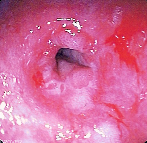
Herpes esophagitis

Endoscopic image of peptic stricture showing n...
Endoscopic image of peptic stricture showing narrowing of the esophagus near the junction with the stomach due to chronic gastroesophageal reflux in the setting of scleroderma. (Photo credit: Wikipedia)

Esophagus is  the tube that carries food from the throat to the stomach. If left untreated, this condition can become very uncomfortable, causing problems with swallowing, ulcers and scarring of the esophagus. In rare instances, a condition known as “Barrett’s esophagus” may develop, which is a risk factor for cancer of the esophagus.

Causes:
Esophagitis is frequently caused by the backflow of acid-containing fluid from the stomach to the esophagus (gastroesophageal reflux). You have a higher risk for esophagitis if you have had excessive vomiting, surgery or radiation to the chest (such as in lung cancer), or if you take medications such as aspirin, ibuprofen, potassium, alendronate, and doxycycline.

Persons with weakened immune systems due to HIV and certain medications (such as corticosteroids) may develop infections that lead to esophagitis. Esophageal infection may be due to viruses such as herpes or cytomegalovirus, and fungi or yeast (especially Candida infections).

The infection or irritation may cause the tissues to become inflamed and occasionally form ulcers. You may have difficulty when swallowing and a burning sensation in the esophagus.

Esophagitis is caused by an infection or irritation in the esophagus. An infection can be caused by bacteria, viruses, fungi or diseases that weaken the immune system. Infections that cause esophagitis include:

*  Candida. This is a yeast infection of the esophagus caused by the same fungus that causes vaginal yeast infections. The infection develops in the esophagus when the body’s immune system is weak (such as in people with diabetes or HIV). It is usually very treatable with antifungal drugs.

* Herpes. Like Candida, this viral infection can develop in the esophagus when the body’s immune system is weak. It is treatable with antiviral drugs.

Irritation causing esophagitis may be caused by any of the following:

* GERD
* Vomiting
* Surgery
* Medications such as aspirin and anti-inflammatories
* Taking a large pill with too little water or just before bedtime
* Swallowing a toxic substance
* Hernias
* Radiation injury (after receiving radiation for cancer treatment)

You may click to see the related topics below:
Gastroesophageal reflux disease
Esophagitis Candida
Esophagitis CMV
Esophagitis herpes
Symptoms:
Symptoms of esophagitis include:

* Difficult and/or painful swallowing
* Heartburn (acid reflux)
* Mouth sores
* A feeling of something of being stuck in the throat
* Nausea
* Vomiting
*Oral lesions (herps)
If you have any of these symptoms, you should contact your health care provider as soon as possible.

Diagnosis:
Once your doctor has performed a thorough physical examination and reviewed your medical history, there are several tests that can be used to diagnose esophagitis. These include:

* Upper endoscopy . A test in which a long, flexible lighted tube, called an endoscope, is used to look at the esophagus.

* Biopsy. During this test, a small sample of the esophageal tissue is removed and then sent to a laboratory to be examined under a microscope.

* Upper GI series (or barium swallow). During this procedure, x-rays are taken of the esophagus after drinking a barium solution. Barium coats the lining of the esophagus and shows up white on an x-ray. This characteristic enables doctors to view certain abnormalities of the esophagus.

Treatment:
Treatment depends on the specific cause. Reflux disease may require medications to reduce acid. Infections will require antibiotics. Possible treatments include:

* Medications that block acid production, like heartburn drugs
* Antibiotics, antifungals or antivirals to treat an infection
* Pain medications that can be gargled or swallowed
* Corticosteroid medication to reduce inflammation
* Intravenous (by vein) nutrition to allow the esophagus to heal, to reduce the likelihood of malnourishment or dehydration
* Endoscopy to remove any lodged pill fragments
* Surgery to remove the damaged part of the esophagus

While being treated for esophagitis, there are certain steps you can take to help limit discomfort.

* Avoid spicy foods such as those with pepper, chili powder, curry and nutmeg.
* Avoid hard foods such as nuts, crackers and raw vegetables.
* Avoid acidic foods and beverages such as tomatoes, oranges, grapefruits and their juices. Instead, try imitation fruit drinks with vitamin C.
* Add more soft foods such as applesauce, cooked cereals, mashed potatoes, custards, puddings and high protein shakes to your diet.
* Take small bites and chew food thoroughly.
* If swallowing becomes increasingly difficult, try tilting your head upward so the food flows to the back of the throat before swallowing.
* Drink liquids through a straw to make swallowing easier.
* Avoid alcohol and tobacco.

Click for Herbal and Alternative Treatment:->.(1).…..(2)…..(3)……(4)

You may click to learn more
Prognosis:-
The disorders that cause esophagitis usually respond to treatment.

Possible Complications :-
If untreated, esophagitis may cause severe discomfort, swallowing difficulty to the extent of causing malnutrition or dehydration, and eventual scarring of the esophagus. This scarring may lead to a stricture of the esophagus, and food or medications may not be able to pass through to the stomach.

A condition called Barrett’s esophagus can develop after years of gastroesophageal reflux. Rarely, Barrett’s esophagus may lead to cancer of the esophagus.

When to Contact a Medical Professional
Call your health care provider if you have symptoms that suggest esophagitis.

Disclaimer: This information is not meant to be a substitute for professional medical advise or help. It is always best to consult with a Physician about serious health concerns. This information is in no way intended to diagnose or prescribe remedies.This is purely for educational purpose.

Resources:
http://www.medicinenet.com/esophagitis/article.htm
http://www.nlm.nih.gov/medlineplus/ency/article/001153.htm

Categories
Diagnonistic Test

Barium Swallow (Upper Gastrointestinal Series or “Upper GI Series”)

[amazon_link asins=’B00GNOQ9R0,B00HX3W0SW,B011DBB5OE,B01FGP9ER8,B01K0SXP2Q,B0012VK3E4,B01FIWVVI4,B01FIWUP8Q,B007Z759CK’ template=’ProductCarousel’ store=’finmeacur-20′ marketplace=’US’ link_id=’704f3253-71bf-11e7-83eb-355c4e3e424d’]


Definition:A barium swallow, or upper GI series, is an x-ray test used to examine the upper digestive tract (the esophagus, stomach, and small intestine). Because these organs are normally not visible on x-rays, you need to swallow barium, a liquid that does show up on x-rays. The barium temporarily coats the lining of the esophagus, stomach, and intestine, making the outline of these organs visible on the xray pictures. This test is useful for diagnosing cancers, ulcers, problems that cause narrowing of the esophagus, some causes of inflammation in the intestine, and some swallowing problems.

CLICK & SEE

An upper gastrointestinal (UGI) series looks at the upper and middle sections of the gastrointestinal tract. The test uses barium contrast material, fluoroscopy, and X-ray. Before the test, you drink a mix of barium (barium contrast material) and water. The barium is often combined with gas-making crystals. Your doctor watches the movement of the barium through your esophagus, stomach, and the first part of the small intestine (duodenum) on a video screen. Several X-ray pictures are taken at different times and from different views.

A small bowel follow-through may be done immediately after a UGI to look at the rest of the small intestine. If just the throat and esophagus are looked at, it is called an esophagram (or barium swallow).

Upper endoscopy is done instead of a UGI in certain cases. Endoscopy uses a thin, flexible tube (endoscope) to look at the lining of the esophagus, stomach, and upper small intestine (duodenum).

Why It Is Done:-
An upper gastrointestinal (UGI) series is done to:

1.Find the cause of gastrointestinal symptoms, such as difficulty swallowing, vomiting, burping up food, belly pain (including a burning or gnawing pain in the center of the stomach), or indigestion. These may be caused by conditions such as hiatal hernia.

*Find narrow spots (strictures) in the upper intestinal tract, ulcers, tumors, polyps, or pyloric stenosis.

*Find inflamed areas of the intestine, malabsorption syndrome, or problems with the squeezing motion that moves food through the intestines (motility disorders).

*Find swallowed objects.

Generally, a UGI series is not used if you do not have symptoms of a gastrointestinal problem. A UGI series is done most often for people who have:

1.A hard time swallowing.
2.A history of Crohn’s disease.
3.A possible blocked intestine (obstruction).
4.Belly pain that is relieved or gets worse while eating.
5.Severe heartburn or heartburn that occurs often.

How To Prepare for the Test:-
Tell your doctor and the x-ray technicians if you :

1.Are taking any medicine.

2.Are allergic to any medicines, barium, or any other X-ray contrast material.

3.Are or might be pregnant. This test is not done during pregnancy because of the risk of radiation to the developing baby (fetus).

4.You may be asked to eat a low-fiber diet for 2 or 3 days before the test. You may also be asked to stop eating for 12 hours before the test. Your doctor will tell you if you need to stop taking certain medicines before the test.

The evening before the test, you may be asked to take a laxative to help clean out your intestines. If your stomach cannot empty well on its own, you may have a special tube put through your nose and down into your stomach just before the test begins. A gentle suction on the tube will drain the stomach contents.

If you are having the small bowel follow-through after the UGI series, you will need to wait between X-rays. The entire small bowel follow-through exam takes up to 6 hours, so bring along a book to read or some other quiet activity.

You may be asked to sign a consent form. Talk to your doctor about any concerns you have regarding the need for the test, its risks, how it will be done, or what the results will mean. To help you understand the importance of this test, fill out the medical test information form (What is a PDF document?) .
How It Is Done
A UGI series is usually done in your doctor’s office, clinic, or X-ray department of a hospital. You do not need to stay overnight in the hospital. The test is done by a radiologist and a radiology technologist.

You will need to take off your clothes and put on a hospital gown. You will need to take out any dentures and take off any jewelry. You may not smoke or chew gum during the test, since the stomach will respond by making more gastric juices and this will slow the movement of the barium through the intestines.

You might also be asked to swallow some tablets that “fizz,” causing air-bubbles to be released in your stomach. This might make you feel like burping, but try not to. You will get better pictures if you can keep yourself from burping.

The x-ray technician may ask you to stand or lie in different positions over the next few minutes, to help spread around the liquid you have swallowed. Most often, the x-ray pictures are taken while you lie on your back on a table. The x-ray machine or the table is moved a few times so it can take pictures of all of the internal structures. You are asked to hold your breath for each picture so that your breathing movement does not blur the image.

You will lie on your back on an X-ray table. The table is tilted to bring you to an upright position with the X-ray machine in front of you. Straps may be used to keep you safely on the table. The technologist will make sure you are comfortable during changes in table position.

You will have one X-ray taken before you drink the barium mix. Then you will take small swallows repeatedly during the series of X-rays that follow. The radiologist will tell you when and how much to drink. By the end of the test, you may have swallowed 1cup to 2.5cups of the barium mixture. See a picture of a barium swallow test.

The radiologist watches the barium pass through your gastrointestinal tract using fluoroscopy and X-ray pictures. The table is tilted at different positions and you may change positions to help spread the barium. Some gentle pressure is put on your belly with a belt or by the technologist’s gloved hand. You may be asked to cough so that the radiologist can see how that changes the barium flow. See an image of a barium swallow.

If you are having an air-contrast study, you will sip the barium liquid through a straw with a hole in it or take pills that make gas in your stomach. The air or gas that you take in helps show the lining of the stomach and intestines in greater detail.

If you are also having a small bowel study, the radiologist watches as the barium passes through your small intestine into your large intestine. X-ray pictures are taken every 30 minutes.

The UGI series 30 to 40 minutes. The UGI series with a small bowel study takes 2 to 6 hours. In some cases, you may be asked to return after 24 hours to have more X-ray pictures taken.

When the UGI series is done, you may eat and drink whatever you like, unless your doctor tells you not to.

You may be given a laxative or enema to flush the barium out of your intestines after the test to prevent constipation. Drink a lot of fluids for a few days to flush out the barium.

How It Feels
The barium liquid is thick and chalky, and some people find it hard to swallow. A sweet flavor, like chocolate or strawberry, is used to make it easier to drink. Some people do not like it when the X-ray table tilts. You may find that pressure on your belly is uncomfortable. After the test, many people feel bloated and a little nauseated.

For 1 to 3 days after the test, your stool (feces) will look white from the barium. Call your doctor if you are not able to have a bowel movement in 2 to 3 days after the test. If the barium stays in your intestine, it can harden and cause a blockage. If you become constipated, you may need to use a laxative to pass a stool.

Risk Factors:
There are no significant risks.

Barium does not move into the blood, so allergic reactions are very rare.

Some people gag while drinking the barium fluid. In rare cases, a person may choke and inhale (aspirate) some of the liquid into the lungs.

There is a small chance that the barium will block the intestine or leak into the belly through a perforated ulcer. A special type of contrast material (Gastrografin) can be used if you have a blockage or an ulcer.

There is always a small chance of damage to cells or tissue from being exposed to any radiation, even the low level of radioactive tracer used for this test.


How long is it before the result of the test is known?

It takes the x-ray department 30 minutes to an hour to develop the pictures from your barium swallow, and it will take additional time for a doctor to examine the x-rays and to decide how they look. Typically you can get the results within a day or two.

Must you do anything special after the test is over
After the test, you can eat normally and do your normal activities. You should drink more water than usual to help clear out the barium and to prevent constipation, which might be a side effect of the test. Your stool may appear light in color for a couple of days.

Results:-
An upper gastrointestinal (UGI) series looks at the upper and middle sections of the gastrointestinal tract. Results are usually ready in 1 to 3 days.

Upper gastrointestinal (UGI) series  Normal: The esophagus, stomach, and small intestine all look normal.

Abnormal: A narrowing (stricture), inflammation, a mass, a hiatal hernia, or enlarged veins (varices) may be seen. Spasms of the esophagus or a backward flow (reflux) of barium from the stomach may occur.

The UGI series may show a stomach (gastric) or intestinal (duodenal) ulcer, a tumor, or something pushing on the intestines from outside the gastrointestinal tract. Narrowing of the opening between the stomach and the small intestine (pyloric stenosis) may be seen.

The small bowel follow-through may show inflammation or changes in the lining that may explain poor absorption of food. This may be caused by Crohn’s disease or celiac disease.

What Affects the Test
Reasons you may not be able to have the test or why the results may not be helpful include:

*Eating before or during the test.
*Too much air in the small intestine.

ABOUT THE TEST:
*A gastrointestinal (GI) motility study may be done if the squeezing motions of the small intestine are not normal during the UGI series and small bowel follow-through. The movement of the barium through the lower intestinal tract is recorded every few hours for up to 24 hours. A barium enema or colonoscopy may be needed to confirm the diagnosis.

*Upper endoscopy is done instead of a UGI test in certain cases. Endoscopy uses a thin, flexible tube (endoscope) to look at the lining of the esophagus, stomach, and upper small intestine (duodenum). For more information, see the medical test Upper Gastrointestinal Endoscopy.

*The UGI series test:

*Cannot show irritation of the stomach lining (gastritis) or esophagus (esophagitis) or ulcers that are smaller than about 0.25in. in diameter.

*Cannot show an infection with the bacteria Helicobacter pylori, which may be a cause of stomach ulcers.

*A biopsy cannot be done during the UGI if a problem is found.

Sources:
http://www.webmd.com/digestive-disorders/upper-gastrointestinal-ugi-series?page=4
https://www.health.harvard.edu/fhg/diagnostics/barium-swallow.shtml

css.php