Categories
Ailmemts & Remedies

Anophthalmia and Microphthalmia (Small Eye Syndrome)

[amazon_link asins=’B010QX24DQ,B00NBUQ366,B00INJ7TWW,B01M6ZKYMV,B00W8FVMNO,B01FIS2T6Q,B01I2788M4,B06XX3B2YM,B00RWG5GNQ’ template=’ProductCarousel’ store=’finmeacur-20′ marketplace=’US’ link_id=’928783e0-7721-11e7-8f1e-01ae0247b770′][amazon_link asins=’B005WJ3H7C,1543270646′ template=’ProductCarousel’ store=’finmeacur-20′ marketplace=’US’ link_id=’6b3aef58-7721-11e7-9741-b1adb6c55a59′]

This information was developed by the National Eye Institute to help patients and their families in searching for general information about anophthalmia and microphthalmia. An eye care professional who has examined the patient’s eyes and is familiar with his or her medical history is the best person to answer specific questions.

Other Names
Anophthalmos and microphthalmos, small eye syndrome.

What are anophthalmia and microphthalmia?
Anophthalmia and microphthalmia are often used interchangeably. Microphthalmia is a disorder in which one or both eyes are abnormally small, while anophthalmia is the absence of one or both eyes. These rare disorders develop during pregnancy and can be associated with other birth defects.

Click to see the pictures

What causes anophthalmia and microphthalmia?
Causes of these conditions may include genetic mutations and abnormal chromosomes. Researchers also believe that environmental factors, such as exposure to X-rays, chemicals, drugs, pesticides, toxins, radiation, or viruses, increase the risk of anophthalmia and microphthalmia, but research is not conclusive. Sometimes the cause in an individual patient cannot be determined.

Can anophthalmia and microphthalmia be treated?
There is no treatment for severe anophthalmia or microphthalmia that will create a new eye or restore vision. However, some less severe forms of microphthalmia may benefit from medical or surgical treatments. In almost all cases improvements to a child’s appearance are possible. Children can be fitted for a prosthetic (artificial) eye for cosmetic purposes and to promote socket growth. A newborn with anophthalmia or microphthalmia will need to visit several eye care professionals, including those who specialize in pediatrics, vitreoretinal disease, orbital and oculoplastic surgery, ophthalmic genetics, and prosthetic devices for the eye. Each specialist can provide information and possible treatments resulting in the best care for the child and family. The specialist in prosthetic diseases for the eye will make conformers, plastic structures that help support the face and encourage the eye socket to grow. As the face develops, new conformers will need to be made. A child with anophthalmia may also need to use expanders in addition to conformers to further enlarge the eye socket. Once the face is fully developed, prosthetic eyes can be made and placed. Prosthetic eyes will not restore vision.

How do conformers and prosthetic eyes look?
A painted prosthesis that looks like a normal eye is usually fitted between ages one and two. Until then, clear conformers are used. When the conformers are in place the eye socket will look black. These conformers are not painted to look like a normal eye because they are changed too frequently. Every few weeks a child will progress to a larger size conformer until about two years of age. If a child needs to wear conformers after age two, the conformers will be painted like a regular prosthesis, giving the appearance of a normal but smaller eye. The average child will need three to four new painted prostheses before the age of 10.

…Click to see the pictures..

How is microphthalmia managed if there is residual vision in the eye?
Children with microphthalmia may have some residual vision (limited sight). In these cases, the good eye can be patched to strengthen vision in the microphthalmic eye. A prosthesis can be made to cap the microphthalmic eye to help with cosmetic appearance, while preserving the remaining sight.

Keeping on Top of Your Condition
Keeping in tune with your disease or condition not only makes treatment less intimidating but also increases its chance of success, and has been shown to lower a patients risk of complications. As well, as an informed patient, you are better able to discuss your condition and treatment options with your physician.

A new service available to patients provides a convenient means of staying informed, and ensures that the information is both reliable and accurate. If you wish to find out more about HealthNewsflash’s innovative service, take the tour.

Resources
The following organizations may be able to provide additional information on anophthalmia and microphthalmia:

National Eye Institute
2020 Vision Place
Bethesda, MD 20892-3655
(301) 496-5248
2020@nei.nih.gov
http://www.nei.nih.gov/

American Society of Ocularists
E-mail: aso@ocularist.org
http://www.ocularist.org/
Represents technicians specializing in making and fitting of custom artificial eyes.

American Society of Ophthalmic Plastic and Reconstructive Surgery
1133 West Morse Blvd. #201
Winter Park, FL 32789
(407) 647-8839
http://www.asoprs.org
Represents ophthalmologists who specialize in reconstructive surgery involving the eye and surrounding structures. Publishes a factsheet on anophthalmos and orbital implants.

International Children’s Anophthalmia Network (ican)
Genetics, Levy 2
Albert Einstein Medical Center
5501 Old York Road
Philadelphia, PA 19141
1-800-580-4226
(215) 456-8722 or
http://www.ioi.com/ican
Provides information on anophthalmia and microphthalmia. Coordinates a patient registry. Offers referrals to local resources. Coordinates gatherings for people with anophthalmia and microphthalmia and their families. Publishes a newsletter, The Conformer.

Additional resources for parents and teachers of children with visual impairments can be found on the National Eye Institute’s website at http://www.nei.nih.gov/health/organizations.htm#resources.

For additional information, you may also wish to contact a local library.

Medical Literature
For information on your topic, you may wish to conduct a search of the medical literature. The National Library of Medicine (NLM) coordinates PubMed, a computerized medical literature database. You can conduct your own free literature search by accessing PubMed through the Internet. For help on how to search PubMed and how to get journal articles, please see PubMed Help. You may also get assistance with a literature search at a local library.

Please keep in mind that articles in the medical literature are usually written in technical language. We encourage you to share articles with a health care professional who can help you understand them.
.

Sources:http://www.nei.nih.gov/health/anoph/

Enhanced by Zemanta
Categories
Ailmemts & Remedies

Chalazion

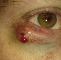
A chalazion immediately after excision
Image via Wikipedia

Alternative Names:Meibomian gland lipogranuloma

Definition:
A chalazion is a small bump in the eyelid caused by a blockage of a tiny oil gland.
It is a cyst in the eyelid that is caused by inflammation of a blocked meibomian gland, usually on the upper eyelid.

..click to see the pictures >....…(01)....`(1)..…....(2)………….(3)..………………

..
…………………………………………...Eyelid affected by Chalazion
Chalazions differ from styes (hordeolums) in that they are usually painless apart from the tenderness caused when they swell up, and in size (chalazia tend to be larger than styes). A chalazion may eventually disappear on its own after a few months, though more often than not, some treatment is necessary.
A large chalazion ca. 20 minutes upon excision. This bipartite chalazion was removed via two separate incisions. Further along the lower eyelid, signs of chronic inflammation are visible.

Causes:

A chalazion develops within the glands that produce the fluid that lubricate the eye. These are called Meibomian glands. The eyelid has approximately 100 of these glands, which are located near the eyelashes.

A chalazion is caused by blockage of the duct that drains one of these glands. A chalazion begins as swelling and tenderness, and later forms a cyst-like growth. Many chalazia disappear without treatment after a few months

Signs and symptoms:

*Painful swelling on the eyelid

*Eyelid tenderness

*Sensitivity to light

*Increased tearing

*Swelling on the eyelid

*Heaviness of the eyelid

Diagnosis:
Exams and Tests

Examination of eyelid confirms the diagnosis.
Rarely, the Meibomian gland duct may be blocked by a skin cancer. If this is suspected, a biopsy may be needed to diagnose the disorder.

Treatment:
A chalazion will often disappear without treatment within a month or so.

The primary treatment is application of warm compresses for 10-15 minutes at least 4 times a day. This may soften the hardened oils blocking the duct and promote drainage and healing. If the chalazion continues to get bigger, it may need to be surgically removed. This is usually done from underneath the eyelid to avoid a scar on the skin.

Topical antibiotic eye drops or ointment (eg chloramphenicol or fusidic acid) are sometimes used for the initial acute infection, but are otherwise of little value in treating a chalazion. Chalazia will often disappear without further treatment within a few months and virtually all will resorb within two years.

If they continue to enlarge or fail to settle within a few months, then smaller lesions may be injected with a corticosteroid or larger ones may be surgically removed using local anesthesia. . If the chalazion is located directly under the eyelid’s outer tissue, however, an excision from above may be more advisable so as not to inflict any unnecessary damage on the lid itself. Eyelid epidermis usually mends well, without leaving any visible traces of cicatrisation. Depending on the chalazion’s texture, the excision procedure varies: while fluid matter can easily be removed under minimal invasivion, by merely puncturing the chalazion and exerting pressure upon the surrounding tissue, hardened matter usually necessitates a larger incision, through which it can be scraped out. Any residual matter should be metabolized in the course of the subsequent healing process, generally aided by regular appliance of dry heat. The excision of larger chalazia may result in visible hematoma around the lid, which will wear off within three or four days, whereas the swelling may persist for longer. Chalazion excision is an ambulant treatment and normally does not take longer than fifteen minutes. Nevertheless, owing to the risks of infection and severe damage to the eyelid, such procedures should only be performed by a doctor.

Rarely chalazia may reoccur and these will be biopsied to help rule out tumors.

Antibiotic eye drops are usually used several days before and after removal of the cyst, but are otherwise of little value in treating a chalazion.

A chalazion that keeps coming back should be biopsied to rule out tumor.

Click for :->Chalazion and its online homeopathic treatment

Prognosis: Chalazia usually heal on their own. The outcome with treatment is usually excellent.

Complications:
A large chalazion can cause astigmatism due to pressure on the cornea. This will resolve with resolution of the chalazion.Complications including, but not limitedtohypopigmentation may occur with corticosteroid injection.The presence of recurring chalazion in the same area sometimes leads to a consideration of sebaceous cell carcinoma.The minor operation is quite painless, the eyelid is injected with a local anesthetic a clamp is put on the eyelid, then the eyelid is turned over and the chalazion is scraped out.

When to contact your Healthcare Provider:
Apply warm compresses and call your health care provider if the swelling progresses or persists longer than 1 month.
Call for an appointment with your health care provider if lumps on the eyelid continue to enlarge despite treatment or are associated with an area of eyelash loss.

Prevention:
Proper cleansing of the eyelid may prevent recurrences in people prone to chalazia. Cleaning the eyelash area with baby shampoo will help reduce clogging of the ducts.

Click to see also:-> Stye
Disclaimer: This information is not meant to be a substitute for professional medical advise or help. It is always best to consult with a Physician about serious health concerns. This information is in no way intended to diagnose or prescribe remedies.This is purely for educational purpose.
Resources:
http://www.nlm.nih.gov/medlineplus/ency/article/001006.htm
http://en.wikipedia.org/wiki/Chalazion

Enhanced by Zemanta
Categories
Ailmemts & Remedies

Nyctalopia(Night Blindness)

[amazon_link asins=’B073QK6B7B,B07439MJKR,B074DJS44T,B073QJVS1S,B073QJVGQQ,B073QJQ6JB,B0742XV6NR,B015KAOYHO,B074CQ3XJM’ template=’ProductCarousel’ store=’finmeacur-20′ marketplace=’US’ link_id=’a21d7a3a-78d4-11e7-8179-9f6a3bdf93c7′]

Night blindness (nyctalopia) is the inability to see well at night or in poor light. It is not a disorder in itself, but rather a symptom of an underlying disorder or problem, especially untreated nearsightedness.

Nyctalopia (Greek for “night blindness”) is a condition making it difficult or impossible to see in relatively low light. It is a symptom of several eye diseases. Night blindness may exist from birth, or be caused by injury or malnutrition (for example, a lack of vitamin A).

Causes:

Night blindness is due to a disorder of the cells in the retina that are responsible for vision in dim light. It has many causes, including:

The most common cause of nyctalopia is retinitis pigmentosa, a disorder in which the rod cells in the retina gradually lose their ability to respond to the light. Patients suffering from this genetic condition have progressive nyctalopia and eventually their daytime vision may also be affected. In X-linked congenital stationary night blindness, from birth the rods either do not work at all, or work very little, but the condition doesn’t get worse.Another cause of night blindness is a deficiency of retinol, or vitamin A, found in fish oils, liver and dairy products. In the Second World War misinformation was spread by the British to cover up the reason for their pilots’ successful night time missions. Their success was, in the misinformation, attributed to improved night vision and pilots flying night missions were encouraged to eat plenty of carrots, which contain carotenoids and can be converted into retinol. The actual reason for their success was their use of advanced radar technologies.

The opposite problem, known as hemeralopia, is much rarer.

The outer area of the retina is made up of more rods than cones. The rod cells are the cells that enable us to see in poor illumination. This is the reason why loss of side vision often results in night blindness. Individuals suffering from night blindness not only see poorly at night, but also require some time for their eyes to adjust from brightly lit areas to dim ones. Contrast vision may also be greatly reduced.

In order to determine what is causing night blindness, the eye doctor will perform a thorough eye exam and may order any of a number of specialized exams .

Historical usage
Aulus Cornelius Celsus, writing ca. 30 AD, described night blindness and recommended an effective dietary supplement: “There is besides a weakness of the eyes, owing to which people see well enough indeed in the daytime but not at all at night; in women whose menstruation is regular this does not happen. But success sufferers should anoint their eyeballs with the stuff dripping from a liver whilst roasting, preferably of a he-goat, or failing that of a she-goat; and as well they should eat some of the liver itself.”

Historically, nyctalopia, also known as moonblink, was a temporary night blindness believed to be caused by sleeping in moonlight in the tropics.
Treatment:

Treatment for night blindness will depend upon its cause. Treatment may be as simple as a new prescription for your eyeglasses or switching glaucoma medications, or it may require surgery in cases of cataracts.

Click for Alternative medication & life style to prevent night blindness

Vitamin A Deficiency, Prevention and Treatment through Ayurveda

CLICK TO SEE :Bilberry Extract Improve Night Blindness

Useful Herbs for Eye Care

Vitamine & mineral guide for eye care against Night Blindness

Night Blindness- Prevention,Treatment & Healing
Disclaimer: This information is not meant to be a substitute for professional medical advise or help. It is always best to consult with a Physician about serious health concerns. This information is in no way intended to diagnose or prescribe remedies.This is purely for educational purpose

Resources:
http://en.wikipedia.org/wiki/Night_blindness
http://www.medicinenet.com/script/main/art.asp?articlekey=43325

Enhanced by Zemanta
Categories
Ailmemts & Remedies

Glaucoma

[amazon_link asins=’0446690627,B004ZRCYGM’ template=’ProductCarousel’ store=’finmeacur-20′ marketplace=’US’ link_id=’4f0a232a-1a11-11e7-9403-1319b0fa24b4′]

[amazon_link asins=’1494265559,1440183201′ template=’ProductCarousel’ store=’finmeacur-20′ marketplace=’US’ link_id=’68716ecb-1a11-11e7-b0ef-1fdf910a249f’]

normal tension glaucom is a serious eye condition that involves an elevation in pressure inside the eye. Increased pressure results from a buildup of excess fluid in the eye. Glaucoma is a dangerous eye condition because it frequently progresses without obvious symptoms. This is why it is frequently referred to as “the sneak thief of sight.”

CLICK & SEE THE PICTURES

Types of Glaucoma
There are several types of glaucoma, for example, congenital, primary, secondary, and normal tension glaucoma. Congenital glaucoma appears in young people; secondary glaucoma is the result of injury or trauma. There are two types of primary glaucoma most frequently associated with aging: acute or closed angle glaucoma, and chronic or open angle glaucoma. The Reference Section at the end of this Fact Sheet provides resources for learning more about each of the types of glaucoma.

Regardless of the type, glaucoma can impair vision by creating pressure that damages the optic nerve, The “cable” of nerve fibers that transmits messages about what we see from the eye to the brain.

It is important to recall the structure of the eye and how it works to understand the dangers posed by glaucoma. Glaucoma can cause damage when the aqueous humor, a fluid that inflates the front of the eye and circulates in a chamber called the anterior chamber, enters the eye but cannot drain properly from the eye. Elevated pressure inside the eye, in turn, can cause damage to the optic nerve or the blood vessels in the eye that nourish the optic nerve. The Human Eye, Its Functions, and Visual Impairment explains how the eye works. When glaucoma begins to affect a person’s vision, the first problems are with peripheral vision, or what can be seen at the sides of the visual field, rather than in the center. If glaucoma progresses, it can destroy all peripheral vision, then impair central vision, and lead to total blindness. Treatments for glaucoma are aimed at bringing down the pressure in the eye to a level that is low enough to prevent harm to the optic nerve. Once the optic nerve is damaged from glaucoma, lowering the pressure in the eye only prevents further damage to the nerve. Damage already done to the optic nerve cannot be reversed.

Diagnosing Glaucoma
When a person receives a diagnosis of glaucoma, it means a diagnosis of a life-long condition. However, early detection of glaucoma, appropriate and ongoing treatment, and the availability of specialized low vision and vision rehabilitation services if vision should become impaired, means that people who have glaucoma can live productive and satisfying lives.

A pressure check for glaucoma should be a routine part of every eye examination at least by the age of 35. A visual field test can also detect glaucoma by indicating the loss of peripheral vision.

How Common Is Glaucoma?
According to the Glaucoma Research Foundation glaucoma affects more than 3 million Americans. It is also reported that glaucoma is the third leading cause of legal blindness in Caucasians, and the leading cause of blindness in African Americans. Although anyone can get glaucoma, some people are at higher risk. Those at risk include:

1.People over the age of 60.

2.African Americans over the age of 40.

3.People with a family history of glaucoma.

Treatment:
Untreated glaucoma can lead to blindness. Eye drops or tablets may be prescribed to reduce fluid production and consequently reduce pressure in the eye.

Laser or surgical treatment may be used when medical treatment isn’t sufficiently effective.

Screening:
People over the age of 40 are advised to have their eyes tested every two years to check for signs of glaucoma. If glaucoma is identified early enough, treatment can be given to prevent further damage and reduce the risk of blindness.

These tests are available at your local optician and should include:

•examination of the optic disc
•measurement of the pressure in the eye
•checking of peripheral vision (by looking for a sequence of spots of light on a screen).
Retaining Independence
People who have experienced vision loss from glaucoma can retain their independence, productivity, and quality of life by learning to use specialized devices and techniques to carry out their daily activities. These may include using special lenses that can help those who have remaining sight make the best use of available vision, and using specialized techniques that enable people to manage home and work responsibilities, travel using mass transportation, and carry out a host of other activities.

Click to learn more about Acute Glaucoma  and its Ayurvedic Remedy

Click to see->:6 Sure-Fire Tips to Prevent Glaucoma Naturally

Disclaimer: This information is not meant to be a substitute for professional medical advise or help. It is always best to consult with a Physician about serious health concerns. This information is in no way intended to diagnose or prescribe remedies.This is purely for educational purpose.

Resources:

http://www.bbc.co.uk/health/physical_health/conditions/glaucoma1.shtml

:www.afb.org

http://www.dwueye.com/glaucoma-eye-care.html

Enhanced by Zemanta
css.php