Categories
Ailmemts & Remedies

Scarlet fever

Alternative Names : Scarlatina

Definition:
Scarlet fever is a disease caused by infection with the group A Streptococcus bacteria (the same bacteria that causes strep throat).Once a major cause of death, it is now effectively treated with antibiotics. The term scarlatina may be used interchangeably with scarlet fever, though it is commonly used to indicate the less acute form of scarlet fever that is often seen since the beginning of the twentieth century.
click to  see the picture
It can affect people of any age. However, it’s most common between the ages of six and 12.

click & see

Symptoms:

The time between becoming infected and having symptoms is short, generally 1 – 2 days. The illness typically begins with a fever and sore throat.

click to see the pictures..>....(01)....(1).……..(2)..……...(3).……………………………

The rash usually first appears on the neck and chest, then spreads over the body. It is described as “sandpapery” in feel. The texture of the rash is more important than the appearance in confirming the diagnosis. The rash can last for more than a week. As the rash fades, peeling (desquamation) may occur around the fingertips, toes, and groin area.

The common signs and symptoms that give scarlet fever are as follows:

*Red rash. The rash looks like a sunburn and feels like sandpaper. It typically begins on the face or neck and spreads to the trunk, arms and legs. If pressure is applied to the reddened skin, it will turn pale.

*Red lines. The folds of skin around the groin, armpits, elbows, knees and neck usually become a deeper red than the surrounding rash.

*Flushed face. The face may appear flushed with a pale ring around the mouth.

*Strawberry tongue. The tongue generally looks red and bumpy, and it’s often covered with a white coating early in the disease.

The rash and the redness in the face and tongue usually last about a week. After these signs and symptoms have subsided, the skin affected by the rash often peels. Other signs and symptoms associated with scarlet fever include:

*Fever of 101 F (38.3 C) or higher, often with chills

*Very sore and red throat, sometimes with white or yellowish patches

*Difficulty swallowing

*Enlarged glands in the neck (lymph nodes) that are tender to the touch

*Nausea or vomiting

*Headache

*Abdominal pain

*Bright red color in the creases of the underarm and groin (Pastia’s lines)

*Chills

*General discomfort (malaise)

*Muscle aches

*Sore throat

*Swollen, red tongue (strawberry tongue)

Causes:
Scarlet fever is caused by the same type of bacteria that cause strep throat. In scarlet fever, the bacteria release a toxin that produces the rash and red tongue.

The infection spreads from person to person via droplets expelled when an infected person coughs or sneezes. The incubation period — the time between exposure and illness — is usually two to four days.

Risk Factors:
Children 6 to 12 years of age are more likely than are other people to get scarlet fever. Scarlet fever germs spread more easily among people in close contact, such as family members or classmates.

Complications:
If scarlet fever goes untreated, the bacteria may spread to the:

*Tonsils
*Sinuses
*Skin
*Blood
*Middle ear

Rarely, scarlet fever can lead to rheumatic fever, a serious condition that can affect the:

*Heart
*Joints
*Nervous system
*Skin

Diagnosis:
Diagnosis of scarlet fever is clinical. The blood test shows marked leukocytosis with neutrophilia and conservated or increased eosinophils, high erythrocyte sedimentation rate (ESR) and C-reactive protein (CRP) (both indications of inflammation), and elevation of antistreptolysin O titer. Blood culture is rarely positive, but the streptococci can usually be demonstrated in throat culture. The complications of scarlet fever include septic complications due to spread of streptococcus in blood and immune-mediated complications due to an aberrant immune response. Septic complications—today rare—include ear and sinus infection, streptococcal pneumonia, empyema thoracis, meningitis and full-blown sepsis, upon which the condition may be called malignant scarlet fever.

Immune complications include acute glomerulonephritis, rheumatic fever and erythema nodosum. The secondary scarlatinous disease, or secondary malignant syndrome of scarlet fever, includes renewed fever, renewed angina, septic ear, nose, and throat complications and kidney infection or rheumatic fever and is seen around the eighteenth day of untreated scarlet fever.

The rash is the most striking sign of scarlet fever. It usually begins looking like a bad sunburn with tiny bumps, and it may itch. The rash usually appears first on the neck and face, often leaving a clear unaffected area around the mouth. It spreads to the chest and back, then to the rest of the body. In body creases, especially around the underarms and elbows, the rash forms classic red streaks (on very dark skin, the streaks may appear darker than the rest of the skin). Areas of rash usually turn white (or paler brown, with dark complected skin) when pressed on. By the sixth day of the infection, the rash usually fades, but the affected skin may begin to peel. Usually there are other symptoms that help to confirm a diagnosis of scarlet fever, including a reddened sore throat, a fever at or above 101 °F (38.3 °C), and swollen glands in the neck. Scarlet fever can also occur with a low fever. The tonsils and back of the throat may be covered with a whitish coating, or appear red, swollen, and dotted with whitish or yellowish specks of pus. Early in the infection, the tongue may have a whitish or yellowish coating. Also, an infected person may have chills, body aches, nausea, vomiting, and loss of appetite.

When scarlet fever occurs because of a throat infection, the fever typically stops within 3 to 5 days, and the sore throat passes soon afterward. The scarlet fever rash usually fades on the sixth day after sore throat symptoms started, and begins to peel (as above). The infection itself is usually cured with a 10-day course of antibiotics, but it may take a few weeks for tonsils and swollen glands to return to normal.

In rare cases, scarlet fever may develop from a streptococcal skin infection like impetigo. In these cases, the person may not get a sore throat.

Treatment:
Other than the occurrence of the diarrhea, the treatment and course of scarlet fever are no different from those of any strep throat. In case of penicillin allergy, clindamycin or erythromycin can be used with success. Patients should no longer be infectious after taking antibiotics for 24 hours. People who have been exposed to scarlet fever should be watched carefully for a full week for symptoms, especially if aged 3 to young adult. It is very important to be tested (throat culture) and if positive, seek treatment.

A drug-resistant strain of scarlet fever has emerged in Hong Kong, accounting for at least two deaths in that city – the first such in over a decade. The mutant strain of the bacterium is about 60% resistant to the antibiotics, says Professor Kwok-yung Yuen, head of Hong Kong University’s microbiology department. This is compared to a previous strain of the disease, which demonstrated a 10-30% resistance. This new strain may have spread to neighboring Macau and mainland China.

Prognosis:
With proper antibiotic treatment, the symptoms of scarlet fever should get better quickly. However, the rash can last for up to 2 – 3 weeks before it fully goes away.

Prevention :
Bacteria are spread by direct contact with infected people, or by droplets exhaled by an infected person. Avoid contact with infected people.

Children should be taught  to practice the following healthy habits:

*Wash  hands. Show your child how to wash his or her hands thoroughly with warm soapy water.

*Don’t share dining utensils or food. As a general rule, your child shouldn’t share drinking glasses or eating utensils with friends or classmates. And that rule applies to food, too.

*Cover your mouth and nose. Tell your child to cover his or her mouth and nose when coughing and sneezing to prevent the potential spread of germs.If your child has scarlet fever, wash his or her drinking glasses, utensils and, if possible, toys in hot soapy water or in a dishwasher.

Disclaimer: This information is not meant to be a substitute for professional medical advise or help. It is always best to consult with a Physician about serious health concerns. This information is in no way intended to diagnose or prescribe remedies.This is purely for educational purpose.

Resources:
http://www.mayoclinic.com/health/scarlet-fever/DS00917
http://en.wikipedia.org/wiki/Scarlet_fever
http://www.bbc.co.uk/health/physical_health/conditions/scarletfever1.shtml
http://www.nlm.nih.gov/medlineplus/ency/article/000974.htm
http://www.umm.edu/imagepages/19082.htm
http://www.healthofchildren.com/S/Scarlet-Fever.html
http://sigma.ontologyportal.org:4010/sigma/Browse.jsp?kb=SUMO&term=ScarletFever

Enhanced by Zemanta
Categories
Herbs & Plants

Coix lacryma-jobi

[amazon_link asins=’B016NGWIEK,B01HKLVN6Q,B00FK1NRZY,B073Z1KQKV,B01A2UKRSM,B004ZRG2V0′ template=’ProductCarousel’ store=’finmeacur-20′ marketplace=’US’ link_id=’2faa5b31-d590-11e7-82b3-b98d9bd24983′]

Botanical name: Coix lacryma-jobi
Family: Gramineae, grass family
Genus :    Coix L. – Job’s tears
Species :Coix lacryma-jobi L. – Job’s tears
Kingdom :Plantae – Plants
Subkingdom :Tracheobionta – Vascular plants
Superdivision: Spermatophyta – Seed plants
Division: MagnoliophytaFlowering plants
Class : Liliopsida – Monocotyledons
Subclass: Commelinidae
Order : Cyperales

Common name: Coix, Job’s tears
Habitat :
Coix lacryma-jobi is  perhaps native  to southeast Asia, but now rather pantropical as cultigen and weed. Listed as a serious weed in Polynesia, a principle weed in Italy and Korea, a common weed in Hawaii, Iran, Japan, Micronesia, and Puerto Rico, also in Australia, Borneo, Burma, Cambodia, China, Congo, Colombia, Costa Rica, Dominican Republic, Fiji, Ghana, Guatemala, Honduras, Hong Kong, India, Iraq, Melanesia, Nepal, Pakistan, Peru, Philippines, Rhodesia, Senegal, South Africa, Sudan, Thailand, United States, and Venezuela (Holm et al, 1979).

Description
Coix lacryma-jobi is an Annual (in the temperate zone) but perennial plant where frost is absent or mild, freely branching upright or ascending herb 1-2 m tall, the cordate clasping leaf blades 20-50 cm long, 1-5 cm broad. Spikelets terminal, and in the upper axils, unisexual, staminate spikelets two-flowered, in twos or threes on the continuous rachis; pistillate spikelets three together, one fertile, and two sterile; glumes of the fertile spikelet several-nerved, all enclosed finally in a bony beadlike involucre, the grain, white to bluish white, or black, globular orvoid, 6-12 mm long.

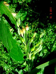
Coix lacryma-jobi L.
Coix lacryma-jobi L. (Photo credit: adaduitokla)

You may click to see the picture

Propagation & Cultivation:
Propagation by seeds, sown during monsoon (in India) at rate of 6-10 kg/ha. Seed dibbled 2.5 cm deep, at spacing of 60 x 60 cm. One intercultivation, before the plants tiller, and shade on ground may be necessary. Sufficient rains in early stage of growth and a dry period when grain is setting are necessary for good yields. Plants respond well to liberal applications of organic manure.

Chemical constituents:
Per 100 g, the seed is reported to contain 380 calories, 11.2 g H2O, 15.4 g protein, 6.2 g fat, 65.3 g total carbohydrate, 0.8 g fiber, 1.9 g ash, 25 mg Ca, 435 mg P, 5.0 mg Fe, 0 ug beta-carotene equivalent, 0.28 mg thiamine, 0.19 mg riboflavin, 4.3 mg niacin, and 0 mg ascorbic acid. According to Hager’s Handbook (List and Horhammer, 1969-1979), there is 50-60% starch 18.7% protein (with glutamic-acid, leucine, tyrosine, arginine, histidine, and lysine) and 5-10% fatty oil with glycerides of myristic- and palmitic-acids.

Uses
Weed to some, necklace to others, staff-of-life to others, job’s tear is a very useful and productive grass increasingly viewed as a potential energy source. Before Zea became popular in South Asia, Coix was rather widely cultivated as a cereal in India. Still taken as a minor cereal, it is pounded, threshed and winnowed, as a cereal or breadstuff. The pounded flour is sometimes mixed with water like barley for barley water. The pounded kernel is also made into a sweet dish by frying and coating with sugar. It is also husked and eaten out of hand like a peanut. Beers and wines are made from the fermented grain. Chinese use the grain, like barley, in soups and broths.

Medicinal Uses:
Folk Medicine
According to Hartwell (1967-1971), the fruits are used in folk remedies for abdominal tumors, esophageal, gastrointestinal, and lung cancers, various tumors, as well as excrescences, warts, and whitlows. This folk reputation is all the more interesting when reading that coixenolide has antitumor activity (List and Horhammer, 1969-1979). Job’s tear is also a folk remedy for abscess, anodyne, anthrax, appendicitis, arthritis, beriberi, bronchitis, catarrh, diabetes, dysentery, dysuria, edema, fever, gotter, halitosis, headache, hydrothorax, metroxenia, phthisis, pleurisy, pneumonia, puerperium, rheumatism, small-pox, splenitis, strangury, tenesmus, and worms (Duke and Wain, 1981). Walker (1971) cites other medicinal uses.

In Chinese medicine, the seeds strengthen the spleen and counteract “damp heat”, and are used for edema, diarrhea, rheumatoid arthritis and difficult urination.  Drains dampness, clears heat, eliminates pus, tonifies the spleen. This herb is added to medicinal formulas to regulate fluid retention and counteract inflammation. It is very good for all conditions and diseases associated with edema and inflammation, including pus, diarrhea, phlegm, edema or abscesses of either the lungs or the intestines, and rheumatic and arthritic conditions. A tea from the boiled seeds is drunk as part of a treatment to cure warts. It is also used in the treatment of lung abscess, lobar pneumonia, appendicitis, rheumatoid arthritis, beriberi, diarrhea, oedema and difficult urination.  The roots have been used in the treatment of menstrual disorders. The FDA has approved testing for cancer therapy. Currently going through testing, the Kanglaite Injection is a new effective diphasic anti-cancer medicine prepared by extracting with modern technology the active anti-cancer component from the Coix Seed, to form an advanced dosage form for intravenous and intra- arterial perfusion. It had been proved experimentally and clinically that the Kanglaite Injection had a broad spectrum of anti-tumor and anti-metastasis action, such as hepatic cancer and pulmonary cancer, along with the action of enhancing host immunity. When used in combined treatment with chemotherapy or radiotherapy, the Kanglaite Injection can increase the sensitivity of tumor cells, reduce the toxicity of chemotherapy and radiotherapy, relieve cancerous pain, improve cachexia, and raise the quality of life in advanced cancer victims. As a fat emulsion, the Kanglaite Injection can provide patients with high-energy nutrients with little toxicity.  It inhibits formation of new blood vessels that promote tumor growth, counteracts weight loss due to cancer.

Some of the latest research also shows that Job’s tears is immunostimulating, induces interferon, Bronchodialates; Lowers blood sugar; Reduces muscle spasms and is anti-convulsant; Stimulates respiration in small doses and inhibits it in higher doses; reduces arterial plaque; Anti-inflammatory, possibly through the suppression of macrophage activity

Disclaimer:
The information presented herein is intended for educational purposes only. Individual results may vary, and before using any supplements, it is always advisable to consult with your own health care provider.

Resources:
http://www.hort.purdue.edu/newcrop/duke_energy/Coix_lacryma-jobi.html
http://www.jadeinstitute.com/herbal-detail-page.php?show=25&order=common_name
http://www.robsplants.com/plants/CoixLacry
http://www.herbnet.com/Herb%20Uses_IJK.htm


Categories
Ailmemts & Remedies

Severe Acute Respiratory Syndrome (SARS)

Definition::
Severe acute respiratory syndrome  is a respiratory disease in humans which is caused by the SARS coronavirus (SARS-CoV). There was one near pandemic, between the months of November 2002 and July 2003, with 8,422 known infected cases and 916 confirmed human deaths (a case-fatality rate of 10.9%) worldwide being listed in the World Health Organization’s (WHO) 21 April 2004 concluding report. Within a matter of weeks in early 2003, SARS spread from Hong Kong to rapidly infect individuals in some 37 countries around the world.

CLICK & SEE THE PICTURES

As of today, the spread of SARS has been fully contained, with the last infected human case seen in June 2003 (disregarding a laboratory induced infection case in 2004). However, SARS is not claimed to have been eradicated (unlike smallpox), as it may still be present in its natural host reservoirs (animal populations) and may potentially return into the human population in the future.

Mortality by age group as of 8 May 2003 is below 1% for people aged 24 or younger, 6% for those 25 to 44, 15% in those 45 to 64 and more than 50% for those over 65. For comparison, the case fatality rate for influenza is usually around 0.6% (primarily among the elderly) but can rise as high as 33% in locally severe epidemics of new strains. The mortality rate of the primary viral pneumonia form is about 70%.

Symptoms:
The main symptoms of SARS are:

•High fever (above 38°C)
•Dry cough
•Breathing difficulties
*Other breathing symptoms
•Headache
•Muscular aches and stiffness
•Loss of appetite
•Malaise or tiredness
•Confusion
•Rash

The most common symptoms are:
*Chills and shaking
*Cough — usually starts 2-3 days after other symptoms
*Fever
*Headache
*Muscle aches

Less common symptoms include:
*Cough that produces phlegm (sputum)
*Diarrhea
*Dizziness
*Nausea and vomiting
*Runny nose
*Sore throat

These symptoms are typical of many severe respiratory infections. There have only ever been a few cases of SARS reported in the UK, so if you’ve similar symptoms, it’s far more likely to be a more typical form of pneumonia. Even if you’ve recently returned from south-east Asia, there’s little risk that you have SARS as the virus has been contained.

Causes:
Coronaviruses are positive-strand, enveloped RNA viruses that are important pathogens of mammals and birds. This group of viruses cause enteric or respiratory tract infections in a variety of animals including humans, livestock and pets.

CLICK & SEE

Initial electron microscopic examination in Hong Kong and Germany found viral particles with structures suggesting paramyxovirus in respiratory secretions of SARS patients. Subsequently, in Canada, electron microscopic examination found viral particles with structures suggestive of metapneumovirus (a subtype of paramyxovirus) in respiratory secretions. Chinese researchers also reported that a Chlamydophila-like disease may be behind SARS. The Pasteur Institute in Paris identified coronavirus in samples taken from six patients, so did the laboratory of Malik Peiris at the University of Hong Kong, which in fact was the first to announce (on 21 March 2003) the discovery of a new coronavirus as the possible cause of SARS after successfully cultivating it from tissue samples and was also amongst the first to develop a test for the presence of the virus. The CDC noted viral particles in affected tissue (finding a virus in tissue rather than secretions suggests that it is actually pathogenic rather than an incidental finding). Upon electron microscopy, these tissue viral inclusions resembled coronaviruses, and comparison of viral genetic material obtained by PCR with existing genetic libraries suggested that the virus was a previously unrecognized coronavirus. Sequencing of the virus genome — which computers at the British Columbia Cancer Agency in Vancouver completed at 4 a.m. Saturday, 12 April 2003 — was the first step toward developing a diagnostic test for the virus, and possibly a vaccine. A test was developed for antibodies to the virus, and it was found that patients did indeed develop such antibodies over the course of the disease, which is highly suggestive of a causative role.

On 16 April 2003, the WHO issued a press release stating that a coronavirus identified by a number of laboratories was the official cause of SARS. Scientists at Erasmus University in Rotterdam, the Netherlands demonstrated that the SARS coronavirus fulfilled Koch’s postulates thereby confirming it as the causative agent. In the experiments, macaques infected with the virus developed the same symptoms as human SARS victims.

An article published in The Lancet identifies a coronavirus as the probable causative agent.

In late May 2003, studies from samples of wild animals sold as food in the local market in Guangdong, China found that the SARS coronavirus could be isolated from palm civets (Paguma sp.), but the animals did not always show clinical signs. The preliminary conclusion was that the SARS virus crossed the xenographic barrier from palm civet to humans, and more than 10,000 masked palm civets were destroyed in Guangdong Province. Virus was also later found in raccoon dogs (Nyctereuteus sp.), ferret badgers (Melogale spp.) and domestic cats. In 2005, two studies identified a number of SARS-like coronaviruses in Chinese bats. Phylogenetic analysis of these viruses indicated a high probability that SARS coronavirus originated in bats and spread to humans either directly, or through animals held in Chinese markets. The bats did not show any visible signs of disease, but are the likely natural reservoirs of SARS-like coronaviruses. In late 2006, scientists from the Chinese Centre for Disease Control and Prevention of Hong Kong University and the Guangzhou Centre for Disease Control and Prevention established a genetic link between the SARS coronavirus appearing in civet cats and humans, bearing out claims that the disease had jumped across species

Viral replication:
Coronavirus (CoV) genome replication takes place in the cytoplasm in a membrane-protected microenvironment and starts with the translation of the genome to produce the viral replicase. CoV transcription involves a discontinuous RNA synthesis (template switch) during the extension of a negative copy of the subgenomic mRNAs. The requirement for base pairing during transcription has been formally demonstrated in arteriviruses and CoVs. The CoV N protein is required for coronavirus RNA synthesis and has RNA chaperon activity that may be involved in template switch. Both viral and cellular proteins are required for replication and transcription. CoVs initiate translation by cap-dependent and cap-independent mechanisms. Cell macromolecular synthesis may be controlled after CoV infection by locating some virus proteins in the host cell nucleus. Infection by different coronaviruses cause in the host alteration in the transcription and translation patterns, in the cell cycle, the cytoskeleton, apoptosis and coagulation pathways, inflammation and immune and stress responses. The balance between genes up- and down-regulated could explain the pathogenesis caused by these viruses. Coronavirus expression systems based on single genome constructed by targeted recombination, or by using infectious cDNAs, have been developed. The possibility of expressing different genes under the control of transcription regulating sequences (TRSs) with programmable strength and engineering tissue and species tropism indicates that CoV vectors are flexible. CoV based vectors have emerged with high potential vaccine development and possibly for gene therapy

Possible Complications:
*Respiratory failure
*Liver failure
*Heart failure
.
Diagnosis:
SARS may be suspected in a patient who has:

1.Any of the symptoms, including a fever of 38 °C (100.4 °F) or higher, and
2.Either a history of:
…..1.Contact (sexual or casual) with someone with a diagnosis of SARS within the last 10 days OR
…..2.Travel to any of the regions identified by the WHO as areas with recent local transmission of SARS (affected regions as of 10 May 2003[13] were parts of China, Hong Kong, Singapore and the province of Ontario, Canada).

A probable case of SARS has the above findings plus positive chest X-ray findings of atypical pneumonia or respiratory distress syndrome.

With the advent of diagnostic tests for the coronavirus probably responsible for SARS, the WHO has added the category of “laboratory confirmed SARS” for patients who would otherwise fit the above “probable” category who do not (yet) have the chest x-ray changes but do have positive laboratory diagnosis of SARS based on one of the approved tests (ELISA, immunofluorescence or PCR).

The chest X-ray (CXR) appearance of SARS is variable. There is no pathognomonic appearance of SARS but is commonly felt to be abnormal with patchy infiltrates in any part of the lungs. The initial CXR may be clear.

White blood cell and platelet counts are often low. Early reports indicated a tendency to relative neutrophilia and a relative lymphopenia — relative because the total number of white blood cells tends to be low. Other laboratory tests suggest raised lactate dehydrogenase and slightly raised creatine kinase and C-Reactive protein levels.

With the identification and sequencing of the RNA of the coronavirus responsible for SARS on 12 April 2003, several diagnostic test kits have been produced and are now being tested for their suitability for use.

Three possible diagnostic tests have emerged, each with drawbacks. The first, an ELISA (enzyme-linked immunosorbent assay) test detects antibodies to SARS reliably but only 21 days after the onset of symptoms. The second, an immunofluorescence assay, can detect antibodies 10 days after the onset of the disease but is a labour and time intensive test, requiring an immunofluorescence microscope and an experienced operator. The last test is a polymerase chain reaction (PCR) test that can detect genetic material of the SARS virus in specimens ranging from blood, sputum, tissue samples and stools. The PCR tests so far have proven to be very specific but not very sensitive. This means that while a positive PCR test result is strongly indicative that the patient is infected with SARS, a negative test result does not mean that the patient does not have SARS.

The WHO has issued guidelines for using these diagnostic tests.  There is currently no rapid screening test for SARS and research is ongoing.

Treatment:
Antibiotics are ineffective as SARS is a viral disease. Treatment of SARS so far has been largely supportive with antipyretics, supplemental oxygen and ventilatory support as needed.

Suspected cases of SARS must be isolated, preferably in negative pressure rooms, with complete barrier nursing precautions taken for any necessary contact with these patients.

There was initially anecdotal support for steroids and the antiviral drug ribavirin, but no published evidence has supported this therapy.

Researchers are currently testing all known antiviral treatments for other diseases including AIDS, hepatitis, influenza and others on the SARS-causing coronavirus.

There is some evidence that some of the more serious damage in SARS is due to the body’s own immune system overreacting to the virus – a cytokine storm. Research is continuing in this area.

In December 2004 it was reported that Chinese researchers had produced a SARS vaccine, it has been tested on a group of 36 volunteers, 24 of whom developed antibodies against the virus.

A 2006 systematic review of all the studies done on the 2003 SARS epidemic found no evidence that antivirals, steroids or other therapies helped patients. A few suggested they caused harm.

The clinical treatment of SARS has been relatively ineffective with most high risk patients requiring artificial ventilation. Currently, corticosteroids and Ribavirin are the most common drugs used for treatment of SARS (Wu et al., 2004). In vitro studies of Ribavirin have yielded little results at clinical, nontoxic concentrations. Better combinations of drugs that have yielded a more positive clinical outcome (when administered early) have included the use of Kaletra, Ribavirin and corticosteroids. The administration of corticosteroids, marketed as Prednisone, during viral infections has been controversial. Lymphopenia can also be a side effect of corticosteroids even further decreasing the immune response and allowing a spike in the viral load; yet physicians must balance the need for the anti-inflammatory treatment of corticosteroids (Murphy 2008). Clinicians have also noticed positive results during the use of human interferon and Glycyrrhizin. No compounds have yielded inhibitory results of any significance. The HIV protease inhibitors Ritonavir and Saquinavir did not show any inhibitory effect at nontoxic levels. Iminocyclitol 7 has been found to have an inhibitory effect on SARS-CoV in that it disrupts the envelope glycoprotein processing. Iminocyclitol 7 specifically inhibits the production of human fucosidase and in vitro trials yielded promising results in the treatment of SARS, yet one problem exists. A deficiency of fucosidase can lead to a condition known as fucosidosis in which there is a decrease in neurological function.

Prognosis:
The death rate from SARS was 9 to 12% of those diagnosed. In people over age 65, the death rate was higher than 50%. The illness was milder in younger patients.

Many more people became sick enough to need breathing assistance. And even more people had to go to hospital intensive care units.

Public health policies have been effective at controlling outbreaks. Many nations have stopped the epidemic in their own countries. All countries must continue to be careful to keep this disease under control. Viruses in the coronavirus family are known for their ability to change (mutate) in order to spread among humans.
.
Prevention:
The WHO set up a network for doctors and researchers dealing with SARS, consisting of a secure web site to study chest x-rays and a teleconference.

A SARS-treating hospital in Taiwan.Attempts were made to control further SARS infection through the use of quarantine. Over 1200 were under quarantine in Hong Kong, while in Singapore and Taiwan, 977 and 1147 were quarantined respectively. Canada also put thousands of people under quarantine.[14] In Singapore, schools were closed for 10 days and in Hong Kong they were closed until 21 April to contain the spread of SARS.

On 27 March 2003, the WHO recommended the screening of airline passengers for the symptoms of SARS.

In Singapore, a single hospital, Tan Tock Seng Hospital, was designated as the sole treatment and isolation centre for all confirmed and probable cases of the disease on 22 March. Subsequently, all hospitals implemented measures whereby all staff members were required to submit to temperature checks twice a day, visitorship was restricted only to pediatric, obstetric and selected other patients, and even then, only one person was allowed to visit at a time. To overcome this inconvenience, videoconferencing was utilised. A dedicated phoneline was designated to report SARS cases, whereupon a private ambulance service was dispatched to transport them to Tan Tock Seng Hospital.

On 24 March, Singapore’s Ministry of Health invoked the Infectious Diseases Act, allowing for a 10-day mandatory home quarantine to be imposed on all who may have come in contact with SARS patients. SARS patients who have been discharged from hospitals were under 21 days of home quarantine, with telephone surveillance requiring them to answer the phone when randomly called up. Discharged probable SARS patients and some recovered cases of suspected SARS patients are similarly required to be home quarantined for 14 days. Security officers from CISCO, a Singaporean auxiliary police force, were utilised to serve quarantine orders to their homes, and installed an electronic picture (ePIC) camera outside the doors of each contact. Sparked in particular by the news surrounding an elderly man who disregarded the quarantine order, flashing it to the public as he strolled to eating outlets and causing a minor exodus of patrons which persisted until the fears over the disease abated, the Singapore government called for an urgent meeting in Parliament on 24 April to amend the Infectious Disease Act and include penalties for violations, revealing at least 11 other violators of quarantine orders. These amendments included:

…*the requirement of suspected persons of infectious diseases to be brought to designated treatment centres, and their prohibition from going to public places;

…*the designation of contaminated areas and the restriction of access to them, and the destruction of suspected sources of infection;

…*the introduction of the power to tag offenders who break home quarantine (persons who failed to be contacted three times by phone consecutively) with electronic wrist tags, and the imposition of fines without court trial;

…*the ability to charge repeated offenders in court which may lead to imprisonment; and

…*the prosecution of anyone caught lying to health officials about their travel to SARS-affected areas or contacts with SARS patients.

Thermal imaging at Taoyuan Airport’s International checkpoint.On 23 April the WHO advised against all but essential travel to Toronto, noting that a small number of persons from Toronto appear to have “exported” SARS to other parts of the world. Toronto public health officials noted that only one of the supposedly exported cases had been diagnosed as SARS and that new SARS cases in Toronto were originating only in hospitals. Nevertheless, the WHO advisory was immediately followed by similar advisories by several governments to their citizens. On 29 April WHO announced that the advisory would be withdrawn on 30 April. Toronto tourism suffered as a result of the WHO advisory, prompting The Rolling Stones and others to organize the massive Molson Canadian Rocks for Toronto concert, commonly known as SARSstock, to revitalize the city’s tourism trade.

Also on 23 April, Singapore instituted thermal imaging scans to screen all passengers departing Singapore from Singapore Changi Airport. It also stepped up screening of travelers at its Woodlands and Tuas checkpoints with Malaysia. Singapore had previously implemented this screening method for incoming passengers from other SARS affected areas but was to include all travelers into and out of Singapore by mid- to late May.

In addition, students and teachers in Singapore were issued with free personal oral digital thermometers. Students took their temperatures daily, usually two or three times a day, but the temperature-taking exercises were suspended with the waning of the outbreak.

Taiwan Taoyuan International Airport also added SARS checkpoints with an infrared screening system similar to Singapore’s Changi Airport.

Disclaimer: This information is not meant to be a substitute for professional medical advise or help. It is always best to consult with a Physician about serious health concerns. This information is in no way intended to diagnose or prescribe remedies.This is purely for educational purpose.

Resources:
http://en.wikipedia.org/wiki/Severe_acute_respiratory_syndrome
http://health.nytimes.com/health/guides/disease/severe-acute-respiratory-syndrome-sars/overview.html
http://www.bbc.co.uk/health/physical_health/conditions/sars1.shtml

http://www.wpro.who.int/NR/rdonlyres/464C8256-9D58-44B3-B292-DB3518117CA8/0/SchematicdrawingsofSARS.jpg

Enhanced by Zemanta
Categories
Herbs & Plants

Copal

[amazon_link asins=’B00TWVI79Y,B00KMD7GHK,B001AYCBMA,B00KMDDWPK,B002U35BDQ,B00TWVN2LC,B00I6XV93U,B0019U0KNC,B00ET0MG2C’ template=’ProductCarousel’ store=’finmeacur-20′ marketplace=’US’ link_id=’711f3f70-53c7-11e7-955e-7fddefc322e7′]

Botanical Name: Protium copal
Common Name: Copal, Pom

Habitat :Protium copal is native to Guatemalan in South America, also grows in several places in Africa

Description:
Copal is a name given to tree resin that is particularly identified with the aromatic resins used by the cultures of pre-Columbian Mesoamerica as ceremonially burned incense and other purposes. More generally, the term copal describes resinous substances in an intermediate stage of polymerization and hardening between “gummier” resins and amber. The word copal is derived from the Nahuatl language word copalli, meaning “incense
CLICK & SEE THE PICTURES
To the pre-Columbian Maya and contemporary Maya peoples it is known in the various Mayan languages as pom (or a close variation thereof), although the word itself has been demonstrated to be a loanword to Mayan from Mixe–Zoquean languages.

Copal is still used by a number of indigenous peoples of Mexico and Central America as an incense and during sweat lodge ceremonies.  It is available in different forms. The hard, amber-like yellow copal is a less expensive version. The white copal, a hard, milky, sticky substance, is a more expensive version of the same resin.

Copal was also grown in East Africa, (the common species there being Hymenaea verrucosa) initially feeding an Indian Ocean demand for incense. By the 18th Century, Europeans found it to be a valuable ingredient in making a good wood varnish. It became widely used in the manufacture of furniture and carriages. By the late 19th and early 20th century varnish manufacturers in England and America were using it on train carriages, greatly swelling its demand.

In 1859 Americans consumed 68 percent of the East African trade, which was controlled through the Sultan of Zanzibar, with Germany receiving 24 percent. The American Civil War and the creation of the Suez Canal led to Germany, India and Hong Kong taking the majority by the end of that century.

East Africa apparently had a higher amount of subfossil copal, which is found one or two meters below living copal trees from roots of trees that may have lived thousands of years earlier. This subfossil copal produces a harder varnish. Subfossil copal is also well-known from New Zealand (Kauri gum), Japan, the Dominican Republic, Colombia and Madagascar. It often has inclusions and is sometimes sold as “young amber”. Copal can be easily distinguished from genuine amber by its lighter citrine colour and its surface getting tacky with a drop of acetone or chloroform

Medicinal Uses:
Chickleros who stayed in the bush for months relied on fresh copal resin to treat painful cavities, a piece of resin was stuffed into the cavity and, in a few days, the tooth broke apart and was easily expelled. The bark is scraped, powdered, and applied to wounds, sores, and infections.  Cut a piece of bark 2.5 cm x 15 cm; boil in 3 cups of water for 10 minutes and drink 1 cup before meals for stomach complaints and intestinal parasites.  It is also used as a remedy for fright and dizziness.

Disclaimer:
The information presented herein is intended for educational purposes only. Individual results may vary, and before using any supplements, it is always advisable to consult with your own health care provider.

Resources:
http://belize.com/copal.html
http://www.marc.ucsb.edu/elpilar/features/trail/documents/plants/copal.htm
http://waynesword.palomar.edu/ecoph22.htm
http://en.wikipedia.org/wiki/File:Copal_with_insects_close-up.jpg

Categories
Herbs & Plants

Crinum asiatica

[amazon_textlink asin=’B000UOG9QK’ text=’Giant Crinum Lily’ template=’ProductLink’ store=’finmeacur-20′ marketplace=’US’ link_id=’cc5daa86-9e8c-11e7-a95b-0fd7e0843d37′]

[amazon_link asins=’B00G124Q42,B00ZLQB962,B017PMKT9W,B06XDXSHX2,B001ECSF6E,B072BGPG8L,B01CUWIQ6U,B00IGHC9YY,B00IGGLYI2′ template=’ProductCarousel’ store=’finmeacur-20′ marketplace=’US’ link_id=’5aa724ce-9e8d-11e7-9ebd-63d6e0e9352c’]

Botanical Name : Crinum asiatica
Family: Amaryllidaceae
Tribe: Amaryllideae
Subtribe: Crininae
Genus: Crinum
Species: C. asiaticum
Kingdom: Plantae
Order: Asparagales

Common names : , Grand Crinum Lily, Grand Crinum Lily, Spider Lily,Kanwal, Nagdaun, Badakanvar, Chindar, Gadambhikanda, Nagadamani, Sudarshan, Poison bulb.


Habitat
:Crinum asiaticum is native to tropical southeastern Asia( China, Hong Kong, India, Ryukyu Islands and Mainland Japan). It is now a favorite landscape plant in Florida, the Gulf Coast, California and other warm climate areas.

Description:
This big crinum lily makes an imposing presence in the garden. The dark green strap-like leaves may be more than 3 ft (1 m) long and 4 in (10 cm) wide. These are held erect and arranged in a spiral rosette to form impressive clumps up to 5 ft (1.5 m) in height by 7 ft (2 m) in width. The leaves emerge from huge bulbs that may weigh 10-20 lbs (5-9 kg)! Flowers are shaped like tubes that flair open into a crown of narrow petals. The flowers are white and are arranged in clusters atop thick, succulent stems.
CLICK & SEE THE PICTURES
Main features
: Grows up to 1.5m, in freshwater or brackish swamps.

Leaves: Long (2m) strap-like, fleshy.

Flowers: Clustered; white; fragrant.

Fruits: Globose; shiny white when ripe; seeds irregularly shape.

Cultivation:
Grand crinum lily is happy in just about any type of well drained soil.
Light: It prefers bright sunny situations but will grow in part shade.

Moisture: Provide average water. Crinum lilies do well in dry soils and are fairly drought tolerant.

Hardiness:USDA Zone 9 – 11. You can grow this crinum in Zone 8, but foliage is killed by freezing winter temperatures. I have several of these growing in Tallahassee; they suffer some degree of cold damage every year but quickly recover their attractiveness in the spring.

Propagation: To propagate crinums, dig up a clump and separate the small offset bulbs from the parent bulb. Plant these in pots or directly in the garden where they will quickly root to form new plants.Grand crinum lily is happy in just about any type of well drained soil.

Medicinal Uses:

Traditional medicinal uses:
It is used as a poultice for aches, sores and chaps. Crushed leaves are used to treat piles, mixed with honey and applied to wounds and abscesses.

Click to see :Tonsilitis Home Remedy Using Crinum

Other Usage:
Use the grand crinum to create a tropical mood near the pool or patio. Use like sculpture to create a focal point in the garden or in an expanse of lawn. This big lily looks great with palm trees and ornamental grasses. Their drought resistance make them useful in xeriscapes. It also does well in a container.

Known Hazards:All parts of crinum lily may cause severe discomfort if ingested, and the sap alone can cause skin irritation.

Disclaimer:
The information presented herein is intended for educational purposes only. Individual results may vary, and before using any supplements, it is always advisable to consult with your own health care provider.

.

Resources:
http://www.floridata.com/ref/c/crin_asi.cfm
http://en.wikipedia.org/wiki/Crinum_asiaticum
http://vaniindia.org.whbus12.onlyfordemo.com/herbal/plantdir.asp
http://www.naturia.per.sg/buloh/plants/crinum_lily.htm

Enhanced by Zemanta
css.php