Botanical Name : Bixa orellana Family: Bixaceae Genus: Bixa Species: B. orellana Kingdom: Plantae Order: Malvales
Synonym(s): Bixa katangensis DelpierreOrellana americana L.Orellana orellana Kuntze
Common Names : Annatto Seed , achiote, lipstick tree. It is also known as Aploppas, and its original Tupi name urucu.(Arabic) : galuga
(Bengali) : latkan
(Creole) : chiót, woukou
(English) : annatto tree, arnato tree, lipstick, lipstick tree
(Filipino) : echuete, sotis
(French) : annato, annatto, chiote, rocouyer, roucou
(Hindi) : latkan
(Indonesian) : jarak belanda, kesumba, kunyit jawa
(Khmer) : châm’-puu, châm-puu chrâluek
(Lao (Sino-Tibetan)) : kh’am, satii, somz phuu
(Malay) : galuga, jarak belanda, kesumba, kunyit jawa
(Portuguese) : urucum
(Spanish) : achiote, anato, bija
(Swahili) : mzingefuri
(Tamil) : japhara
(Thai) : kam set, kam tai
(Vietnamese) : diêù nhuôm, siêm phung
Habitat :Bixa orellana is native to tropical Central and South America but now widely distributed throughout the tropics. Introduced to the Philippines during its Spanish period. It is cultivated there and in Southeast Asia, where it was introduced by the Spanish in the 17th century.
Description:
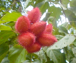
Bixa orellana is an evergreen shrub or small tree, 2-8 m high; trunk up to 10 cm in diameter; bark light to dark brown, tough, smooth, sometimes fissured, lenticellate; inner bark pinkish towards the outside with orange sap, slightly bitter; twigs green with minute, rusty, reddish-brown scales, becoming dark brown. Leaves spirally arranged, simple, stipulate, ovate, 7.5-24 x 4-16 cm, shallowly cordate to truncate at base, longly acuminate at apex, green or dark green above, grey or brownish-green beneath; scaly when young, glabrous; petiole terete, thickened at both ends, 2.5-12 cm long. Flowers in terminal branched panicles, 8-50 flowered, fragrant, 4-6 cm across; pedicel scaly, thickened at the apex, bearing 5-6 large glands; sepals 4-5, free, obovate, 1-1.2 cm long, caducous, covered with reddish-brown scales; petals 4-7, obovate, 2-3 x 1-2 cm, pinkish, whitish or purplish tinged; stalks scaly; stamens numerous, 1.6 cm long; anthers violet; pistil 1.6 cm long, composed of bristly 1-celled, superior ovary; style thickened upwards, 12-15 mm long; a short, 2-lobed stigma. Fruit a spherical or broadly elongated ovoid capsule, 2-4 x 2-3.5 cm, flattened, 2 valved, more or less densely cloaked with long bristles, green, greenish-brown or red when mature; seeds numerous, obovoid and angular, 4.5 mm long, with bright orange-red fleshy coats. ‘Bixa’ is derived from a local South American name. click to see the pictures>…..(01)..…..(1)..…...(2).……..(3).……....(4).……..(5)..…....(6).……..(7)………...(8)..…….(9)..……..(10)..…...(11)
The inedible fruit is harvested for its seeds, which contain annatto, also called bixin. It can be extracted by stirring the seeds in water. It is used to color food products, such as cheeses, fish, and salad oil. Sold as a paste or powder for culinary use, mainly as a color, it is known as “achiote,” “annatto,” “bijol,” or “pimentão doce.” It is a main ingredient in the Yucatecan spice mixture recado rojo, or “achiote paste.” The seeds are ground and used as a subtly flavored and colorful additive in Latin American, Jamaican, Chamorro and Filipino cuisine. Annatto is growing in popularity as a natural alternative to synthetic food coloring compounds. While it has a distinct flavor of its own, it can be used to color and flavor rice instead of the much more expensive saffron. It is an important ingredient of cochinita pibil, the spicy pork dish popular in Mexico. It is also a key ingredient in the drink tascalate from Chiapas, Mexico.
In several European countries (e.g. Great Britain, Denmark, Sweden, Norway) the pigment, extracted by solvent or boiling the seeds in oil, have been and often still is used as color in margarines and several other foods. The pigment has E-number E160b. The seeds are collected from wild-growing bushes or from plantations, in Latin America, Africa (e.g. Kenya) and Asia. However, since there is no strong organization promoting the use of annatto, the color beta carotene, which is more expensive, has pushed the natural pigment out of many applications
Edible Uses: Achiote paste, favored in Yucatán, Oaxacan, and Belizean cuisine, is made from the slightly bitter, earthy flavored, red annatto seeds, mixed with other spices and ground into a paste. Achiote is a distinctly colored and flavored mainstay of Mexican and Belizean kitchens.
The paste is dissolved in either lemon juice, water, oil or vinegar to create a marinade, and marinated or rubbed directly upon meat. The meat is then grilled, baked, barbecued or broiled. Sometimes it is added to corn dough to create a zesty flavor and color in empanadas and red tamales.
Medicinal Uses:
The achiote has long been used by American Indians to make body paint, especially for the lips, which is the origin of the plant’s nickname, lipstick tree. The use of the dye in the hair by men of the Tsáchila of Ecuador is the origin of their usual Spanish name, the Colorados.
In developing countries, particularly in Colombia, people with low income and less access to modern medicine resources use folk medicine and natural remedies for the treatment of common infections. Achiote is among those herbs used in Colombian folk medicine to treat infections of microbial origin. Adding to the known health benefits exerted by carotenoids, a bioactive sesquiterpene from achiote exhibited moderate anti-fungal activity. Extracts of the leaves of achiote possess antimicrobial activity against Gram positive microorganisms, with maximum activity against Bacillus pumilus. Achiote leaves have been employed to treat malaria and Leishmaniasis.
Disclaimer : The information presented herein is intended for educational purposes only. Individual results may vary, and before using any supplement, it is always advisable to consult with your own health care provider
Habitat : The Florida fishpoison tree grows in coastal zones. It prefers well-drained sandy soils, with a top layer of humus. The tree has some tolerance to short-term storm surges of brackish water or seawater. Although it grows in coastal conditions, the tree is usually protected from direct salt spray by adjoining vegetation. Established trees are highly tolerant of drought. Its sensitivity to the cold limits Florida fishpoison tree to areas no colder than plant hardiness zone11.
Description: Florida fishpoison tree, is a medium-sized, deciduous, tropical tree endemic to southern Florida, the Florida Keys, Texas, Caribbean, and Latin America.Native Americans of the West Indies discovered extracts from the tree could sedate fish, allowing them to be caught by hand. This practice led to the tree’s common names—fishpoison and fishfuddle. The tree has medicinal value as an analgesic and sedative.
The generic name is Latin for “fish killer”, and the specific name is Latin for “little fish,
Size: The Florida fishpoison tree attains medium size with heights of 12 to 15 m and bole diameters of 46 to 118 cm. An irregular, open crown develops with stout, erect branches.
Foliage: Its deciduous leaves (9 to 23 cm long) are alternate and pinnately compound. Five to 11 leaflets (each 4 to 8 cm long) are present in an opposite arrangement. Leaflets are dark green above and distinctly paler grayish-green below with pubescence.
Flowers/Fruit: The white flowers are tinged with red or pink. They appear in pea-like clusters in May and are attractive to bees. Trees will potentially bloom when about 4 m tall and 4 years old. Flowers develop into a light brown, bean-like pod (8 to 10 cm long) with four papery wings. Ripening in July and August, the pods contain red-brown seeds with oval shapes.
Bark: Stem bark is thin and olive gray in color with irregular dark patches and many smaller scales. The bark has an unpleasant odor and a distinctly acrid and bitter taste, causing a burning sensation in the mouth.
Propagation: After removal from the ripe pod, seeds will germinate in 8 to 10 days when sown about 6 mm deep in moist soil. Until seedlings become well established, they should be fertilized and watered. Cuttings placed in moist soil quickly sprout roots. In fact, rooting has been observed to occur so readily that posts made from fresh timber occasionally take root unintentionally.
Medicinal Uses:
Florida fishpoison tree has long been used in traditional medicine for treating nervous conditions and pain. Recent scientific studies in animals suggest that bark extracts have potential for their anti-inflammatory, sedative, and anti-spasmodic effects.
Other Uses:e Ornamental: In areas with a suitable climate and soils, Florida fishpoison tre makes a hardy, medium-sized shade tree with attractive seasonal flowers. It is ideal for yards and along fence rows. The species is shade intolerant, requiring full sunlight for maximum development.
Wood: The yellow-brown wood of fishpoison tree is resistant to decay, making its timber suitable for outdoor usage, such as boat building, fence posts, and poles. The dense, tight-grained wood is also used as a fuel, to make charcoal, and as a good carving material.
Fish poison: Indigenous peoples all over the world used local poisonous plants to aid in catching fish, and because of this many plants bear common names descriptive of this use. Within its natural range, Native Americans used an extract from the bark, roots, twigs, and leaves of Florida fishpoison tree to sedate fish, making them easier to catch. A number of chemicals are present in the tree’s tissues that are toxic to fish, the principal one being the well-known Rotenone.
Disclaimer:The information presented herein is intended for educational purposes only. Individual results may vary, and before using any supplements, it is always advisable to consult with your own health care provider.
Alternative Names:Hepatic amebiasis; Extraintestinal amebiasis; Abscess – amebic liver Definition : Amebic liver abscess is a collection of pus in the liver caused by an intestinal parasite.Organisms that carry disease can travel through the blood stream into the liver and form an abscess, a collection of infected tissue and pus.
YOU MAY CLICK TO SEE THE PICTURES
….
Amebic liver abscess is an extraintestinal manifestation caused by a protozoa Entamoeba histolytica. The disease spreads through the ingestion of cysts in fecally-contaminated food or water. The infection is worldwide, most common in tropical areas usually in developing countries where poor sanitation exist.
This is an Abscess (walled area of infection/pus) in the liver that is caused by the organism Entamoeba histolytica. It is common in tropical and subtropical areas (southeast Asia, Africa, India, Latin America). In the U.S., it is most common in young Hispanic adults. It is also seen in those with recent travels to tropical areas, homosexuals, and among the inhabitants of institutions for the mentally ill.
Causes :-
Travel to endemic areas (as above), where the cysts of the parasite may be ingested by consuming water or food contaminated by fecal matter.
Amebic liver abscess is caused by Entamoeba histolytica, the same organism that causes amebiasis, an intestinal infection. The organism is carried by the blood from the intestines to the liver.
The disease spreads through ingestion of cysts in fecally-contaminated food or water, use of human waste as fertilizer, and person-to-person contact.
The infection occurs worldwide, but is most common in tropical areas where crowded living conditions and poor sanitation exist. Africa, Latin America, Southeast Asia, and India have significant health problems associated with this disease.
Risk Factors :-
*Malnutrition
*Old age
*Pregnancy
*Steroid use
*Cancer *Immunosuppression
*Alcoholism
*Recent travel to a tropical region
*Homosexuality, particularly in males
Symptoms:- There may or may not be symptoms of intestinal infection. Symptoms may include:
*Right upper abdominal pain
*Right sided chest pain worse on a deep breath
*Intense, continuous, or stabbing pain
*Chills
*Diarrhea precedes infection in 20% of patients
*Fever
*General discomfort, uneasiness, or ill feeling (malaise) *Jaundice *Joint pain
*Loss of appetite
*Sweating
*Weight loss
*malaise
Nature of the disease:-
Most infected people, perhaps 90%, are asymptomatic, but this disease has the potential to make the sufferer dangerously ill. It is estimated by the World Health Organization that about 70,000 people die annually worldwide.
Infections can sometimes last for years. Symptoms take from a few days to a few weeks to develop and manifest themselves, but usually it is about two to four weeks. Symptoms can range from mild diarrhoea to dysentery with blood and mucus. The blood comes from amoebae invading the lining of the intestine. In about 10% of invasive cases the amoebae enter the bloodstream and may travel to other organs in the body. Most commonly this means the liver, as this is where blood from the intestine reaches first, but they can end up almost anywhere.
Onset time is highly variable and the average asymptomatic infection persists for over a year. It is theorised that the absence of symptoms or their intensity may vary with such factors as strain of amoeba, immune response of the host, and perhaps associated bacteria and viruses.
In asymptomatic infections the amoeba lives by eating and digesting bacteria and food particles in the gut, a part of the gastrointestinal tract. It does not usually come in contact with the intestine itself due to the protective layer of mucus that lines the gut. Disease occurs when amoeba comes in contact with the cells lining the intestine. It then secretes the same substances it uses to digest bacteria, which include enzymes that destroy cell membranes and proteins. This process can lead to penetration and digestion of human tissues, resulting first in flask-shaped ulcers in the intestine. Entamoeba histolytica ingests the destroyed cells by phagocytosis and is often seen with red blood cells inside when viewed in stool samples. Especially in Latin America, a granulomatous mass (known as an amoeboma) may form in the wall of the ascending colon or rectum due to long-lasting immunological cellular response, and is sometimes confused with cancer.
Theoretically, the ingestion of one viable cyst can cause an infection.
Treatment
A medicine called metronidazole (Flagyl) is the usual treatment for liver abscess. Medications such as paromomycin must also be taken to remove intestinal amebiasis to prevent recurrence of the disease.
In rare cases, the abscess may need to be drained to help relieve some of the abdominal pain.
Prognosis:
Without treatment, the abscess may rupture and spread into other organs, leading to death. Persons who receive treatment have a very high chance of a complete cure or having only minor complications. Possible Complications :In the majority of cases, amoebas remain in the gastrointestinal tract of the hosts. Severe ulceration of the gastrointestinal mucosal surfaces occurs in less than 16% of cases. In fewer cases, the parasite invades the soft tissues, most commonly the liver. Only rarely are masses formed (amoebomas) that lead to intestinal obstruction.
The abscess may rupture into the abdominal cavity, the lining of the lungs, the lungs, or the sac around the heart. The infection can also spread to the brain.
When to Contact a Medical Professional
Call your health care provider if symptoms develop after travel to an area where the disease is known to occur.
Prevention: To help prevent the spread of amoebiasis around the home :
*Wash hands thoroughly with soap and hot running water for at least 10 seconds after using the toilet or changing a baby’s diaper, and before handling food.
*Clean bathrooms and toilets often; pay particular attention to toilet seats and taps.
*Avoid sharing towels or face washers.
To help prevent infection:
*Avoid raw vegetables when in endemic areas, as they may have been fertilized using human feces.
*Boil water or treat with iodine tablets.
*When traveling in tropical countries where poor sanitation exists, drink purified water and do not eat uncooked vegetables or unpeeled fruit.
Disclaimer: This information is not meant to be a substitute for professional medical advise or help. It is always best to consult with a Physician about serious health concerns. This information is in no way intended to diagnose or prescribe remedies.This is purely for educational purpose.
Definition:
Yellow fever (also called yellow jack, black vomit or sometimes American Plague) is an acute viral disease. It is an important cause of hemorrhagic illness in many African and South American countries despite existence of an effective vaccine. The yellow refers to the jaundice symptoms that affect some patients.It is a viral infection transmitted by mosquitoes.
Yellow fever is a viral hemorrhagic fever caused by the yellow fever virus. The yellow fever virus is a single-stranded enveloped virus that belongs to the flavivirus group. The disease can result in mild symptoms or severe illness and death (mortality rate 5-70%). Yellow fever derives its name from the yellowing of the skin and whites of the eyes (jaundice) that occur in some people infected with the virus. Jaundice is caused by the presence of bile pigment (bilirubin) in the bloodstream and results from damage to liver cells (hepatocytes) during severe infection.…click & see
The yellow fever virus infects mainly monkeys and humans: monkeys are the animal reservoir. Infection is transmitted from human to human, monkey to monkey, monkey to human, and human to monkey by daytime-biting mosquitos. Several species of Aedes and Haemoagogus mosquitos can serve as vectors, transmitting the virus during a blood meal.
Three types of transmission cycles exist for yellow fever: sylvatic (jungle), intermediate and urban. Although all three transmission cycles occur in Africa, only sylvatic and urban transmission cycles occur in South America.
* Occurs in monkeys infected by wild mosquitos in tropical rainforests
* Infected monkeys pass the virus to mosquitos during feeding
* Infected wild mosquitos bite humans entering the rainforest (accidental infection)
Intermediate yellow fever (monkey to human; human to monkey)
* Small-scale epidemics that occur in humid or semi-humid grasslands of Africa
* Separate villages experience simultaneous infections transmitted by semi-domestic mosquitos that infect both monkey and human hosts
* Most common type of outbreak in Africa
Urban yellow fever (human to human)
* Large epidemics occurring when the virus is introduced into high human population areas by migrants
* Domestic mosquitos of one species (Aedes aegypti) transmit the virus from person to person
* Monkeys are not involved in transmission
* Outbreaks spread from one source to cover a wide area
Yellow fever has been a source of several devastating epidemics. Yellow fever epidemics broke out in the 1700s in Italy, France, Spain, and England. 300,000 people are believed to have died from yellow fever in Spain during the 19th century. French soldiers were attacked by yellow fever during the 1802 Haitian Revolution; more than half of the army perished from the disease. Outbreaks followed by thousands of deaths occurred periodically in other Western Hemisphere locations until research, which included human volunteers (some of whom died), led to an understanding of the method of transmission to humans (primarily by mosquitos) and development of a vaccine and other preventive efforts in the early 20th century.
Despite the costly and sacrificial breakthrough research by Cuban physician Carlos Finlay, American physician Walter Reed, and many others over 100 years ago, unvaccinated populations in many developing nations in Africa and Central and South America continue to be at risk. As of 2001, the World Health Organization (WHO) estimates that yellow fever causes 200,000 illnesses and 30,000 deaths every year in unvaccinated populations.
Causes :-
Yellow fever is caused by a small virus that is spread by the bite of mosquitoes. This disease is common in South America and in sub-Saharan Africa.
Anyone can get yellow fever, but the elderly have a higher risk of severe infection. If a person is bitten by an infected mosquito, symptoms usually develop 3 – 6 days later.
Yellow fever has three stages:
1.Early stage: Headache, muscle aches, fever, loss of appetite, vomiting, and jaundice are common. After approximately 3 – 4 days, often symptoms go away briefly (remission).
2.Period of remission: After 3 – 4 days, fever and other symptoms go away. Most people will recover at this stage, but others may move onto the third, most dangerous stage (intoxication stage) within 24 hours.
3.Period of intoxication: Multi-organ dysfunction occurs. This includes liver and kidney failure, bleeding disorders/hemorrhage, and brain dysfunction including delirium, seizures, coma, shock, and death.
Although viral replication begins in cells at the site of the mosquito bite, symptoms of infection are not usually noted for a period of three to six days when the acute phase of infection presents. Acute yellow fever infection is characterized by high fever, muscle pain, backache, headache, shivers, loss of appetite, nausea and/or vomiting. Most people infected improve after three to four days.
However, within 24 hours of the disappearance of symptoms, up to 15% of those infected enter a toxic phase during which fever resumes, and the yellow fever virus quickly spreads to the kidneys, lymph nodes, spleen, bone marrow and liver. Liver invasion of one of the last stages to occur: as the liver is increasingly damaged, patients develop jaundice as bilirubin is released from damaged liver cells, experience abdominal pain and vomiting, and develop coagulopathies (inability of the blood to clot) characterized by bleeding from the mouth, nose, eyes and stomach, and presence of blood in vomit and stool. Up to 50% of people who enter the toxic phase die within two weeks of infection.
Diagnosis:-
Yellow fever may be difficult to diagnose, especially during the early stages, and may be confused with malaria, typhoid, other hemorrhagic fevers (dengue, Rift Valley, Venezuelan, Bolivian, Argentine, Lassa, Crimean-Congo, Marburg and Ebola), rickettsial infection, leptospirosis, viral hepatitis, other causes of liver failure and toxic hepatitis (e.g. carbon-tetrachloride poisoning).
Exams and Tests
A person with advanced yellow fever may show signs of liver failure, renal failure, and shock.
If you have symptoms of yellow fever, tell your doctor if you have traveled to areas where the disease is known to thrive. Blood tests can confirm the diagnosis.
Treatment :-
There is no specific treatment for yellow fever. Treatment for symptoms can include:
*Blood products for severe bleeding
*Dialysis for kidney failure
*Fluids through a vein (intravenous fluids)
The treatment for yellow fever is supportive: control of fever, fluids to treat dehydration, and intensive support related to organ damage.
The World Health Organization estimates 200,000 cases of yellow fever per year with approximately 30,000 deaths.
Yellow fever ranges in severity. Severe infections with internal bleeding and fever (hemorrhagic fever) are deadly in up to half of cases.
Historical reports have claimed a mortality rate of between 1 in 17 (5.8%) and 1 in 3 (33%). CDC has claimed that case-fatality rates from severe disease range from 15% to more than 50%. The WHO factsheet on yellow fever, updated in 2001, states that 15% of patients enter a “toxic phase” and that half of that number die within ten to fourteen days, with the other half recovering
If you will be traveling to an area where yellow fever is common:
*Sleep in screened housing
*Use mosquito repellents
*Wear clothing that fully covers your body
*There is an effective vaccine against yellow fever. Ask your doctor at least 10 – 14 days before traveling if you should be *vaccinated against yellow fever.
In 1937, Max Theiler, working at the Rockefeller Foundation, developed a safe and highly efficacious vaccine for yellow fever that gives a ten-year or more immunity from the virus. The vaccine consists of a live, but attenuated, virus called 17D. The 17D vaccine has been used commercially since the 1950s. The mechanisms of attenuation and immunogenicity for the 17D strain are not known. However, this vaccine is very safe, with few adverse reactions having been reported and millions of doses administered, and highly effective with over 90% of vaccinees developing a measurable immune response after the first dose.
Although the vaccine is considered safe, there are risks involved. The majority of adverse reactions to the 17D vaccine result from allergic reaction to the eggs in which the vaccine is grown. Persons with a known egg allergy should discuss this with their physician prior to vaccination. In addition, there is a small risk of neurologic disease and encephalitis, particularly in individuals with compromised immune systems and very young children. The 17D vaccine is contraindicated in infants, pregnant women, and anyone with a diminished immune capacity, including those taking immunosuppressant drugs.
According to the travel clinic at the University of Utah Hospital, the vaccine presents an increased risk of adverse reaction in adults aged 60 and older, with the risk increasing again after age 65, and again after age 70. The reaction is capable of producing multiple organ failure and should be evaluated carefully by a qualified health professional before being administered to the elderly.
Finally, there is a very small risk of more severe yellow fever-like disease associated with the vaccine. This reaction occurs in 1~3 vaccinees per million doses administered. This reaction, called YEL-AVD, causes a fairly severe disease closely resembling yellow fever caused by virulent strains of the virus. The risk factor/s for YEL-AVD are not known, although it has been suggested that it may be genetic. The 2`-5` oligoadenylate synthetase (OAS) component of the innate immune response has been shown to be particularly important in protection from Flavivirus infection. In at least one case of YEL-AVD, the patient was found to have an allelic mutation in a single nucleotide polymorphism (SNP) of the OAS gene. People most at risk of contracting the virus should be vaccinated. Woodcutters working in tropical areas should be particularly targeted for vaccination. Insecticides, protective clothing, and screening of houses are helpful, but not always sufficient for mosquito control; people should always use an insecticide spray while in certain areas. In affected areas, mosquito control methods have proven effective in decreasing the number of cases.
Recent studies have noted the increase in the number of areas affected by a number of mosquito-borne viral infections and have called for further research and funding for vaccines
Current research:-
In the hamster model of yellow fever, early administration of the antiviral ribavirin is an effective early treatment of many pathological features of the disease. Ribavirin treatment during the first five days after virus infection improved survival rates, reduced tissue damage in target organs (liver and spleen), prevented hepatocellular steatosis, and normalized alanine aminotransferase (a liver damage marker) levels. The results of this study suggest that ribavirin may be effective in the early treatment of yellow fever, and that its mechanism of action in reducing liver pathology in yellow fever virus infection may be similar to that observed with ribavirin in the treatment of hepatitis C, a virus related to yellow fever. Because ribavirin had failed to improve survival in a virulent primate (rhesus) model of yellow fever infection, it had been previously discounted as a possible therapy.
In 2007, the World Community Grid launched a project whereby computer modelling of the yellow fever virus (and related viruses), thousands of small molecules are screened for their potential anti-viral properties in fighting yellow fever. This is the first project to utilize computer simulations in seeking out medicines to directly attack the virus once a person is infected. This is a distributed process project similar to SETI@Home where the general public downloads the World Community Grid agent and the program (along with thousands of other users) screens thousands of molecules while their computer would be otherwise idle. If the user needs to use the computer the program sleeps. There are several different projects running, including a similar one screening for anti-AIDS drugs. The project covering yellow fever is called “Discovering Dengue Drugs – Together.” The software and information about the project can be found at: World Community Grid web site
Disclaimer: This information is not meant to be a substitute for professional medical advise or help. It is always best to consult with a Physician about serious health concerns. This information is in no way intended to diagnose or prescribe remedies.This is purely for educational purpose. Resources:
http://en.wikipedia.org/wiki/Yellow_fever
http://www.nlm.nih.gov/medlineplus/ency/article/001365.htm
http://microbiology.suite101.com/article.cfm/yellow_fever


_seeds.jpg)