Categories
Diagnonistic Test

Pleural Fluid Sampling (or Thoracentesis)

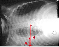
Pleural effusion Chest x-ray of a pleural effu...
Image via Wikipedia

[amazon_link asins=’B01J8WALE0,B00GNOPG8S,1331951720,1893441776,B01MXOPZIB,B01J2IC12U,B01J37411E,B009KT2GWM,B01J2GLUVU’ template=’ProductCarousel’ store=’finmeacur-20′ marketplace=’US’ link_id=’316e2df4-5fab-11e7-aab1-4db380a805de’]


Definition:
Thoracentesis is a procedure used to obtain a sample of fluid from the space around the lungs. Normally, only a thin layer of fluid is present in the area between the lungs and chest wall (show radiograph 1). However, some conditions can cause a large amount of fluid to accumulate. This collection of fluid is called a pleural effusion (show radiograph 2). Thoracentesis is done to collect a sample of the fluid, which can help determine why the pleural effusion developed.
CLICK & SEE
Some infections and diseases cause fluid to accumulate in the space between the lung and the rib cage or between the lung and the diaphragm. This collection of fluid is called a pleural effusion. A pleural effusion might be detected on a chest x-ray. Sampling this fluid is important because it enables doctors to understand what caused the fluid to collect and how to treat the problem. The fluid can be sampled with a needle.

Reasons for Thoracentesis: — A thoracentesis is performed to determine the cause of a pleural effusion. In some cases, a physician may perform thoracentesis to relieve symptoms caused by the pleural effusion, including shortness of breath and low blood oxygen levels. A pleural effusion may be detected during a physical examination or on a chest x-ray.

Pleural effusion can be caused by many different conditions, including infections, heart failure, cancer, or tuberculosis. In some cases, blood or other fluid may be leaking into the pleural space from another part of the body, causing the effusion. By examining the fluid and the types of cells it contains, the cause of the effusion can usually be determined.

In general, there is no reason a thoracentesis cannot be performed. The procedure is more easily performed and complications are fewer when the pleural effusion is large. Special consideration may be necessary in patients who are on respirators.

Patients who have a bleeding disorder or who are on medications that affect blood clotting may need extra care to minimize the risk of bleeding. Patients should tell their healthcare provider if they have a history of bleeding problem or if they are taking medicine that decreases blood clotting. In some cases, a blood test will be taken prior to the procedure to exclude any blood clotting abnormalities caused by disease or medications.

Procedure: A thoracentesis involves the following steps:

*The patient will be placed in a position that allows the doctor to easily access the effusion. Usually, the patient is asked to sit upright during the procedure. It is important to remain still during the procedure so that the fluid does not shift.

*The skin is cleaned with an antibacterial solution in the area where the needle will be inserted.

*A small amount of numbing medicine (a local anesthetic, similar to novocaine) is injected into the area. This medicine helps minimize discomfort during the procedure.

*A slightly larger needle is inserted in the same location. A syringe is attached to this needle and is used to withdraw fluid from around the lung. Patients who have symptoms from the effusion (eg, shortness of breath) may have a large amount of fluid removed, which allows the lung to re-expand.

*The needle is removed and pressure is briefly applied to the insertion site.

How do you prepare for the test?
You will need to sign a consent form giving your doctor permission to perform this test. Some patients have this test done in a doctor’s office, while others are admitted to the hospital for it. Generally your doctor will decide whether you need to be in the hospital based on your medical condition. A chest x-ray or an ultrasound is done before the procedure.

Tell your doctor if you have ever had an allergic reaction to lidocaine or the numbing medicine used at the dentist’s office. If you take aspirin, nonsteroidal anti-inflammatory drugs, or other medicines that affect blood clotting, talk with your doctor. It may be necessary to stop or adjust the dose of these medicines before your test.

What happens when the test is performed?
You wear a hospital gown and sit on a bed or table leaning forward against some pillows. The doctor listens to your lungs with a stethoscope and may tap on your back to find out how much fluid has collected.

Soap is used to disinfect an area of skin on one side of your back. A small needle is used to numb a patch of skin between two of your lower ribs. The numbing medicine usually stings for a second. A needle on an empty syringe is then inserted into the fluid pocket. Usually this pocket is around one inch below the skin surface. You might feel some minor pressure as the needle is inserted. Depending on the quantity of fluid that the doctor plans to remove, either the syringe itself is filled or soft plastic tubing is used to remove fluid into a collection bag or jar. While the doctor is attaching the tubing, he or she might ask you to hum out loud. This humming is for your safety: It prevents you from taking a deep breath, which could expand your lung, causing it to touch the needle.

It sometimes takes 15 minutes or longer to remove the necessary amount of fluid. Most patients feel no discomfort during this time, although a few patients feel some chest pain at the end of the procedure as their lung expands and touches the chest wall. After the fluid is removed, a bandage is placed on your back.

Risk Factors:
This procedure carries a few serious risks, but most patients have no complications. If the needle touches the lung it may create an air leak, which is seen on the x-ray and might require you to stay in the hospital for a few days. Some patients with this complication need to have a plastic tube (called a chest tube) inserted between two ribs. The tube uses vacuum pressure to keep the lung expanded until it has healed.

In most cases, a thoracentesis is performed without complications. Most complications are minor and resolve on their own or are easily treated. Potential complications include the following:

*Pain — Some discomfort may occur when the needle is inserted. Using a local anesthetic helps to reduce the pain. Pain generally resolves once the needle is removed.

*Bleeding — A blood vessel may be nicked as the needle is inserted through the skin and chest wall, causing bleeding. The bleeding is usually minor and stops on its own, although it may cause bruising around the puncture site. In rare cases, bleeding into or around the lung may occur, requiring drainage or surgery.

*Infection
Infection can occur if bacteria are introduced by the needle puncture. Using disinfectant solution to clean the area and using sterile technique during the procedure minimizes this risk.

*Pneumothorax or collapsed lung Occasionally, the needle used to obtain a fluid sample can puncture the lung. The hole created by the puncture usually seals quickly on its own. If it does not, air can build up around the lung, causing the lung to collapse. This is called a pneumothorax. When a pneumothorax occurs, a chest tube may be used to drain the air and allow the lung to re-expand.

A pneumothorax may also occur if the lung fails to expand when fluid is withdrawn. This is considered to be a drainage-related pneumothorax, and is the most common type of pneumothorax to occur when ultrasound is used for needle placement. Drainage-related pneumothorax is most commonly caused by disorders of the surface lining of the lung and not by the puncture needle. Treatment is rarely needed.

Pneumothorax occurs in less than 12 percent of procedures. Those that do occur are usually small and resolve on their own. A chest tube to helps re-expand the lung is necessary only if the pneumothorax is large, continues to expand, or causes symptoms.

*Liver or spleen puncture — In very rare cases, the liver or spleen may be punctured during thoracentesis. Sitting upright and remaining still during the procedure helps to keep the liver and spleen away from the insertion area and minimizes the risk of this complication.

Must you do anything special after the test is over?
You will need to have an x-ray taken after the sampling is completed. Your breathing should feel the same (or better) after the procedure.

How long is it before the result of the test is known?
The fluid may be tested for a variety of things, including infection and cancer. Cells in the fluid will be examined. It may be several days before full results are available.

Where you may get more information:-Your healthcare provider is the best source of information for questions and concerns related to your medical problem. Because no two patients are exactly alike and recommendations can vary from one person to another, it is important to seek guidance from a provider who is familiar with your individual situation.

This discussion will be updated as needed every four months on our web site (www.uptodate.com/patients). Additional topics as well as selected discussions written for healthcare professionals are also available for those who would like more detailed information.

Some of the most pertinent include:
Professional  Level Information:-
Diagnostic thoracentesis 
An overview of medical thoracoscopy
Diagnostic evaluation of a pleural effusion in adults
Imaging of pleural effusions in adults
Management of malignant pleural effusions

A number of web sites have information about medical problems and treatments, although it can be difficult to know which sites are reputable. Information provided by the National Institutes of Health, national medical societies and some other well-established organizations are often reliable sources of information, although the frequency with which they are updated is variable.

*American Thoracic Society
(www.thoracic.org)

*American Lung Association
(lungusa.org)

*National Heart Lung & Blood Institute
(www.nhlbi.nih.gov/index.htm)

*National Library of Medicine
(www.nlm.nih.gov/medlineplus/healthtopics.html)

Resources:
https://www.health.harvard.edu/diagnostic-tests/pleural-fluid-sampling.htm
http://www.uptodate.com/patients/content/topic.do?topicKey=~0aPG4xpnulisDf

Reblog this post [with Zemanta]
Categories
Herbs & Plants

Hyocyamus Niger

Botanical Name :Hyocyamus nigar
Family: Solanaceae
Genus: Hyoscyamus
Species:H. niger
Kingdom:Plantae
Order: Solanales

Common Name : HENBANE, NIGER SEED, BIRD FEED, BLACK HENBANE, COMMON HENBANE
Popular Name(s): Henbane Henbane, Niger Seed, Bird Feed, Black Henbane, and Common Henbane
Part Used : SEEDS

Habitat: Low-lying ground near the sea and Lower Mountain slopes.Found in sandhills, sandy open areas and waste ground in seven counties in Ireland.

Description: Annual/Biennial plant growing to a height of 1m. The plant prefers light (sandy), medium (loamy) and heavy (clay) soils and requires a well-drained soil. The plant prefers acid, neutral and basic (alkaline) soils. It cannot grow in the shade. It requires dry or moist soil. The plant can tolerate maritime exposure. The plant flowers from June to August, and the seeds ripen from August to September. The scented flowers are hermaphrodite (have both male and female organs) and are pollinated by Insects.

CLICK & SEE THE PICTURES

Cultivation details:
Prefers a sunny position and a dry soil. Succeeds in ordinary garden soil[1] but prefers an alkaline soil. Plants succeed in sandy spots near the sea.

Cultivated commercially as a medicinal plant, only the biennial form is considered officinal.

Grows well in maritime areas, often self-sowing freely. Older plants do not transplant well due to a brittle taproot.

Propagation:
Seed – sow summer in a cold frame and pot on as soon as possible before the taproot is too long.

Scent:
Flowers: Fresh
The flowers emit a sickly fishy smell.

Uses : It is widely used as a nutritious Bird feed.

Medicinal Uses: Gastric, or intestinal cramps, diarhhoea, neuralgia, cough hysteria, manis, skin inflammation and boils. Niger seeds has anodyne, narcotic and mydriatic properties, employed as a sedative in nervous infections. In veterinary practice used as urnary sedative.

Henbane has a very long history of use as a medicinal herb, and has been widely cultivated to meet the demand for its use. It is used extensively as a sedative and pain killer and is specifically used for pain affecting the urinary tract, especially when due to kidney stones. Its sedative and antispasmodic effect makes it a valuable treatment for the symptoms of Parkinson’s disease, relieving tremor and rigidity during the early stages of the disease. This species is the form generally considered best for external use, whilst the white henbane (H. albus) is considered the most appropriate for internal use.

All parts of the plant, but especially the leaves and the seeds, can be used – they are anodyne, antispasmodic, mildly diuretic, hallucinogenic, hypnotic, mydriatic, narcotic and sedative. The plant is used internally in the treatment of asthma, whooping cough, motion sickness, Meniere’s syndrome, tremor in senility or paralysis and as a pre-operative medication. Henbane reduces mucous secretions, as well as saliva and other digestive juices. Externally, it is used as an oil to relieve painful conditions such as neuralgia, dental and rheumatic pains.The leaves should be harvested when the plant is in full flower and they can then be dried for later use. There is an annual and a biennial form of this species, both can be used medicinally but the biennial form is considered to be superior. This is a very poisonous plant that should be used with great caution, and only under the supervision of a qualified practitioner.

The seed is used in the treatment of asthma, cough, epilepsy, myalgia and toothache.

The seeds are used in Tibetan medicine, they are said to have a bitter, acrid taste with a neutral and poisonous potency. Anthelmintic, antitumor and febrifuge, they are used in the treatment of stomach/intestinal pain due to worm infestation, toothache, inflammation of the pulmonary region and tumours.

Other Uses:
Repellent.
The leaves scattered about a house will drive away mice.

Known Hazards:
Henbane can be toxic, even fatal, to animals in low doses. Not all animals are susceptible; for example, the larvae of some Lepidoptera species, including cabbage moths, eat henbane.

It was sometimes one of the ingredients in gruit, traditionally used in beers as a flavouring, until replaced by hops in the 11th to 16th centuries (for example, the Bavarian Purity Law of 1516 outlawed ingredients other than barley, hops, yeast, and water).

Henbane is thought to have been the “hebenon” poured into the ear of Hamlet’s father, although other candidates for hebenon exist

Disclaimer:The information presented herein is intended for educational purposes only. Individual results may vary, and before using any supplements, it is always advisable to consult with your own health care provider.

Resources:
http://www.iloveindia.com/indian-herbs/hyocyamus-niger.html
http://www.ibiblio.org/pfaf/cgi-bin/arr_html?Hyoscyamus+niger
http://www.tcd.ie/Botany/GHI/slideshow6.html
Hyocyamus niger 'Black Henbane'
http://en.wikipedia.org/wiki/Hyoscyamus_niger

Reblog this post [with Zemanta]
Categories
Ailmemts & Remedies

Gynecomastia

Definition:

Gynecomastia, or gynaecomastiais the development of abnormally large mammary glands in males resulting in breast enlargement, which can sometimes cause secretion of milk. The term comes from the Greek gyne (stem gynaik-) meaning “woman” and masto meaning “breast”. The condition can occur physiologically in neonates (due to female hormones from the mother; this is called witches’ milk), in adolescence, and in the elderly. In adolescent boys the condition is often a source of distress, but for the large majority of boys whose pubertal gynecomastia is not due to obesity, the breast development shrinks or disappears within a couple of years. The causes of common gynecomastia remain uncertain, although it has generally been attributed to animbalance of sex hormones or the tissue responsiveness to them; a root cause is rarely determined for individual cases.

…....CLICK & SEE

Breast prominence can result from hypertrophy of breast tissue, chest adipose tissue and skin, and is typically acombination. Breast prominence due solely to excessive adipose is often termed pseudogynecomastia or sometimes lipomastia.

Gynecomastia should be distinguished from work hypertrophy of the pectoralis muscles caused by much exercise, e.g. swimming,bench press.

Description of Gynecomastia

Gynecomastia is fairly common. It is a physiologic phenomenon that occurs during puberty, when at least half of males experience enlargement of one or both breasts. Pubertal hypertrophy is characterized by a tender discoid enlargement of the breast tissue beneath the areola and usually subsides spontaneously within a year.

Gynecomastia also is common among elderly men, particularly when there is associated weight gain.

This condition is usually temporary and benign. It may be caused by hormonal imbalance, medication with estrogens or steroidal compounds, or failure of the liver to inactivate circulating estrogen, as in alcoholic cirrhosis.

It tends to remit spontaneously but, if marked, may be corrected surgically for cosmetic or psychological reasons.

It can be the first sign of a serious disorder such as a testicular tumor. Medical evaluation is always indicated when breast enlargement occurs.

Less commonly, gynecomastia may be caused by a hormone-secreting tumor of the breast, lung, or other organ. Biopsy may be performed to rule out the presence of cancer.

It is more common, however, in patients with Klinefelter’s syndrome.

Pseudogynecomastia is breast enlargement due to fat accumulation.

Pseudogynecomastia can be distinguished by physical examination. The examiner places the thumb and forefinger at opposite margins of the breast. The fingers are then brought slowly together along the nipple line. Enlarged glandular tissue can be recognized as a rubbery to firm disk of tissue concentric to and beneath the areolar area. The tissue often is freely mobile and may be exquisitely tender to palpation during the acute phase of development of gynecomastia.

Causes

Physiologic gynecomastia (also called Turcios Disease) occurs in neonates, at or before puberty and with aging. Many cases of gynecomastia are idiopathic, meaning they have no clear cause. Potential pathologic causes of gynecomastia are: medications including hormones, increased serum estrogen, decreased testosterone production, androgen receptor defects, chronic kidney disease, chronic liver disease, HIV treatment, and other chronic illness. Gynecomastia as a result of spinal cord injury and refeeding after starvation has been reported. In 25% of cases, the cause of the gynecomastia is not known.

Medications cause 10-20% of cases of gynecomastia in post-adolescent adults. These include cimetidine, omeprazole, spironolactone, imatinib mesylate, finasteride and certain antipsychotics. Some act directly on the breast tissue, while others lead to increased secretion of prolactin from the pituitary by blocking the actions of dopamine (prolactin-inhibiting factor/PIF) on the lactotrope cell groups in the anterior pituitary. Androstenedione, used as a performance enhancing food supplement, can lead to breast enlargement by excess estrogen activity. Medications used in the treatment of prostate cancer such as antiandrogens and GnRH analogs can also cause gynecomastia. Marijuana use is also thought by some to be a possible cause; however, published data is contradictory.

Increased estrogen levels can also occur in certain testicular tumors, and in hyperthyroidism. Certain adrenal tumors cause elevated levels of androstenedione which is converted by the enzyme aromatase into estrone, a form of estrogen. Other tumors that secrete hCG can increase estrogen. A decrease in estrogen clearance can occur in liver disease, and this may be the mechanism of gynecomastia in liver cirrhosis. Obesity tends to increase estrogen levels.

Decreased testosterone production can occur in congenital or acquired testicular failure, for example in genetic disorders such as Klinefelter Syndrome. Diseases of the hypothalamus or pituitary can also lead to low testosterone. Abuse of anabolic androgenic steroids (AAS) has a similar effect. Mutations to androgen receptors, such as those found in Kennedy disease can also cause gynecomastia.

Although stopping these medications can lead to regression of the gynecomastia, surgery is sometimes necessary to eliminate the condition.

Repeated topical application of products containing lavender and tea tree oils among other unidentified ingredients to three prepubescent males coincided with gynecomastia; it has been theorised that this could be due to their estrogenic and antiandrogenic activity. However, other circumstances around the study are not clear, and the sample size was insignificant so serious scientific conclusions cannot be drawn.

Diagnosis

The condition usually can be diagnosed by examination by a physician. Occasionally, imaging by X-rays or ultrasound is needed to confirm the diagnosis. Blood tests are required to see if there is any underlying disease causing the gynecomastia.

Prognosis
Gynecomastia is not physically harmful, but in some cases can be an indicator of other more dangerous underlying conditions.

Growing glandular tissue, typically from some form of hormonal stimulation, is often tender or painful. Furthermore, it can frequently present social and psychological difficulties for the sufferer. Weight loss can alter the condition in cases where it is triggered by obesity, but losing weight will not reduce the glandular component and patients cannot target areas for weight loss. Massive weight loss can result in sagging tissues about the chest, chest ptosis.

Questions To Ask Your Doctor About Gynecomastia
*Is it gynecomastia or pseudogynecomastia?

*What is the cause?

*Is it a hormonal problem?

*Can you rule out a serious disorder such as testicular or breast cancer?

*Is it related to male hypogonadism or hyperthyroidism?

*Is the gynecomastia drug-related?

*Under what circumstances would surgical correction be indicated?

Treatment

Treating the underlying cause of the gynecomastia may lead to improvement in the condition. Patients should talk with their doctor about revising any medications that are found to be causing gynecomastia; often, an alternative medication can be found that avoids gynecomastia side-effects, while still treating the primary condition for which the original medication was found not to be suitable due to causing gynecomastia side-effects (e.g., in place of taking spironolactone the alternativeeplerenone can be used.) Selective estrogen receptor modulator medications, such as tamoxifen and clomiphene, or androgens or aromatase inhibitors such as Letrozole are medical treatment options, although they are not universally approved for the treatment of gynecomastia. Endocrinological attention may help during the first 2-3 years. After that window, however, the breast tissue tends to remain and harden, leaving surgery (either liposuction, gland excision, skin sculpture, reduction mammoplasty, or a combination of these surgical techniques) the only treatment option. Many American insurance companies deny coverage for surgery for gynecomastia treatment on the grounds that it is a cosmetic procedure. Radiation therapy is sometimes used to prevent gynecomastia in patients with prostate cancer prior to estrogen therapy. Compression garments can camouflage chest deformity and stabilize bouncing tissue bringing emotional relief to some. There are also those who choose to live with the condition

Click to see:->Gynecomastia Treatment Alternatives – What Really Works?

Herbal treatment for Gynecomastia.(1) :…(2)…..(3)

Homeopathic Medication for Gynecomastia.…….(1)….(2)

You may click to see->:Just what is it about moobs?

Disclaimer: This information is not meant to be a substitute for professional medical advise or help. It is always best to consult with a Physician about serious health concerns. This information is in no way intended to diagnose or prescribe remedies.This is purely for educational purpose.

Resources:
http://en.wikipedia.org/wiki/Gynecomastia
http://www.healthscout.com/ency/68/323/main.html

Enhanced by Zemanta
Categories
Diagnonistic Test

Bronchoscopy

[amazon_link asins=’034096832X,1455703206,1107449529,3805593104,0781700957,0521711096,9350903407,1517270898,B015UWKSPE’ template=’ProductCarousel’ store=’finmeacur-20′ marketplace=’US’ link_id=’164a9041-894a-11e7-aff1-f166518e5c19′]

Definition;
Bronchoscopy is a technique of visualizing the inside of the airways for diagnostic and therapeutic purposes. An instrument (bronchoscope) is inserted into the airways, usually through the nose or mouth, or occasionally through a tracheostomy. This allows the practitioner to examine the patient’s airways for abnormalities such as foreign bodies, bleeding, tumors, or inflammation. Specimens may be taken from inside the lungs: biopsies, fluid (bronchoalveolar lavage), or endobronchial brushing. The construction of bronchoscopes ranges from rigid metal tubes with attached lighting devices to flexible fibreoptic instruments with realtime video equipment.
CLICK & SEE THE PICTURES
A bronchoscope is a long snakelike instrument with a tiny video camera and biopsy instruments on one end. It can be maneuvered through your mouth and directly into the airways of your lungs. Bronchoscopy is usually done to obtain a sample of deep lung mucus or lung tissue to help diagnose cancer, pneumonia, or other lung disease.

Why it is done?
Bronchoscopy is usually done to find the cause of a lung problem. Samples of mucus or tissue may be taken from the patient’s lungs during the procedure to test in a lab.

Bronchoscopy may show a tumor, signs of an infection, excess mucus in the airways, the site of bleeding, or something blocking the airway, like a piece of food.

Sometimes bronchoscopy is used to treat lung problems. It may be done to insert a stent in an airway. An airway stent is a small tube that holds the airway open. It is used when a tumor or other condition blocks an airway.

In children, the procedure is most often used to remove something blocking the airway. In some cases, it is used to find out what’s causing a cough that has lasted for at least a few weeks.

How do you prepare for the test?
You will need to sign a consent form giving your doctor permission to perform this test. Some patients have this test done in a clinic procedure area, while others are admitted to the hospital for it. Generally your doctor will decide whether you need to be in the hospital based on your medical condition. If you are not staying in the hospital afterward, you should arrange for a ride home.

Talk with your doctor ahead of time if you are taking insulin, or if you take aspirin, nonsteroidal antiinflammatory drugs, or other medicines that affect blood clotting. It may be necessary to stop or adjust the dose of these medicines before your test. Most people need to have a blood test done some time before the procedure to make sure they are not at high risk for bleeding complications. Also tell your doctor if you have ever had an allergic reaction to the medicine lidocaine or the numbing medicine used at the dentist’s office.

Usually you will be told not to eat anything after midnight on the night before the test. This is so you will have an empty stomach in case you experience nausea from anti-anxiety medicines (sedatives) or have a choking sensation or nausea when the camera is first lowered past your throat.

What happens when the test is performed?
You wear a hospital gown during the procedure. You have an IV (intravenous) line inserted into a vein in case you need medicines or fluid during the procedure.

Bronchoscopy can be performed in a special room designated for such procedures, operating room, intensive care unit, or other location with resources for the management of airway emergencies. The patient will often be given antianxiety and antisecretory medications (to prevent oral secretions from obstructing the view), generally atropine, and sometimes an analgesic such as morphine. During the procedure, sedatives such as midazolam or propofol may be used. A local anesthetic is often given to anesthetise the mucous membranes of the pharynx, larynx, and trachea. The patient is monitored during the procedure with periodic blood pressure checks, continuous ECG monitoring of the heart, and pulse oximetry.

During the procedure, a thin, flexible tube called a bronchoscope is passed through the patient’s nose (or sometimes the mouth), down the throat, and into the airways. If the patient has a breathing tube, the bronchoscope can be passed through it to the airways.

At the bronchoscope’s tip are a light and a mini-camera, so the doctor can see your windpipe and airways. The patient will be given medicine to make them relaxed and sleepy during the procedure.

In some cases, your doctor decides that this procedure would be safer or easier if you were intubated before the test and for a short time afterward. This means having a plastic tube placed through your mouth into your main airway. If you are intubated, you are able to breathe, but you cannot speak while the tube is in place, as it passes between your vocal cords in your voice box. Intubation is always done with the assistance of an anesthesiologist, who gives you medicines to relax your throat muscles and make you unconscious for a minute or two while the tube is placed. Most patients do not require intubation.

If you are not intubated, your doctor or nurse sprays a numbing medicine onto the back of your throat just before the procedure. This medicine makes it easier for you to have the bronchoscope placed. Most patients are also given some medicine through the IV to relax them.

You lie on a hospital bed for the procedure. Your doctor (usually a pulmonary specialist) moves one end of the bronchoscope through your mouth and throat and into your trachea (windpipe). Some patients cough or gag briefly when this is done. The bronchoscope is much narrower than your trachea, so you are able to breathe easily during the procedure.

The doctor can see into your lungs by watching a TV screen that shows the view from the camera on the end of the bronchoscope. Your doctor can control a miniature vacuum at the end of the scope that allows him or her to take a sample of mucus from inside the lung. It is also possible for the doctor to take a biopsy sample of the lung tissue using a needle that can be moved through the scope. At the end of the test, the bronchoscope is pulled out, and you might cough forcefully a few times, possibly coughing out some phlegm.

Bronchoscopy usually takes 30 minutes to an hour, including setup time. The camera is usually in place for less than 20 minutes.

What risks are there from the test?
Besides the risks associated with the drug used, there are also specific risks of the procedure. Although the rigid bronchoscope can scratch or tear airway or damage the vocal cords, the risk of bronchoscopy is limited. Complications from fiberoptic bronchoscopy remain extremely low. Common complications include excessive bleeding following biopsy. A lung biopsy also may cause leakage of air called pneumothorax. Pneumothorax occurs in less than 1% of cases requiring lung biopsy. Laryngospasm is a rare complication but may sometimes require intubation. Patients with tumors or significant bleeding may experience increased difficulty breathing after a bronchoscopic procedure, sometimes due to swelling of the mucous membranes of the airways.

The risks of bronchoscopy are primarily associated with the needle biopsy procedure that is sometimes done through the bronchoscope. If a biopsy is done, the risks include bleeding in the lung or the formation of an air leak. If a patient vomits during the procedure and stomach contents leak down around the bronchoscope, this can irritate the lung and cause a type of pneumonia called aspiration pneumonia. Some patients have a hoarse voice or a sore throat for a day or two after bronchoscopy. Most people have no side effects from the procedure.

The other risks include:

*A drop in a patient’s oxygen level during the procedure. Oxygen will be administered if this happens.
*A slight risk of minor bleeding and developing a fever or pneumonia.

A rare but more serious side effect is a pneumothorax. A pneumothorax is a condition in which air or gas collects in the space around the lungs. This can cause the lung(s) to collapse.

This condition is easily treated and may go away on its own. If it interferes with breathing, a tube may need to be placed in the space around the lungs to remove the air.

A chest X-ray may be done after bronchoscopy to check for problems

Must you do anything special after the test is over?
You will probably feel sleepy after the procedure for a few hours, due to the anti-anxiety medicines. Generally, patients either spend a few hours in a recovery room or stay overnight in the hospital after bronchoscopy. If you do go home the same day, you should not drive or drink alcohol.

What does bronchoscopy show?
Bronchoscopy may show a tumor, signs of an infection, excess mucus in the airways, the site of bleeding, or something blocking the airway.

The doctor will use the procedure results to decide how to treat any lung problems that were found. Other tests may be needed.
Recovery and recuperation :
Patients will be advised by their doctors about when they can return to their normal activities, such as driving, working, and physical activity. For the first few days, a sore throat, cough, and hoarseness are common. The doctor should be called right away if the patient:

*Develops a fever
*Has chest pain
*Has trouble breathing
*Coughs up more than a few tablespoons of blood

How long is it before the result of the test is known?
Your doctor can tell you what the airways in your lungs look like as soon as the test is over. If a sample of mucus or lung tissue was obtained, analysis will require anywhere from a few hours to a few days.

Resources:
https://www.health.harvard.edu/diagnostic-tests/bronchoscopy.htm
http://www.daviddarling.info/encyclopedia/B/bronchoscopy.html
http://en.wikipedia.org/wiki/Bronchoscopy

Reblog this post [with Zemanta]
Categories
News on Health & Science

The Evidence of Stoping Hair Loss by Laser Comb is Thin

[amazon_link asins=’B015EX2NVQ,B01B29PCUU,B00KU8FYN0,B01MG3TRZP,B004WFFKVI,B01M4MLIXS,B01FVRJQ2O,B01H554NUA,B004H7GB9G’ template=’ProductCarousel’ store=’finmeacur-20′ marketplace=’US’ link_id=’777aa990-2971-11e7-8e11-bbc5d8f5f184′]

Popular hand-held laser device LaserComb might revive follicles for some men. Maybe.

Americans spend billions on hair-care products each year, a remarkable investment for a part of the body with no real function. We clean it, nourish it and style it — and we definitely mourn its loss.

Lots of products and procedures promise to restore thinning or disappearing hair. One especially intriguing option is the HairMax LaserComb, a hand-held laser device that supposedly revives hair follicles. Hailed on TV news programs as a potential “cure for baldness,” the device received FDA clearance for men in 2007. Unlike drugs, most medical devices can be approved without rigorous testing. A company must merely persuade the Food and Drug Administration that the new device is “substantially equivalent” to other products already on the market. In this case, the makers of the LaserComb told the agency that their product was roughly as safe and effective as a wide range of other laser devices, including a gadget intended to kill lice. They also claimed to be in the same league as the Evans Vacuum Cap, an early 20th century hair-growth contraption that’s pretty much what it sounds like.

The LaserComb is sold online and through the SkyMall catalog for about $500.

Users are instructed to slowly move the comb back and forth through their remaining hair for 10 to 15 minutes at a time, three days a week.

The claims
According to the HairMax website, “90% of HairMax users notice positive benefits starting in as little as 8 weeks. These results include: increased hair growth, cessation of hair loss, faster growing hair, more manageability and more vibrant color.”

David Michaels, the managing director of Lexington International, the company behind the LaserComb, says it works by “transferring light energy to cellular energy” in the follicles. The device can’t restore hair to a bald spot, he says, but it can make any remaining hair grow “faster, thicker, heavier and stronger.”

The bottom line
Lasers can undoubtedly encourage hair growth, says Dr. Marc Avram, a clinical associate professor of dermatology at Weill Cornell Medical College in New York City.

In fact, a small percentage of people who undergo laser hair removal end up with more hair than they had to start with. As Avram and colleagues noted in a 2007 issue of the Journal of Cosmetic and Laser Therapy, many hair-loss centers offer treatment with low-level laser devices, and some patients really do seem to benefit. Nobody knows why hair responds to lasers, he explains, although it’s possible that the beams somehow encourage blood flow to the follicles.

Still, according to Avram, there’s no good evidence that the LaserComb works any better than more-established treatments such as the prescription medications Rogaine or Propecia. For his patients who are unwilling or unable to use the medications, he says that the device could be worth a try. The LaserComb is safe, he says, and it just might help. “But I set low expectations for it.”

Avram recently tested the HairMax LaserComb on a handful of patients in his office over six months. (Contrary to claims made for the LaserComb, Avram says, it takes at least six months to see real results from any hair-loss treatment.)

“In 20% of the subjects, it seemed to maybe have an effect” on the appearance of hair, Avram says. The study hasn’t been published yet, and it didn’t include a control group for the sake of comparison. Avram readily admits his study “isn’t definitive,” but he hopes it might encourage more research in the future.

By contrast, Rogaine and Propecia have already been tested in multiple high-quality studies and have been shown to stop hair loss in 80% to 90% of patients, Avram says.

Uncertainty aside, the LaserComb has clearly captured the public‘s imagination. Patients ask about it “all the time,” says Dr. Paradi Mirmirani, a dermatologist with the Kaiser Permanente Vallejo Medical Center and a member of the North American Hair Research Society. Mirmirani says the device could potentially stimulate hair growth. “But I don’t have any evidence. If patients want to spend $500 on this device, it’s their choice. But I wouldn’t recommend it. They should save it for something that we know actually works.”

Last May, the FDA issued a warning letter against Lexington International for illegally marketing the device to women when it had been officially cleared only for men. The HairMax website now says that the device is intended for men only, but recorded messages for callers on hold to customer service still say that it “works equally well on both men and women” and that “anyone of any age, male or female, can benefit.”

Michaels says the company has asked the FDA for approval to market the device to women and expects a decision soon.

Is there a consumer product you’d like the Healthy Skeptic to examine? E-mail the details to health@latimes.com.

Sources: Los Angles Times

Reblog this post [with Zemanta]
css.php