Categories
Ailmemts & Remedies

Osteoarthritis (OA)

[amazon_link asins=’1591026156,B00Q08M4UA,B0112M60KI,0312990634,1541100182,1457513838,B01AJKWX2S,B015X283P8,0684814390′ template=’ProductCarousel’ store=’finmeacur-20′ marketplace=’US’ link_id=’d5c0db4e-8fe8-11e7-a343-595a702e7a5d’]

Alternative Names : Hypertrophic osteoarthritis; Osteoarthrosis; Degenerative joint disease; DJD; OA; Arthritis – osteoarthritis.

Definition : Osteoarthritis (OA) is the most common joint disorder.
Osteoarthritis (previously called degenerative arthritis, degenerative joint disease) is a chronic disorder of joint cartilage and surrounding tissues that is characterized by pain, stiffness, and loss of function.

CLICK  & SEE THE PICTURES

Osteoarthritis also occurs in almost all animals with a backbone—including fish, amphibians, and birds. Because the disorder is so widespread in the animal kingdom, some authorities believe that osteoarthritis may have evolved from an ancient method of cartilage repair.

Many myths about osteoarthritis persist—for example, that it is an inevitable part of aging, like gray hair and skin changes; that it results in little disability; and that treatment is not effective. Although osteoarthritis is more common in older people, it is not caused simply by the wear and tear that occurs with years of use. Instead, microscopic changes in the structure and composition of cartilage appear to be responsible. Most people who have the disorder, especially younger people, have few if any symptoms; however, some older people develop significant disabilities.
Causes:
Most of the time, the cause of OA is unknown. It is mainly related to aging, but metabolic, genetic, chemical, and mechanical factors can also lead to OA.

Normally, joints have such a low friction level that they are protected from wearing out, even after years of use. Osteoarthritis probably begins most often with an abnormality of the cells that synthesize the components of cartilage, such as collagen (a tough, fibrous protein in connective tissue) and proteoglycans (substances that provide resilience). Next, the cartilage may swell because of water retention, become soft, and then develop cracks on the surface. Tiny cavities form in the bone beneath the cartilage, weakening the bone. Bone can overgrow at the edges of the joint, producing bumps (osteophytes) that can be seen and felt. Ultimately, the smooth, slippery surface of the cartilage becomes rough and pitted, so that the joint can no longer move smoothly and absorb impact. All the components of the joint—bone, joint capsule (tissues that enclose most joints), synovial tissue (tissue lining the joint), tendons, ligaments, and cartilage—fail in various ways, thus altering the joint.

The symptoms of osteoarthritis usually appear in middle age and almost everyone has them by age 70. Before age 55, the condition occurs equally in both sexes. However, after 55 it is more common in women.

The disease causes the cushioning (cartilage) between the bone joints to wear away, leading to pain and stiffness. As the disease gets worse, the cartilage disappears and the bone rubs on bone. Bony spurs usually form around the joint.

OA can be primary or secondary.

Primary OA occurs without any type of injury or obvious cause.

Secondary OA is osteoarthritis due to another disease or condition. The most common causes of secondary OA are metabolic conditions, such as acromegaly, problems with anatomy (for example, being bow-legged), injury, or inflammatory disorders such as septic arthritis.

Some people who repetitively stress one joint or a group of joints, such as foundry workers, coal miners, and bus drivers, are particularly at risk. Much of the risk for osteoarthritis of the knee comes from occupations that involve bending of the joint. Curiously, long-distance running champions appear not to be at higher risk of developing the disorder. However, once osteoarthritis develops, this type of exercise often makes the disorder worse. Obesity may be a major factor in the development of osteoarthritis, particularly of the knee and especially in women.
Symptoms :

The symptoms of osteoarthritis include:

*Deep aching joint pain that gets worse after exercise or putting weight on it and is relieved by rest.

*Grating of the joint with motion

*Joint pain in rainy weather

*Joint swelling

*Limited movement

*Morning stiffness

Some people might not have symptoms.

Usually, symptoms develop gradually and affect only one or a few joints at first. Joints of the fingers, base of the thumbs, neck, lower back, big toes, hips, and knees are commonly affected. Pain, usually made worse by activities that involve weight bearing (such as standing), is the first symptom. In some people, the joint may be stiff after sleep or some other inactivity, but the stiffness usually subsides within 30 minutes of moving the joint.

As the condition causes more symptoms, the joint may become less movable and eventually may not be able to fully straighten or bend. The attempt of the tissues to repair may lead to new growth of cartilage, bone, and other tissue, which can enlarge the joints. The irregular cartilage surfaces cause joints to grind, grate, or crackle when they are moved. Bony growths commonly develop in the joints at the ends or middle of the fingers (called Heberden’s or Bouchard’s nodes).

Osteoarthritis
In some joints (such as the knee), the ligaments, which surround and support the joint, stretch so that the joint becomes unstable. Alternatively, the hip or knee may become stiff, losing its range of motion. Touching or moving the joint (particularly when standing, climbing stairs, or walking) can be very painful.

Osteoarthritis often affects the spine. Back pain is the most common symptom. Usually, damaged disks or joints in the spine cause only mild pain and stiffness. However, osteoarthritis in the neck or lower back can cause numbness, pain, and weakness in an arm or leg if the overgrowth of bone presses on nerves. The overgrowth of bone may be within the spinal canal, pressing on nerves before they exit the canal to go to the legs. This may cause leg pain after walking, suggesting incorrectly that the person has a reduced blood supply to the legs (intermittent claudication (see Peripheral Arterial Disease: Arteries of the Legs and Arms). Rarely, bony growths compress the esophagus, making swallowing difficult.

Osteoarthritis may be stable for many years or may progress very rapidly, but most often it progresses slowly after symptoms develop. Many people develop some degree of disability.

Diagnosis:

Exams and Tests

A physical exam can show limited range of motion, grating of a joint with motion, joint swelling, and tenderness.

An x-ray of affected joints will show loss of the joint space, and in advanced cases, wearing down of the ends of the bone and bone spurs.
The doctor makes the diagnosis based on the characteristic symptoms, physical examination, and the x-ray appearance of joints (such as bone enlargement and narrowing of the joint space). By age 40, many people have some evidence of osteoarthritis on x-rays, especially in weight-bearing joints such as the hip and knee, but only half of these people have symptoms. However, x-rays are not very useful for detecting osteoarthritis early because they do not show changes in cartilage, which is where the earliest abnormalities occur. Also, changes on the x-ray correlate poorly with symptoms. For example, an x-ray may show only a minor change while the person is having severe symptoms, or an x-ray may show numerous changes while the person is having very few, if any, symptoms.

Magnetic resonance imaging (MRI) can reveal early changes in cartilage, but it is rarely needed for the diagnosis. Also, MRI is too expensive to justify routine use. There are no blood tests for the diagnosis of osteoarthritis, although blood tests may help rule out other disorders

Treatment :
The goals of treatment are to relieve pain, maintain or improve joint movement, increase the strength of the joints, and reduce the disabling affects of the disease. The treatment depends on which joints are involved.

MEDICATIONS:

The most common medications used to treat osteoarthritis are nonsteroidal anti-inflammatory drugs (NSAIDs). They are pain relievers that reduce pain and swelling. Types include aspirin, ibuprofen, and naproxen.

Although NSAIDs work well, long-term use of these drugs can cause stomach problems, such as ulcers and bleeding. Manufacturers of NSAIDs include a warning label on their products that alerts users to an increased risk for cardiovascular events (heart attacks and strokes) and gastrointestinal bleeding.

Other medications used to treat OA include:

  • COX-2 inhibitors (coxibs). Coxibs block a substance called COX-2 that causes swelling. This class of drugs was first thought to work as well as other NSAIDs, but with fewer stomach problems. However, reports of heart attacks and stroke have led the FDA to re-evaluate the risks and benefits of the COX-2s. Celecoxib (Celebrex) is still available at the time of this report, but labeled with strong warnings and a recommendation that it be prescribed at the lowest possible dose for the shortest possible period of time. Ask your doctor whether the drug is right and safe for you.
  • Steroids. These medications are injected right into the joint. They can also be used to reduce inflammation and pain.
  • Supplements. Many people are helped by over-the-counter remedies such as glucosamine and chondroitin sulfate. There is some evidence that these supplements can help control pain, although they do not seem to grow new cartilage.
  • Artificial joint fluid (Synvisc, Hyalgan). These medications can be injected into the knee. They may relieve pain for up to 6 months.

LIFESTYLE CHANGES
Exercise helps maintain joint and overall movement. Ask your health care provider to recommend an appropriate home exercise routine. Water exercises, such as swimming, are especially helpful.

Applying heat and cold, protecting the joints, using self-help devices, and rest are all recommended.

Good nutrition and careful weight control are also important. If you’re overweight, losing weight will reduce the strain on the knee and ankle joints.

PHYSICAL THERAPY

Physical therapy can help improve muscle strength and the motion at stiff joints. Therapists have many techniques for treating osteoarthritis. If therapy does not make you feel better after 3-6 weeks, then it likely will not work at all.

BRACES
Splints and braces can sometimes support weakened joints. Some prevent the joint from moving; others allow some movement. You should use a brace only when your doctor or therapist recommends one. Using a brace the wrong way can cause joint damage, stiffness, and pain.

SURGERY
Severe cases of osteoarthritis might need surgery to replace or repair damaged joints. Surgical options include:

  • Total or partial replacement of the damaged joint with an artificial joint (knee arthroplasty,hip arthroplasty)
  • Arthroscopic surgery to trim torn and damaged cartilage and wash out the joint
  • Cartilage restoration to replace the damaged or missing cartilage in some younger patents with arthritis
  • Change in the alignment of a bone to relieve stress on the bone or joint (osteotomy)
  • Surgical fusion of bones, usually in the spine (arthrodesis)

Prognosis:
Your movement may become very limited. Treatment generally improves function.

Possible Complications :
Decreased ability to walk
Decreased ability to perform everyday activities, such as personal hygiene, household chores, or cooking
Adverse reactions to drugs used for treatment
Surgical complications
When to Contact a Medical Professional

Complementary or alternative therapies for osteoarthritis

.Alternative Therapies

Use of Alternative Therapy, Quality of Life, And Healthcare Spending in Chinese Patients with Osteoarthritis.
Acupuncture Therapy , methods

Call your health care provider if you have symptoms of osteoarthritis.

Prevention :
Weight loss can reduce the risk of knee osteoarthritis in overweight women.
How to Live With Osteoarthritis :

*Exercise affected joints gently (in a pool, if possible)

*Massage at and around affected joints (this measure should preferably be performed by a trained therapist)

*Apply a heating pad or a damp and warm towel to affected joints

*Maintain an appropriate weight (so as not to place extra stress on joints)

*Use special equipment as necessary (for example, cane, crutches, walker, neck collar, or elastic knee support to protect joints from overuse; a fixed seat placed in a bathtub to enable less stretching while washing)

*Wear well-supported shoes or athletic shoes


Disclaimer:
This information is not meant to be a substitute for professional medical advise or help. It is always best to consult with a Physician about serious health concerns. This information is in no way intended to diagnose or prescribe remedies.This is purely for educational purpose

Resources:
http://www.nlm.nih.gov/medlineplus/ency/article/000423.htm
http://www.merck.com/mmhe/sec05/ch066/ch066a.html


Enhanced by Zemanta
Categories
Ailmemts & Remedies

Frozen Shoulder

[amazon_link asins=’B017AD9B9K,1612436439,B071149H43,1452510725,1569757380,157224447X,B01MQUF0O4,B000BF70RA,B06XY58F5T’ template=’ProductCarousel’ store=’finmeacur-20′ marketplace=’US’ link_id=’8492b58a-e549-11e7-868d-157bdd34ede3′]

Frozen shoulder, medically referred to as adhesive capsulitis, is a disorder in which the shoulder capsule, the connective tissue surrounding the glenohumeral joint of the shoulder, becomes inflamed and stiff. Movement of the shoulder is severely restricted. The condition is sometimes caused by injury that leads to lack of use due to pain but also often arises spontaneously with no obvious preceding trigger factor. These seemingly spontaneous cases are usually referred to as Idiopathic Frozen Shoulder. Rheumatic disease progression and recent shoulder surgery can also cause a pattern of pain and limititation similar to frozen shoulder. Intermittent periods of use may cause inflammation.

You may click  to see the pictures


Abnormal bands of tissue (adhesions) grow between the joint surfaces, restricting motion. There is also a lack of synovial fluid, which normally helps the shoulder joint move by lubricating the gap between the humerus (upper arm bone) and the socket in the scapula (shoulder blade). It is this restricted space between the capsule and ball of the humerus that distinguishes adhesive capsulitis from a less complicated, painful, stiff shoulder. People with diabetes, stroke, lung disease, rheumatoid arthritis, and heart disease, or who have been in an accident, are at a higher risk for frozen shoulder. Adhesive capsulitis has been indicated as a possible adverse effect of some forms of highly active antiretroviral therapy (HAART). The condition rarely appears in people under 40 years old and (at least in its idiopathic form) is much more common in women than in men. Frozen shoulder in diabetic patients is generally thought to be a more troublesome condition than in the non-diabetic population. If a diabetic patient develops frozen shoulder then the time to full recovery is often prolonged.

Causes:
There are several different causes of a frozen shoulder. Some are obvious, whereas the others are difficult to find. A history of a fracture, a previous dislocated shoulder, or other trauma to the shoulder, can often aggravate the process of scar tissue formation. This is often made much worse by a period of prolonged immobilization in which the arm is held in a sling — a measure that is often necessary as a fracture heals or because pain from the original trauma limits motion. Loss of motion can also commonly occur as the result of a prior shoulder surgery for the treatment of other conditions — such as fractures or a torn rotator cuff.

The term “idiopathic adhesive capsulitis” is used to describe the gradual loss of shoulder motion which has no obvious cause or explanation. The reasons why this process occurs are unknown and are still the subject of debate among orthopedic surgeons. It is known that people with diabetes, neurologic illnesses, and other forms of inflammatory arthritis are at increased risk of developing a frozen shoulder. In general, this is a self-limiting disease, which means that over the course of several years it should run its course and then resolve itself. When there is no objective evidence of an obvious cause of a frozen shoulder (such as a prior fracture), then the first step in restoring motion is a program of supervised physical therapy in combination with a home program of maintenance exercises.

Signs and diagnosis:
With a frozen shoulder, one sign is that the joint becomes so tight and stiff that it is nearly impossible to carry out simple movements, such as raising the arm. People complain that the stiffness and pain worsen at night. Pain due to frozen shoulder is usually dull or aching. It can be worsened with attempted motion. A doctor, or therapist (occupational, massage or physical), may suspect the patient has a frozen shoulder if a physical examination reveals limited shoulder movement. Frozen shoulder can also be diagnosed if limits to the active range of motion (range of motion from active use of muscles) are the same or almost the same as the limits to the passive range of motion (range of motion from a person manipulating the arm and shoulder). An arthrogram or an MRI scan may confirm the diagnosis – although in practice this is rarely required. Most orthopaedic specialists make the diagnosis of frozen shoulder by recognising the typical pattern of signs and symptoms.

Physicians have described the normal course of a frozen shoulder as having three stages:

Stage one: In the “freezing” or painful stage, which may last from six weeks to nine months, the patient has a slow onset of pain. As the pain worsens, the shoulder loses motion.

Stage two: The “frozen” or adhesive stage is marked by a slow improvement in pain, but the stiffness remains. This stage generally lasts four months to nine months.

Stage three: The “thawing” or recovery, during which shoulder motion slowly returns toward normal. This generally lasts five months to 26 months.

Modern Treatment:
The natural course of a frozen shoulder can be separated into a few different phases. The first phase can be considered the “inflammatory” phase, during which the shoulder is painful and becomes less mobile. In the second phase, the shoulder is stiff, but the pain gradually decreases. In the third phase of “resolution”, the motion gradually improves in the shoulder. The natural course of this disorder can be very long, and the goals of treatment are to speed you towards the “resolution” phase faster.

A supervised physical therapy program can be successful in helping improve the shoulder pain and limited motion of the frozen shoulder. Physical therapy is more often successful in people who suffer from “idiopathic” adhesive capsulitis, and it allows them to gain a lot more use and function from their shoulder. Unfortunately, this approach is less successful in the treatment of shoulder problems after a previous surgery or a bad injury.

Because there is some scientific evidence that inflammation of the shoulder joint is one of the causes of a frozen shoulder, many doctors will inject the shoulder joint in order to calm the inflammation down. This injection uses a long acting local anesthetic like the Xylocaine® that the dentist uses and a powerful anti-inflammatory steroid as well. This is a very safe procedure, it is not terribly painful, and sometimes a single shot can make a very significant difference in the amount of shoulder pain and motion. The reason that an injection is more effective than oral medications is that it allows doctors to deliver a higher concentration of a more powerful anti-inflammatory medication to the inflamed tissues. If these treatments prove to be unsuccessful after a period of several months, then there are several options. The first option after these measures have failed is for a doctor to perform manipulation under general anesthesia.

Manipulation under anesthesia and What happens if this doesn’t work?

Shoulder Surgery Explained

Alternative Medication:

Zenotin: Simple time tested natural medicine as remedy for Frozen Shoulder

Treatment of Frozen Shoulder Using Chinese Medicine

Exercise program by physical therapist to treat frozen shoulder

Self-Treatment of Frozen Shoulder

Prevention:
To prevent the problem, a common recommendation is to keep the shoulder joint fully moving to prevent a frozen shoulder. Often a shoulder will hurt when it begins to freeze. Because pain discourages movement, further development of adhesions that restrict movement will occur unless the joint continues to move full range in all directions (adduction, abduction, flexion, rotation, and extension). Therapy will help one continue movement to discourage freezing and warm it. A medical doctor referral is needed before occupational or physical therapy can begin under law in most US states. Medical referral is not required for physical or occupational therapy in most Canadian provinces.

Management:
Management of this disorder focuses on restoring joint movement and reducing shoulder pain. Usually, it begins with nonsteroidal anti-inflammatory drugs (NSAIDs) and the application of heat, followed by gentle stretching exercises. These stretching exercises, which may be performed in the home with the help of a physical, massage or occupational therapist, are the treatment of choice. In some cases, transcutaneous electrical nerve stimulation (TENS) with a small battery-operated unit may be used to reduce pain by blocking nerve impulses.

One of the most successful treatments for frozen shoulder has been shown to be The Bowen Technique with average range of motion improvement of 23° during controlled trials.

If these measures are unsuccessful, the doctor may recommend manipulation of the shoulder under general anesthesia to break up the adhesions. Surgery to cut the adhesions is only necessary in some cases.

Disclaimer: This information is not meant to be a substitute for professional medical advise or help. It is always best to consult with a Physician about serious health concerns. This information is in no way intended to diagnose or prescribe remedies.This is purely for educational purpose.

Resources:
http://en.wikipedia.org/wiki/Frozen_shoulder
http://www.shouldersolutions.com/frozen_2.php


Enhanced by Zemanta
Categories
Ailmemts & Remedies

Ankylosing Spondylitis

In ankylosing spondylitis, chronic joint inflammation particularly affects the sacroiliac joints at the back of the pelvis and the vertebrae. If the spine is severely diseased, new bone starts to grow between the vertebrae, which eventually fuse together.

Click to see the pictures…….(1)……….(2)……….(3)

spon-1.jpgspon-2.jpg

What are the causes?
The cause of ankylosing spondylitis is unknown, but about 9 in 10 people with the condition have a particular antigen called hla-b27 on the surface of most cells. This antigen is inherited, which helps explain why ankylosing spondylitis runs in families. Most people with hla-b27 do not develop the condition, and a bacterial infection is thought to trigger ankylosing spondylitis in those who are predisposed.

What are the symptoms?
The symptoms of ankylosing spondylitis usually appear in late adolescence or early adulthood and develop gradually over a period of months or even years. Men are usually more severely affected. the main symptoms include:

· Lower back pain, which may spread down into the buttocks and thighs.
· Lower back stiffness that may be worse in the morning and improves with exercise.
· Pain in other joints, such as the hips, knees, and shoulders.
· Pain and tenderness in the heels.
· Fatigue, weight loss, and mild fever.

If left untreated, ankylosing spondylitis can distort the spine, resulting in a stooped posture. if the joints between the spine and the ribs are affected, expansion of the chest becomes restricted. in some people, ankylosing spondylitis causes inflammation or damage to the tissues in the areas other than the joints, such as the eyes.

How is it diagnosed?
Your doctor may suspect that you have ankylosing spondylitis from your symptoms. he or she will perform a physical examination and may arrange for an x-ray to look for evidence of fusion in the joints of the pelvis and the spine. Your doctor may also arrange for you to have blood tests to measure the level of inflammation and look for the hla-b27 antigen.

What is the treatment?
Treatment of ankylosing spondylitis is aimed at relieving symptoms and preventing spinal deformity. Your doctor may prescribe a nonsteroidal anti-inflammatory drug to control pain and inflammation. he or she may also refer you for physical therapy, which may include breathing exercises and daily exercises to help improve posture, strengthen muscles, and prevent deformities of the spine. You may also benefit from regular, gentle physical activity, such as swimming which may help relieve pain and stiffness. If a joint such as a hip is affected, you may eventually need to have it replaced surgically. If your mobility becomes severely reduced, you may need occupational therapy and that therapist may suggest that you use specially designed equipment and furniture to help make your life easier.

What is the prognosis?
Although the condition is not curable, most people with ankylosing spondylitis are only mildly affected, causing minimum disruption of their everyday lives. Even in those people with more severe symptoms, the condition tends to become less severe with age.In many cases, early treatment and regular exercise help relieve pain and stiffness of the back and prevent deformity of the spine. However, about 1 in 20 people with ankylosing spondylitis eventually becomes disabled and has difficulty in carrying out many routine activities.

Ayurvedic Recommended Product: Rymanyl
Ayurvedic Recommended Therapy: Virechan , Basti

Click to learn more about Ankylosing Spondylitis ……………………..(1).…….(2)…….(3).

Click for Ayurvedic treatment………………………………………………………….(1)

Disclaimer: This information is not meant to be a substitute for professional medical advise or help. It is always best to consult with a Physician about serious health concerns. This information is in no way intended to diagnose or prescribe remedies.

Source:

http://www.charak.com/DiseasePage.asp?thx=1&id=179

Enhanced by Zemanta
Categories
Ailmemts & Remedies

Neck Pain or Neck stiffness

[amazon_link asins=’B01FBFIQ0E,B06X3W2QBF,B00ZDOAVHA,B00IKKVZBA,B0049Q0P9M,B01IW61F34,B00GCIENY8,B0026HDURA,B00SA5IPK4′ template=’ProductCarousel’ store=’finmeacur-20′ marketplace=’US’ link_id=’b51d1711-94a0-11e7-8d94-1776e78770d6′]

Alternative Names
Pain – neck; Neck stiffness

What is neck pain?

Neck pain can be so mild that it is merely annoying and distracting. Or it can be so severe that it is unbearable and incapacitating.

CLICK & SEE THE PICTURES

Most instances of neck pain (and stiffness) are minor and commonly caused by something you did. That is, if you keep your head in an awkward position for too long the joints in your neck can “lock” and the neck muscles can become painfully fatigued. The price you pay for carelessness in how you position your head and neck (say, while working, watching TV, using a computer, reading a book, or talking on the phone with the receiver held against your shoulder and under your chin), is a pain in the neck. You may be one of the many unfortunates who, after a long and tiring day, has “harmlessly” fallen asleep in a chair or in bed with your head propped up, only to awake with a stiff and painful neck. Fortunately, most minor, posture-induced neck pain episodes clear up on their own after rest and efforts not to repeat the offending stresses on the neck.

Neck pain afflicts almost three-quarters of adults at some point; for nearly one in six, pain is chronic. If you suffer from neck pain, many different forms of therapy are available, including spinal manipulation, drug regimens and exercises – but which form is best?

But neck pain that just won’t go away after a day or so is a more serious matter. Neck pain that lasts for many days or keeps coming back is a signal that something isn’t right. Disease, an injury (such as whiplash in an auto accident), a congenital malformation, or progressive degeneration that can come with age may be responsible for the more significant pain you experience. An expert must determine the underlying causes of such neck pain. Examination, diagnosis and treatment by a doctor of chiropractic can relieve your mind and may quickly relieve your pain.

Who suffers from neck pain?
Almost everyone experiences some sort of neck pain or stiffness at one time or another. Because you are human and walk upright, your head is “balanced” atop your spinal column. If the muscles that support your head are not kept strong and in good condition, the upper part of your spinal column is vulnerable to strains and injuries.

Older people, whose joints have been worn by much use over time, are subject to osteoarthritis (also known as degenerative joint disease or DJD). When this form of arthritis hits your back and neck, you feel it as neck pain that gets worse over time. The pain may radiate into your shoulders and arms, and you may feel numbness or tingling in hands and fingers. Arthritis can also involve symptoms including headaches, dizziness, and even a grating/grinding feeling when you move your head. It is very important for your chiropractor to examine you to rule out osteoarthritis or identify it and see that it is properly treated.

To compare the effectiveness of three forms of neck pain therapy, researchers followed approximately 200 people suffering from chronic neck pain over 11 weeks of treatment, and recorded their progress over the next two years. The patients were randomly divided to receive 20 one-hour treatments, in one of the methods listed below:

1.spinal manipulation and light soft-tissue massage from experienced chiropractic clinicians;

2.chiropractic spinal manipulation plus rehabilitative exercise from trained exercise therapists, including stretching and dynamic neck exercises;

3. rehabilitative neck exercises using a variable resistance, neck extension and rotation machine.

Patient-rated pain was lower for both exercise groups than for manipulation alone, and the exercise groups benefited more regarding pain, disability, improvement and health status. Spinal manipulation plus exercise provided greater satisfaction than manipulation alone or rehabilitative exercises, however. The advantage of both manipulation plus exercise and machine exercises over manipulation alone continued over the two-year follow-up period.

If you suffer from chronic neck pain and don’t know where to turn, your local chiropractor is the best place to start. Cervical manipulations along with regular neck exercises can help end the pain and get you headed in the right direction.

Between 10-15% of people suffer from neck pain, which is most commonly seen in middle-aged individuals and women. Chiropractors often provide a form of manual therapy called “mobilization,” in addition to cervical adjustments, intended to increase neck flexibility and reduce pain.

In a recent study from the Netherlands, 183 patients with neck pain lasting at least two weeks were divided into three groups and received either manual therapy, physical therapy, or continued care from a general practitioner. Manual therapy involved weekly “hands-on” techniques in which “experienced manual therapists” sought to decrease restrictions in neck range of motion; physical therapy focused primarily on exercise in 30-minute sessions twice per week; and general practitioner care involved advice on recovery, self-care, and ergonomics.

After seven weeks of treatment, the success rate was nearly twice as high in the manual therapy group as in the group receiving care from a general practitioner. The recovery rates were 68%, 51%, and 36% for the manual therapy, physical therapy, and general care groups, respectively. The manual therapy patients had half the absences from work due to pain during the study as the other two groups. Also, manual therapy proved better than physical therapy in all outcome measures in this study in the Annals of Internal Medicine.

The fundamental objective of manual therapy is restoration of normal joint motion. This goal was attained in the study, with a “relatively large” increase in neck range of motion. If you are suffering from neck pain, your chiropractor can treat your symptoms with manual therapy, adjustments, and neck exercises to address not just the pain, but also range of motion and strength.
Considerations:
Neck pain may originate from any of the structures in the neck. These include muscles and nerves as well as spinal vertebrae and the cushioning discs in between. Neck pain may also come from regions near the neck, like the shoulder, jaw, head, and upper arms.

When your neck is sore, you may have difficulty moving it, especially to one side. Many people describe this as having a stiff neck.

If neck pain involves nerves (for example, significant muscle spasm pinching on a nerve or a slipped disc pressing on a nerve), you may feel numbness, tingling, or weakness in your arm, hand, or elsewhere.

Common Causes:
A common cause of neck pain is muscle strain or tension. Usually, everyday activities are to blame. Such activities include bending over a desk for hours, having poor posture while watching TV or reading, placing your computer monitor too high or too low, sleeping in an uncomfortable position, or twisting and turning the neck in a jarring manner while exercising.

Traumatic accidents or falls can cause severe neck injuries like vertebral fractures, whiplash, blood vessel injury, and even paralysis.

Other causes include herniated disc, fibromyalgia (pain syndrome throughout the body), and arthritis. Meningitis, although much less common, can cause significant neck stiffness.

Click for the knowledge Other causes and remedy :1.Whiplash 2.Herniated nucleus pulposus (slipped disk) 3.Heart attack . 4.Spinal stenosis 5.Osteoporosis 6.Sprains and Strains 7.Torticollis 8.Vertebrobasilar circulatory disorders 9.Cervical spondylosis
Home Care

For minor, common causes of neck pain:
Take acetaminophen or ibuprofen.
Apply heat or ice to the painful area. One good method is to use ice for the first 48 – 72 hours, then use heat after that. Heat may be applied with hot showers, hot compresses, or a heating pad. Be careful not to fall asleep with a heating pad on.
Perform slow range-of-motion exercises — up-and-down, side-to-side, and from ear-to-ear — to gently stretch the neck muscles.
Have a partner gently massage the sore or painful areas.
Try sleeping on a firm mattress without a pillow or with a special neck pillow.

Call your health care provider if:
One week of self care hasn’t helped.
You have a fever and headache, and your neck is so stiff that you cannot touch your chin to your chest. THIS MAY BE MENINGITIS — CALL 911 or get to a hospital.
You have numbness, tingling, or weakness in your arm or hand.
Your neck pain was caused by a fall, blow, or injury (if you cannot move your arm or hand, have someone call 911).
You have swollen glands or a lump in your neck.
Your pain does not respond to standard doses of over-the-counter pain medication.

What can chiropractic do?
Doctors of chiropractic have the training and skills to relieve your neck pain, overcome stiffness, and restore the mobility and range of motion of any frozen neck vertebrae. They are devoted to helping you get back to your normal pursuits and start feeling like yourself again.

Perhaps their most important contribution is their ability to bring their specialized diagnostic skills, techniques, and equipment to bear in assessing what is causing your neck problems. Your chiropractor can determine if you have a relatively minor and treatable condition or a more serious underlying condition (from disease, degeneration, or trauma) that may require more intensive, extended treatment or referral to a specialist.

Your chiropractor will ask you for detailed information on your behavior, posture, physical condition, and work and home environment He or she will obtain x-rays and other diagnostic images to pinpoint which of many possible causes is a responsible for your discomfort. Only then will the appropriate treatment be recommended.

Once your normal feeling and function is restored, your chiropractor will be available to keep the muscles and joints of your neck and back in optimum condition to prevent recurrence of neck pain and related life-restricting symptoms.


Natural Neck Pain Remedy

Ayurvedic remedy is useful for hemiplegia stiff-neck facial

Disclaimer: This information is not meant to be a substitute for professional medical advise or help. It is always best to consult with a Physician about serious health concerns. This information is in no way intended to diagnose or prescribe remedies.

Help taken from :www,chirofind.com and healthline.com

Reblog this post [with Zemanta]
Categories
Ailmemts & Remedies

Knee pain

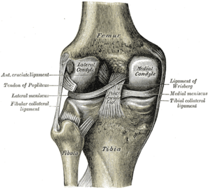
Left knee-joint from behind, showing interior ...
Image via Wikipedia

Common Causes

Knee pain usually results from overuse,but in several cases poor form during physical activity, not warming up or cooling down, or inadequate stretching. Simple causes of knee pain often clear up on their own with self care. Being overweight can put you at greater risk for knee problems.

Other Knee pain causes :-

  • Arthritis — including rheumatoid, osteoarthritis, and gout, or other connective tissue disorders like lupus.
  • Bursitis — inflammation from repeated pressure on the knee (like kneeling for long periods of time, overuse, or injury).
  • Tendinitis — a pain in the front of your knee that gets worse when going up and down stairs or inclines. Happens to runners, skiers, and cyclists.
  • Baker’s cyst — a fluid-filled swelling behind the knee that may accompany inflammation from other causes, like arthritis. If the cyst ruptures, pain in the back of your knee can travel down your calf.
  • Torn cartilage (a meniscus tear) — can cause pain on the inside or outside of the knee joint.
  • Torn ligament (ACL tear) — can cause pain and instability of the knee.
  • Strain or sprain — minor injuries to the ligaments caused by sudden or unnatural twisting.
  • Dislocation of the kneecap.
  • Infection in the joint.
  • Knee injuries — can cause bleeding into your knee, which worsens the pain.
  • Hip disorders — may cause pain that is felt in the knee. For example, iliotibial band syndrome is injury to the thick band that runs from your hip to the outside of your knee.

Less common conditions that can lead to knee pain include the following:

Home Care

Many causes of knee pain, especially those related to overuse or physical activity, respond well to self-care:

  • Rest and avoid activities that aggravate the pain, especially weight bearing activities.
  • Apply ice. First, apply it every hour for up to 15 minutes. After the first day, apply it at least 4 times per day.
  • Keep your knee elevated as much as possible to bring any swelling down.
  • Gently compress the knee by wearing an ace bandage or elastic sleeve. Either can be purchased at most pharmacies. This may reduce swelling and provide support.
  • Take acetaminophen for pain or ibuprofen for pain and swelling.
  • Sleep with a pillow underneath or between your knees

YOGA EXERCISE

Several kind of knee pain problem can be cured through yoga exercise under some Yoga Expart.

css.php