Categories
Human Organ Transplantation

Liver Transplantation

[amazon_link asins=’1455702684,3319072080,0323528449,1118277384,0781720397,B009QOL4UQ,3798512566,8184487703,0323320163′ template=’ProductCarousel’ store=’finmeacur-20′ marketplace=’US’ link_id=’6f1f2b71-7d87-11e7-bcd0-7568b2e27733′]


Introduction:Your liver helps fight infections and cleans your blood. It also helps digest food and stores energy for when you need it. You cannot live without a liver that works.

If your liver fails, your doctor may put you on a waiting list for a liver transplant. Doctors do liver transplants when other treatments cannot keep a damaged liver working.
Liver transplantation or hepatic transplantation is the replacement of a diseased liver with a healthy liver allograft. The most commonly used technique is orthotopic transplantation, in which the native liver is removed and the donor organ is placed in the same anatomic location as the original liver. Liver transplantation nowadays is a well accepted treatment option for end-stage liver disease and acute liver failure.

CLICK & SEE THE PICTURES

During a liver transplantation, the surgeon removes the diseased liver and replaces it with a healthy one. Most transplant livers come from a donor who has died. Sometimes a healthy person donates part of his or her liver for a specific patient. In this case the donor is called a living donor. The most common reason for transplantation in adults is cirrhosis. This is a disease in which healthy liver cells are killed and replaced with scar tissue. The most common reason in children is biliary atresia, a disease of the bile ducts.

People who have transplants must take drugs for the rest of their lives to keep their bodies from rejecting their new livers.

Liver transplantation is usually done when other medical treatment cannot keep a damaged liver functioning.

History:-
The first human liver transplant was performed in 1963 by a surgical team led by Dr. Thomas Starzl of Denver, Colorado, United States. Dr. Starzl performed several additional transplants over the next few years before the first short-term success was achieved in 1967 with the first one-year survival posttransplantation. Despite the development of viable surgical techniques, liver transplantation remained experimental through the 1970s, with one year patient survival in the vicinity of 25%. The introduction of cyclosporine by Sir Roy Calne markedly improved patient outcomes, and the 1980s saw recognition of liver transplantation as a standard clinical treatment for both adult and pediatric patients with appropriate indications. Liver transplantation is now performed at over one hundred centres in the USA, as well as numerous centres in Europe and elsewhere. One year patient survival is 80-85%, and outcomes continue to improve, although liver transplantation remains a formidable procedure with frequent complications. Unfortunately, the supply of liver allografts from non-living donors is far short of the number of potential recipients, a reality that has spurred the development of living donor liver transplantation.

Indications:-
Liver transplantation is potentially applicable to any acute or chronic condition resulting in irreversible liver dysfunction, provided that the recipient does not have other conditions that will preclude a successful transplant. Metastatic cancer outside liver, active drug or alcohol abuse and active septic infections are absolute contraindications. While infection with HIV was once considered an absolute contraindication, this has been changing recently. Advanced age and serious heart, pulmonary or other disease may also prevent transplantation (relative contraindications). Most liver transplants are performed for chronic liver diseases that lead to irreversible scarring of the liver, or cirrhosis of the liver.

Techniques
:-
Before transplantation liver support therapy might be indicated (bridging-to-transplantation). Artificial liver support like liver dialysis or bioartificial liver support concepts are currently under preclinical and clinical evaluation. Virtually all liver transplants are done in an orthotopic fashion, that is the native liver is removed and the new liver is placed in the same anatomic location. The transplant operation can be conceptualized as consisting of the hepatectomy (liver removal) phase, the anhepatic (no liver) phase, and the postimplantation phase. The operation is done through a large incision in the upper abdomen. The hepatectomy involves division of all ligamentous attachments to the liver, as well as the common bile duct, hepatic artery, hepatic vein and portal vein. Usually, the retrohepatic portion of the inferior vena cava is removed along with the liver, although an alternative technique preserves the recipient’s vena cava (“piggyback” technique).

The donor’s blood in the liver will be replaced by an ice-cold organ storage solution, such as UW (Viaspan) or HTK until the allograft liver is implanted. Implantation involves anastomoses (connections) of the inferior vena cava, portal vein, and hepatic artery. After blood flow is restored to the new liver, the biliary (bile duct) anastomosis is constructed, either to the recipient’s own bile duct or to the small intestine. The surgery usually takes between five and six hours, but may be longer or shorter due to the difficulty of the operation and the experience of the surgeon.

The large majority of liver transplants use the entire liver from a non-living donor for the transplant, particularly for adult recipients. A major advance in pediatric liver transplantation was the development of reduced size liver transplantation, in which a portion of an adult liver is used for an infant or small child. Further developments in this area included split liver transplantation, in which one liver is used for transplants for two recipients, and living donor liver transplantation, in which a portion of healthy person’s liver is removed and used as the allograft. Living donor liver transplantation for pediatric recipients involves removal of approximately 20% of the liver (Couinaud segments 2 and 3).

Immunosuppressive management:-
Like all other allografts, a liver transplant will be rejected by the recipient unless immunosuppressive drugs are used. The immunosuppressive regimens for all solid organ transplants are fairly similar, and a variety of agents are now available. Most liver transplant recipients receive corticosteroids plus a calcinuerin inhibitor such as tacrolimus or Cyclosporin plus a antimetabolite such as Mycophenolate Mofetil.

Liver transplantation is unique in that the risk of chronic rejection also decreases over time, although recipients need to take immunosuppresive medication for the rest of their lives. It is theorized that the liver may play a yet-unknown role in the maturation of certain cells pertaining to the immune system. There is at least one study by Dr. Starzl’s team at the University of Pittsburgh which consisted of bone marrow biopsies taken from such patients which demonstrate genotypic chimerism in the bone marrow of liver transplant recipients.

Results:-
About 80 to 90 percent of people survive liver transplantation. Survival rates have improved over the past several years because of drugs like cyclosporine and tacrolimus that suppress the immune system and keep it from attacking and damaging the new liver.

Prognosis is quite good. However those with certain illnesses may differ.  There is no exact model to predict survival rates however those with transplant have a 58% chance of surviving 15 years.

Living donor transplantation:-
Living donor liver transplantation (LDLT) has emerged in recent decades as a critical surgical option for patients with end stage liver disease, such as cirrhosis and/or hepatocellular carcinoma often attributable to one or more of the following: long-term alcohol abuse, long-term untreated Hepatitis C infection, long-term untreated Hepatitis B infection. The concept of LDLT is based on (1) the remarkable regenerative capacities of the human liver and (2) the widespread shortage of cadaveric livers for patients awaiting transplant. In LDLT, a piece of healthy liver is surgically removed from a living person and transplanted into a recipient, immediately after the recipient’s diseased liver has been entirely removed.

Historically, LDLT began as a means for parents of children with severe liver disease to donate a portion of their healthy liver to replace their child’s entire damaged liver. The first report of successful LDLT was by Dr. Silvano Raia at the Universidade de São Paulo (USP) Medical School in 1986. Surgeons eventually realized that adult-to-adult LDLT was also possible, and now the practice is common in a few reputable medical institutes. It is considered more technically demanding than even standard, cadaveric donor liver transplantation, and also poses the ethical problems underlying the indication of a major surgical operation (hepatectomy) on a healthy human being. In various case series the risk of complications in the donor is around 10%, and very occasionally a second operation is needed. Common problems are biliary fistula, gastric stasis and infections; they are more common after removal of the right lobe of the liver. Death after LDLT has been reported at 0% (Japan), 0.3% (USA) and <1% (Europe), with risks likely to improve further as surgeons gain more experience in this procedure.

In a typical adult recipient LDLT, 55% of the liver (the right lobe) is removed from a healthy living donor. The donor’s liver will regenerate to 100% function within 4-6 weeks and will reach full volumetric size with recapitulation of the normal structure soon thereafter. It may be possible to remove 70% to 75% of the liver from a healthy living donor without harm in most cases. The transplanted portion will reach full function and the appropriate size in the recipient as well, although it will take longer than for the donor.

For More Information:-

American Liver Foundation
75 Maiden Lane, Suite 603
New York, NY 10038
Phone: 1–800–GO–LIVER (465–4837)
Email: info@liverfoundation.org
Internet: www.liverfoundation.org

Hepatitis Foundation International (HFI)
504 Blick Drive
Silver Spring, MD 20904–2901
Phone: 1–800–891–0707 or 301–622–4200
Fax: 301–622–4702
Email: hepfi@hepfi.org
Internet: www.hepfi.org

United Network for Organ Sharing (UNOS)
P.O. Box 2484
Richmond, VA 23218
Phone: 1–888–894–6361 or 804–782–4800
Internet: www.unos.org

Additional Information on Liver Transplantation :-

The National Digestive Diseases Information Clearinghouse collects resource information on digestive diseases for National Institute of Diabetes and Digestive and Kidney Diseases (NIDDK) Reference Collection. This database provides titles, abstracts, and availability information for health information and health education resources. The NIDDK Reference Collection is a service of the National Institutes of Health.

To provide you with the most up-to-date resources, information specialists at the clearinghouse created an automatic search of the NIDDK Reference Collection. To obtain this information, you may view the results of the automatic search on Liver Transplantation.

If you wish to perform your own search of the database, you may access and search the NIDDK Reference Collection database online.

National Digestive Diseases Information Clearinghouse
2 Information Way
Bethesda, MD 20892–3570
Phone: 1–800–891–5389
TTY: 1–866–569–1162
Fax: 703–738–4929
Email: nddic@info.niddk.nih.gov
Internet: www.digestive.niddk.nih.gov

You may click to see->

Recent Developments in Transplantation Medicine

What I need to know about Liver Transplantation

Liver Transplantation at UCLA: One of the largest liver transplant centers in the world

You may click to see the external links:-
*Official organ sharing network of U.S.
*Official organ procurement center of the U.S.
*American Liver Foundation: Comprehensive information about Hepatitis C, Liver Transplant and other liver diseases, including links to chapters for finding local resources
*Management of HBV Infection in Liver Transplantation Patients
*Management of HCV Infection and Liver Transplantation
*Antiviral therapy of HCV in the cirrhotic and transplant candidate
*Living Donors Online
*Liver Transplantation Guide and Liver Transplant Surgery in India
*History of pediatric liver transplantation
*ABC Salutaris: Living Donor Liver Transplant
*Organ Donation Awareness and former potential donor blog
*All You Need to Know about Adult Living Donor Liver Transplantation
*Children’s Liver Disease Foundation
*A Liver Donor’s Blog

Resources:
http://www.nlm.nih.gov/medlineplus/livertransplantation.html
http://en.wikipedia.org/wiki/Liver_transplantation
http://digestive.niddk.nih.gov/ddiseases/pubs/livertransplant/

Reblog this post [with Zemanta]
Categories
Human Organ Transplantation

Pancreas Transplant

[amazon_link asins=’0199565864,0071761217,0199768897,0323340628,0192632558,935152339X,1620231158,3319230530,0387005897′ template=’ProductCarousel’ store=’finmeacur-20′ marketplace=’US’ link_id=’6b31253e-0970-11e8-adc1-3154197f16f9′]

Definition  :
A pancreas transplant is surgery to implant a healthy pancreas from a donor into a patient with diabetes. Pancreas transplants give the patient a chance to become independent of insulin injections.

click to see the pictures—> (01).....(1)..…....(2)....…..

A pancreas transplant is an organ transplant that involves implanting a healthy pancreas (one that can produce insulin) into a person who usually has diabetes. Because the pancreas is a vital organ, performing functions necessary in the digestion process, the recipient’s native pancreas is left in place, and the donated pancreas is attached in a different location. In the event of rejection of the new pancreas which would quickly cause life-threatening diabetes, the recipient could not survive without the native pancreas still in place. The healthy pancreas comes from a donor who has just died or it may be a partial pancreas from a living donor.  Whole pancreas transplants from living donors are not possible, again because the pancreas is a necessary organ for digestion. At present, pancreas transplants are usually performed in persons with insulin-dependent diabetes, who have severe complications that are usually of a renal nature. Patients with pancreatic cancer are not eligible for valuable pancreatic transplantations, since the condition has a very high mortality rate and the disease, being highly malignant, could and probably would soon return.

Description :
The healthy pancreas is obtained from a donor who has suffered brain-death, but remains on life-support. The donor pancreas must meet numerous criteria to make sure it is suitable.

In addition to insulin, the pancreas produces other secretions, such as digestive enzymes, which drain through the pancreatic duct into the duodenum. Therefore, a portion of the duodenum is removed with the donor pancreas. The healthy pancreas is transported in a cooled solution that preserves the organ for up to 20 hours.

The patient’s diseased pancreas is not removed during the operation. The donor pancreas is usually inserted in the right lower portion of the patient’s abdomen and attachments are made to the patient’s blood vessels. The donor duodenum is attached to the patient’s intestine or bladder to drain pancreatic secretions.

The operation is usually done at the same time as a kidney transplant in diabetic patients with kidney disease.

Types:
There are three main types of pancreas transplantation:

*Simultaneous pancreas-kidney transplant (SPK), when the pancreas and kidney are transplanted simultaneously from the same deceased donor....CLICK & SEE

*Pancreas-after-kidney transplant (PAK), when a cadaveric, or deceased, donor pancreas transplant is performed after a previous, and different, living or deceased donor kidney transplant....CLICK & SEE

*Pancreas transplant alone, for the patient with type 1 diabetes who usually has severe, frequent hypoglycemia, but adequate kidney function…..CLICK & SEE

Indications:
In most cases, pancreas transplantation is performed on individuals with type 1 diabetes with end-stage renal disease The majority of pancreas transplantations (>90%) are simultaneous pancreas-kidney transplantions.

Why the Procedure is Performed  :
A pancreas transplant may be recommended for people with pancreatic disease, especially if they have type 1 diabetes and poor kidney function.

Pancreas transplant surgery is not recommended for patients who have:

*Heart or lung disease
*Other life-threatening diseases

Solitary pancreas transplant for diabetes, without simultaneous kidney transplant, remains controversial.

Risks Factor:

The risks for any anesthesia are:

*Heart attack
*Reactions to medications
*Problems breathing

The risks for any surgery are:
*Bleeding
*Infection
*Scar formation

The body’s immune system considers the transplanted organ foreign, and fights it accordingly. Thus, to prevent rejection, organ transplant patients must take drugs (such as cyclosporine and corticosteroids) that suppress the immune response of the body. The disadvantage of these drugs is that they weaken the body’s natural defense against various infections.

Preservation until implantation:
The donor’s blood in the pancreatic tissue will be replaced by an ice-cold organ storage solution, such as UW (Viaspan) or HTK until the allograft pancreatic tissue is implanted.

Complications:
Complications immediately after surgery include rejection, thrombosis, pancreatitis and infection.

Prognosis:
The prognosis after pancreas transplantation is very good. Over the recent years, long-term success has improved and risks have decreased. One year after transplantation more than 95% of all patients are still alive and 80-85% of all pancreases are still functional. After transplantation patients need lifelong immunosuppression. Immunosuppression increases the risk for a number of different kinds of infection and cancer.

The main problem, as with other transplants, is graft rejection. Immunosuppressive drugs, which weaken your body’s ability to fight infections, must be taken indefinitely. Normal activities can resume as soon as you are strong enough, and after consulting with the doctor. It is possible to have children after a transplant.

The major problems with all organ transplants are:

*Finding a donor
*Preventing rejection
*Long-term immunosuppression

Recovery :
It usually takes about 3 weeks to recover. Move your legs often to reduce the risk of blood clots or deep vein thrombosis. The sutures or clips are removed about two to three weeks after surgery. Resume normal activity as soon as possible, after consulting with the physician. A diet will be prescribed.

Resources:
http://www.nlm.nih.gov/medlineplus/ency/article/003007.htm
http://en.wikipedia.org/wiki/Pancreas_transplantation

Enhanced by Zemanta
Categories
Human Organ Transplantation

Kidney Transplantation

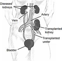
Kidney location after transplantation.
Image via Wikipedia

[amazon_link asins=’1496326156,1455740969,0781753228,1493903411,1416033432,144191689X,1841843164,0062418173,0967038448′ template=’ProductCarousel’ store=’finmeacur-20′ marketplace=’US’ link_id=’11a01150-0970-11e8-81ed-39a60e701c07′]

Alternative Names:Renal transplant; Transplant – kidney

Definition:

A kidney transplant is surgery to place a healthy kidney into a person with kidney failure. Kidney transplantation or renal transplantation is the organ transplant of a kidney in a patient with end-stage renal disease. Kidney transplantation is typically classified as deceased-donor (formerly known as cadaveric) or living-donor transplantation depending on the source of the recipient organ. Living-donor renal transplants are further characterized as genetically related (living-related) or non-related (living-unrelated) transplants, depending on whether a biological relationship exists between the donor and recipient.

..CLICK & SEE THE PICTURES

Description :
Kidney transplants are one of the most common transplant operations in the United States.

A donated kidney is needed to perform a kidney transplant.

The donated kidney may be from:

*Living related donor — related to the recipient, such as a parent, sibling, or child
*Living unrelated donor — such as a friend or spouse

Indications:
The indication for kidney transplantation is end-stage renal disease (ESRD), regardless of the primary cause. This is defined as a drop in the glomerular filtration rate (GFR) to 20-25% of normal. Common diseases leading to ESRD include malignant hypertension, infections, diabetes mellitus and glomerulonephritis; genetic causes include polycystic kidney disease as well as a number of inborn errors of metabolism as well as autoimmune conditions including lupus and Goodpasture’s syndrome. Diabetes is the most common cause of kidney transplant, accounting for approximately 25% of those in the US. The majority of renal transplant recipients are on some form of dialysis – hemodialysis, peritoneal dialysis, or the similar process of hemofiltration – at the time of transplantation. However, individuals with chronic renal failure who have a living donor available often elect to undergo transplantation before dialysis is needed.

Sources of kidneys:
Since medication to prevent rejection is so effective, donors need not be genetically similar to their recipient. Most donated kidneys come from deceased donors, with some coming from living donors. However, the utilization of living donors in the United States is on the rise. In the year 2006, 47% of donated kidneys were actually from living donors (Organ Procurement and Transplantation Network, 2007). It is important to note that this varies by country: for example, only 3% of transplanted kidneys during 2006 in Spain came from living donors (Organización Nacional de Transplantes (ONT), 2007).

Living donors:
Potential donors are carefully evaluated on medical and psychological grounds. This ensures that the donor is fit for surgery and has no kidney disease whilst confirming that the donor is purely altruistic. Traditionally, the donor procedure has been through a single, 4-7 inch incision but live donation is being increasingly performed by laparoscopic surgery. This reduces pain and accelerates recovery for the donor. Excellent results have been demonstrated with laparoscopic donor nephrectomy, for both donor and recipient outcomes. Overall, recipients of kidneys from live donors do extremely well, in comparison to deceased donor recipients.

In 2004 the FDA approved the Cedars-Sinai High Dose IVIG therapy which reduces the need for the living donor to be the same blood type (ABO compatible) or even a tissue match. The therapy reduced the incidence of the recipient’s immune system rejecting the donated kidney in highly-sensitized patients

PROCEDURE FOR A LIVING KIDNEY DONOR:-
If you are donating a kidney, you will be placed under general anesthesia before surgery. This means you will be asleep and pain-free. The surgeon makes a cut in the side of your abdomen, removes the proper kidney, and then closes the wound. The procedure used to require a long surgical cut. However, today surgeons can use a short surgical cut (mini-nephrectomy) or laparoscopic techniques.

Deceased donors:-
Deceased donors can be divided in two groups:

Brain-dead (BD) donors
Donation after Cardiac Death (DCD) donors
Although brain-dead (or “heart-beating”) donors are considered dead, the donor’s heart continues to pump and maintain the circulation. This makes it possible for surgeons to start operating while the organs are still being perfused. During the operation, the aorta will be cannulated, after which the donor’s blood will be replaced by an ice-cold storage solution, such as UW (Viaspan), HTK, or Perfadex. [Depending on which organs are transplanted, more than one solution may be used simultaneously.] Due to the temperature of the solution (and since large amounts of cold NaCl-solution are poured over the organs for a rapid cooling of the organs), the heart will stop pumping.

Donation after Cardiac Death”
donors are patients who do not meet the brain-dead criteria, but due to the small chance of recovery have elected, via a living will or through family, to withdraw support. In this procedure, treatment is discontinued (mechanical ventilation is shut off). Usually, a certain amount of minutes after death has been pronounced, the patient is rushed to the operating theatre, where the organs are recovered, after which the storage solution is flushed through the organs itself. Since the blood is no longer being circulated, coagulation must be prevented with relatively large amounts of anti-coagulation agents, such as heparin. It is important to note that several ethical and procedural guidelines must be followed, chief of which is that the organ recovery team should not participate in the patient’s care in any manner until after death has been declared.

Kidneys from brain-dead donors are generally of a superior quality, since they have not been exposed to warm ischemia (the time between the heart stopping and the kidney being cooled).

Compatibility:
If plasmapheresis or IVIG is not performed, the donor and recipient have to be ABO blood group compatible. Also, they should ideally share as many HLA and “minor antigens” as possible. This decreases the risk of transplant rejection and the need for another transplant. The risk of rejection may be further reduced if the recipient is not already sensitized to potential donor HLA antigens, and if immunosuppressant levels are kept in an appropriate range. In the United States, up to 17% of all deceased donor kidney transplants have no HLA mismatch. However, it is important to note that HLA matching is a relatively minor predictor of transplant outcomes. In fact, living non-related donors are now almost as common as living (genetically)-related donors.

In the 1980s, experimental protocols were developed for ABO-incompatible transplants using increased immunosuppression and plasmapheresis. Through the 1990s these techniques were improved and an important study of long-term outcomes in Japan was published. . Now, a number of programs around the world are routinely performing ABO-incompatible transplants.

In 2004 the FDA approved the Cedars-Sinai High Dose IVIG protocol which eliminates the need for the donor to be the same blood type.

Procedure:
Since in most cases the barely functioning existing kidneys are not removed because this has been shown to increase the rates of surgical morbidities, the kidney is usually placed in a location different from the original kidney (often in the iliac fossa), and as a result it is often necessary to use a different blood supply:

*The renal artery of the kidney, previously branching from the abdominal aorta in the donor, is often connected to the external iliac artery in the recipient.

*The renal vein of the new kidney, previously draining to the inferior vena cava in the donor, is often connected to the external iliac vein in the recipient.

Why the Procedure is Performed :

A kidney transplant may be recommended if you have kidney failure caused by:

*Diabetes
*Glomerulonephritis
*Severe, uncontrollable high blood pressure
*Certain infections

A kidney transplant alone may NOT be recommended if you have:

*Certain infections, such as TB or osteomyelitis
*Difficulty taking medications several times each day for the rest of your life
*Heart, lung, or liver disease
*Other life-threatening diseases

Risks  Factor:

The risks for any anesthesia are:

*Problems breathing
*Reactions to medications

The risks for any surgery are:
*Bleeding
*Infection

Other risks include:
Infection due to medications that suppress the immune response that must be taken to prevent transplant rejections

Post operation:
The transplant surgery lasts about three hours. The donor kidney will be placed in the lower abdomen and its blood vessels connected to arteries and veins in the recipient’s body. When this is complete, blood will be allowed to flow through the kidney again, so the ischemia time is minimized. In most cases, the kidney will soon start producing urine. Since urine is sterile, this has no effect on the operation. The final step is connecting the ureter from the donor kidney to the bladder.

Depending on its quality, the new kidney usually begins functioning immediately. Living donor kidneys normally require 3-5 days to reach normal functioning levels, while cadaveric donations stretch that interval to 7-15 days. Hospital stay is typically for four to seven days. If complications arise, additional medicines may be administered to help the kidney produce urine.

Medicines are used to suppress the immune system from rejecting the donor kidney. These medicines must be taken for the rest of the patient’s life. The most common medication regimen today is : tacrolimus, mycophenolate, and prednisone. Some patients may instead take cyclosporine, rapamycin, or azathioprine. Cyclosporine, considered a breakthrough immunosuppressive when first discovered in the 1980’s, ironically causes nephrotoxicity and can result in iatrogenic damage to the newly transplanted kidney. Blood levels must be monitored closely and if the patient seems to have a declining renal function, a biopsy may be necessary to determine if this is due to rejection or cyclosporine intoxication.

Acute rejection occurs in 10% to 25% of people after transplant during the first sixty days. Rejection does not necessarily mean loss of the organ, but may require additional treatment.

Complications:
Problems after a transplant may include:

*Transplant rejection (hyperacute, acute or chronic)

*Infections and sepsis due to the immunosuppressant drugs that are required to decrease risk of rejection

*Post-transplant lymphoproliferative disorder (a form of lymphoma due to the immune suppressants)

*Imbalances in electrolytes including calcium and phosphate which can lead to bone problems amongst other things

*Other side effects of medications including gastrointestinal inflammation and ulceration of the stomach and esophagus, hirsutism (excessive hair growth in a male-pattern distribution), hair loss, obesity, acne, diabetes mellitus (type 2), hypercholesterolemia, and others.

*The average lifetime for a donor kidney is ten to fifteen years. When a transplant fails a patient may opt for a second transplant, and may have to return to dialysis for some intermediary time.

Prognosis:
Kidney transplantation is a life-extending procedure. The typical patient will live ten to fifteen years longer with a kidney transplant than if kept on dialysis. The years of life gained is greater for younger patients, but even 75 year-old recipients (the oldest group for which there is data) gain an average four more years’ life. People generally have more energy, a less restricted diet, and fewer complications with a kidney transplant than if they stay on conventional dialysis.

Some studies seem to suggest that the longer a patient is on dialysis before the transplant, the less time the kidney will last. It is not clear why this occurs, but it underscores the need for rapid referral to a transplant program. Ideally, a kidney transplant should be pre-emptive, i.e. take place before the patient starts on dialysis.

At least three professional athletes have made a comeback to their sport after receiving a transplant: NBA players Sean Elliott and Alonzo Mourning; and New Zealand rugby union player Jonah Lomu as well as the German-Croatian Soccer Player Ivan Klasni?.

Recovery
The recovery period is 4 – 6 weeks for people who donate a kidney. If you’ve done so, you should avoid heavy activity during this time. Your doctor removes the stitches after a week or so.

If you received a donated kidney, you will need to stay in the hospital for about a week. Afterwards, you will need close follow-up by a doctor and regular blood tests.

Resources:
http://en.wikipedia.org/wiki/Kidney_transplantation
http://www.nlm.nih.gov/medlineplus/ency/article/003005.htm

Reblog this post [with Zemanta]
Categories
News on Health & Science

Now, ‘Smart Scaffold’ to Help Heart Heal Itself

[amazon_link asins=’B0015WLYY8,B019O4PMWI,3642268129,B014881T2C,B001JL18XC,B00BXQSMQ4,B00BXQSORQ,B00BGHYMFK,B010A1W6MI’ template=’ProductCarousel’ store=’finmeacur-20′ marketplace=’US’ link_id=’4d5a4a8d-76c4-11e7-8161-cded53339a46′]

New treatments are being developed that heal broken hearts or muscle loss by prompting the body to repair damaged tissues.

Canadian researchers, for the first time, have developed an organic substance (scaffold) that attracts and supports cells necessary for tissue repair and can be directly injected into problem areas.

This development is a major step toward treatments that allow people to more fully recover from injury and disease rather than having to live with chronic health problems. It may even help reduce the need for organ transplantation by allowing physicians to save organs that would have been previously damaged beyond repair.

These “smart scaffolds”, developed by Erik Suuronen from the University of Ottawa and Ottawa Heart Research Institute, contain a protein that allows progenitor cells to adhere to the damaged tissue and survive long enough to promote healing. These cells emit homing signals that summon other cells to join in the process and give off chemical signals that order cells to grow blood vessels necessary for healing to occur.

“Ultimately, we envision a scaffold material that can be taken off the shelf and injected into the hearts of patients suffering from blocked arteries,” he said. “The scaffold materials would direct the repair process, and restore blood flow and function to the heart

Sources: The Times Of India

Reblog this post [with Zemanta]
Categories
Ailmemts & Remedies

Aspergillosis

[amazon_link asins=’B00CMOJKKI,B00DQR99S8′ template=’ProductCarousel’ store=’finmeacur-20′ marketplace=’US’ link_id=’68dbd97c-eed2-11e6-8d87-99954932bf15′]

[amazon_link asins=’B00014CZV2,1780661754,B0015CMCDA,B000W6QK6K,B00JMI7APO,B01CF5XQWG’ template=’ProductCarousel’ store=’finmeacur-20′ marketplace=’US’ link_id=’8b092493-ffbe-11e6-9d25-bf387163bb36′]

Other Names:Brooder Pneumonia; Mycotic Pneumonia: Pneumomycosis

Definition:Aspergillosis is the name given to a wide variety of diseases caused by fungi of the genus Aspergillus. The most common forms are allergic bronchopulmonary aspergillosis, pulmonary aspergilloma and invasive aspergillosis. Most humans inhale Aspergillus spores every day; aspergillosis develops mainly in immunocompromised individuals. The most common cause is Aspergillus fumigatus.
The aspergillus species includes more than 150 types of mold that occur widely in the indoor and outdoor environment. Although most of the molds are harmless, a few can cause serious illnesses in people with a weakened immune system, underlying lung disease or asthma. These illnesses, collectively called aspergillosis, range from allergic responses to severe and sometimes fatal infections.

Aspergillosis begins when susceptible people inhale mold spores into their lungs. In some people, the spores trigger an allergic reaction. Other people develop mild to serious lung infections. The most deadly form of aspergillosis — invasive aspergillosis — occurs when the infection spreads beyond the lungs to other organs. Even when discovered and treated early, invasive aspergillosis is often fatal.

CLICK & SEE THE PICTURES

Aspergillosis infections are treated with antifungal agents, though not always successfully and often with serious side effects. In 2005, British researchers cracked the genetic code of aspergillus molds. The hope is that this knowledge will lead to better ways of diagnosing and treating aspergillosis.

Symptoms:A fungus ball in the lungs may cause no symptoms and may be discovered only with a chest x-ray. Or it may cause repeated coughing up of blood and occasionally severe, even fatal, bleeding. A rapidly invasive Aspergillus infection in the lungs often causes cough, fever, chest pain, and difficulty breathing.
The signs and symptoms of aspergillosis vary with the type of infection.

Allergic bronchopulmonary aspergillosis
Some people with asthma or cystic fibrosis have an allergic reaction to aspergillus mold. Signs and symptoms of this condition, known as allergic bronchopulmonary aspergillosis, include:

*Fever
*A general feeling of being unwell (malaise)
*A cough that may bring up blood or plugs of mucus
*Wheezing
*Worsening asthma

Aspergilloma
People whose lungs have been damaged by emphysema, tuberculosis or other diseases that leave air spaces (cavities) in the lungs may develop a pulmonary aspergilloma — a tangled ball of fungus fibers that forms in these spaces. Initially, an aspergilloma may not produce symptoms, but over time, it can cause:

*A cough that often brings up blood
*Chest pain
*Wheezing
*Shortness of breath
*Unintentional weight loss

Invasive pulmonary aspergillosis
The most severe form of aspergillosis, invasive pulmonary aspergillosis, occurs when the infection spreads rapidly through your bloodstream to your brain, heart, kidneys or skin. Signs and symptoms depend on which organs are affected, but in general, invasive aspergillosis can cause:

*Fever and chills
*Headaches
*Bloody cough
*Shortness of breath
*Chest or joint pain
*Massive bleeding from your lungs

Other aspergillus infections
Aspergillus can invade your sinuses and ear canals as well as your lungs. In your sinuses, it can cause a stuffy nose, drainage, inflammation, fever, facial pain and headache. Ear canal infections can cause itching, drainage and pain.

Causes:-
Aspergillus plays an indispensable role in the ecosystem by breaking down organic matter. It’s also virtually unavoidable. Outdoors, it’s found in decaying leaves and compost and on plants, trees and grain crops. Inside, the spores — the reproductive parts of mold — thrive in air conditioning and heating ducts, insulation, carpeting, ornamental plants, tap water, dust and food — especially ground pepper and other spices.

Everyday exposure to aspergillus is rarely a problem for people with healthy immune systems. When mold spores are inhaled, immune system cells simply surround and destroy them. But people who have a weakened immune system from illness or medications have fewer infection-fighting cells. This allows aspergillus to take hold, invading the lungs, and in the most serious cases, other parts of the body.

Risk factors:-
Your risk of developing aspergillosis depends on your overall health and the extent of your exposure to mold, but in general, these factors make you more vulnerable to infection:

*Weakened immune system. This is the greatest risk factor for invasive aspergillosis. People taking immune-suppressing drugs after undergoing transplant surgery, especially bone marrow or stem cell transplants, are the most severely affected. In fact, aspergillosis is the leading cause of death among people who have received a transplant. People with later-stage AIDS also may be at increased risk.

*Low white blood cell level. White blood cells called neutrophils play a key role in fighting fungal infections. Having a very low level of these cells (neutropenia) due to chemotherapy, an organ transplant or leukemia makes you much more susceptible to invasive aspergillosis. So does having chronic granulomatous disease — an inherited disorder that affects immune system cells.

*Lung cavities. An aspergilloma develops when mold spores germinate in a healed air space (cavity) in your lungs. Cavities are areas that have been damaged by serious lung diseases such as tuberculosis or sarcoidosis — an illness that causes inflammation in your lungs and other organs. The larger the cavity, the greater your chance of developing an aspergilloma. Most often, aspergillomas don’t spread beyond the cavity, but when they do, they can cause a cough that brings up blood, and the bleeding may be severe.

*Asthma or cystic fibrosis. About 7 percent of people with asthma and cystic fibrosis have an allergic response to aspergillus mold. This is more likely to occur in people whose lung problems are long-standing or hard to control.

*Long-term corticosteroid therapy. Because corticosteroids suppress your immune system, they increase the risk of aspergillosis. Infections that result from corticosteroid use tend to be severe and to progress rapidly.

*A hospital stay. Aspergillus mold is found on many hospital surfaces — bedrails, plants, surgical instruments, air conditioning ducts, insulation and in tap water. Though healthy people aren’t likely to be affected, people with a weakened immune system or serious illness are highly susceptible to infection. Most major hospital outbreaks have been traced to hospital construction and renovation projects, and to contaminated air filters and carpeting.

*Genetics. Some researchers speculate that genetic factors may make certain people more prone to aspergillosis infection.

Diagnosis:
Diagnosing aspergillosis can be difficult. Aspergillus is common in the environment and is sometimes found in the saliva and sputum of healthy people. What’s more, it’s hard to distinguish aspergillus from other molds under the microscope, and symptoms of the infection are similar to those of conditions such as tuberculosis.

To arrive at an accurate diagnosis, your doctor is likely to use one or more of the following tests:

*Imaging tests. A chest X-ray or computerized tomography (CT) scan — a type of X-ray that produces more detailed images than conventional X-rays do — can usually reveal an aspergilloma as well as characteristic signs of invasive and allergic aspergillosis.

*Sputum stain and culture. In this test, a sample of your sputum is stained with a dye and checked for the presence of aspergillus filaments. The specimen is then placed on a medium that encourages the mold to grow.

*Skin and blood tests. Diagnosing allergic bronchopulmonary aspergillosis usually requires skin and blood tests. For the skin test, a small amount of aspergillus antigen is injected into your forearm. If you have antibodies to the mold in your bloodstream, you’ll develop a hard, red bump at the injection site. Blood tests look for elevated levels of certain antibodies, indicating an allergic response.

*Biopsy. Examining a sample of tissue from your lungs or sinuses under a microscope is usually necessary to confirm a diagnosis of invasive aspergillosis.

Complications:-
Depending on the type of infection, aspergillosis can cause a variety of serious complications:

*Bone loss and spread of infection. An aspergillus infection in your sinuses can destroy facial bones. It can also spread beyond your sinuses, and may be life-threatening if you have a severely impaired immune system.

*Bleeding. Both aspergillomas and invasive aspergillosis can cause massive, and sometimes fatal, bleeding in your lungs.

*Systemic infection. The most devastating complication of aspergillosis is the spread of the infection to other parts of your body, especially your brain, heart and kidneys. Invasive aspergillosis spreads rapidly and is often fatal in spite of early treatment.

Modern Treatments :-
There’s no universally effective therapy for aspergillosis. Available treatments vary with the type of disease:

The drugs amphotericin B, caspofungin, flucytosine, itraconazole, voriconazole [2] are used to treat this fungal infection. For severe cases of invasive aspergillosis a combination therapy of voriconazole and caspofungin is suggested as a first line treatment.

*Oral corticosteroids. The goal in treating allergic bronchopulmonary aspergillosis is to prevent existing asthma or cystic fibrosis from becoming worse. The best way to do this is with oral corticosteroids. Antifungal medications by themselves aren’t helpful for allergic aspergillosis, but they may be used in combination with corticosteroids to reduce the dose of steroids and improve lung function.

*Antifungal medications. These drugs are the standard treatment for invasive pulmonary aspergillosis. Historically, the drug of choice has been amphotericin B, but the newer medication voriconazole is now preferred because it appears more effective and may have fewer side effects. All antifungals can cause serious problems, however, including kidney and liver damage, and they frequently interact with other medications given to people who have weakened immune systems.

*Watchful waiting. Aspergillomas often don’t need treatment, and may simply be closely monitored. When they cause life-threatening bleeding, the options are limited. Because antifungal medications aren’t effective against aspergillomas, surgery is the first choice. The surgery is risky, however, and your doctor may suggest another option, which involves threading a small catheter into the artery that supplies blood to the cavity containing the fungus ball. Though this procedure can stop massive bleeding, it doesn’t prevent it from recurring.

Click for Alternative Treatment of Aspergillosis..(1).…...(2)…….(3)

Outbreaks:-
Albeit relatively rare in humans, aspergillosis is a common and dangerous infection in birds, particularly in pet parrots. Mallards and other ducks are particularly susceptible as they will often resort to poor food sources during bad weather.

Aspergillosis has been the culprit in several recent rapid die-offs among waterfowl. From 8 December until 14 December 2006 over 2,000 Mallards died in the Burley, Idaho area, an agricultural community approximately 150 miles southeast of Boise. Moldy waste grain from the farmland and feedlots in the area is the suspected source. A similar aspergillosis outbreak caused by moldy grain killed 500 Mallards in Iowa in 2005.

While there is no connection between aspergillosis and the H5N1 strain of Avian Influenza (commonly called “bird flu”), rapid die-offs caused by aspergillosis can spark fears of bird flu outbreaks. Laboratory analysis is the only way to distinguish bird flu and aspergillosis.

Prevention:-
It’s nearly impossible to avoid aspergillus entirely, but if you’ve had a transplant or are undergoing chemotherapy, try to stay away from the most obvious sources of mold such as construction sites, compost piles and stored grain.

Hospitals, for their part, are taking increasingly aggressive measure to protect patients, including using barriers around areas under construction, monitoring air and air filters for spores, minimizing exposure to carpet cleaning and vacuuming, and carefully maintaining ventilation systems.

Disclaimer: This information is not meant to be a substitute for professional medical advise or help. It is always best to consult with a Physician about serious health concerns. This information is in no way intended to diagnose or prescribe remedies.This is purely for educational purpose.

Resources:
http://www.mayoclinic.com/health/aspergillosis/DS00950
http://cal.vet.upenn.edu/projects/poultry/Syllabus/page30_31.htm
http://en.wikipedia.org/wiki/Aspergillosis

css.php