Categories
Diagnonistic Test

Pleural Fluid Sampling (or Thoracentesis)

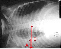
Pleural effusion Chest x-ray of a pleural effu...
Image via Wikipedia

[amazon_link asins=’B01J8WALE0,B00GNOPG8S,1331951720,1893441776,B01MXOPZIB,B01J2IC12U,B01J37411E,B009KT2GWM,B01J2GLUVU’ template=’ProductCarousel’ store=’finmeacur-20′ marketplace=’US’ link_id=’316e2df4-5fab-11e7-aab1-4db380a805de’]


Definition:
Thoracentesis is a procedure used to obtain a sample of fluid from the space around the lungs. Normally, only a thin layer of fluid is present in the area between the lungs and chest wall (show radiograph 1). However, some conditions can cause a large amount of fluid to accumulate. This collection of fluid is called a pleural effusion (show radiograph 2). Thoracentesis is done to collect a sample of the fluid, which can help determine why the pleural effusion developed.
CLICK & SEE
Some infections and diseases cause fluid to accumulate in the space between the lung and the rib cage or between the lung and the diaphragm. This collection of fluid is called a pleural effusion. A pleural effusion might be detected on a chest x-ray. Sampling this fluid is important because it enables doctors to understand what caused the fluid to collect and how to treat the problem. The fluid can be sampled with a needle.

Reasons for Thoracentesis: — A thoracentesis is performed to determine the cause of a pleural effusion. In some cases, a physician may perform thoracentesis to relieve symptoms caused by the pleural effusion, including shortness of breath and low blood oxygen levels. A pleural effusion may be detected during a physical examination or on a chest x-ray.

Pleural effusion can be caused by many different conditions, including infections, heart failure, cancer, or tuberculosis. In some cases, blood or other fluid may be leaking into the pleural space from another part of the body, causing the effusion. By examining the fluid and the types of cells it contains, the cause of the effusion can usually be determined.

In general, there is no reason a thoracentesis cannot be performed. The procedure is more easily performed and complications are fewer when the pleural effusion is large. Special consideration may be necessary in patients who are on respirators.

Patients who have a bleeding disorder or who are on medications that affect blood clotting may need extra care to minimize the risk of bleeding. Patients should tell their healthcare provider if they have a history of bleeding problem or if they are taking medicine that decreases blood clotting. In some cases, a blood test will be taken prior to the procedure to exclude any blood clotting abnormalities caused by disease or medications.

Procedure: A thoracentesis involves the following steps:

*The patient will be placed in a position that allows the doctor to easily access the effusion. Usually, the patient is asked to sit upright during the procedure. It is important to remain still during the procedure so that the fluid does not shift.

*The skin is cleaned with an antibacterial solution in the area where the needle will be inserted.

*A small amount of numbing medicine (a local anesthetic, similar to novocaine) is injected into the area. This medicine helps minimize discomfort during the procedure.

*A slightly larger needle is inserted in the same location. A syringe is attached to this needle and is used to withdraw fluid from around the lung. Patients who have symptoms from the effusion (eg, shortness of breath) may have a large amount of fluid removed, which allows the lung to re-expand.

*The needle is removed and pressure is briefly applied to the insertion site.

How do you prepare for the test?
You will need to sign a consent form giving your doctor permission to perform this test. Some patients have this test done in a doctor’s office, while others are admitted to the hospital for it. Generally your doctor will decide whether you need to be in the hospital based on your medical condition. A chest x-ray or an ultrasound is done before the procedure.

Tell your doctor if you have ever had an allergic reaction to lidocaine or the numbing medicine used at the dentist’s office. If you take aspirin, nonsteroidal anti-inflammatory drugs, or other medicines that affect blood clotting, talk with your doctor. It may be necessary to stop or adjust the dose of these medicines before your test.

What happens when the test is performed?
You wear a hospital gown and sit on a bed or table leaning forward against some pillows. The doctor listens to your lungs with a stethoscope and may tap on your back to find out how much fluid has collected.

Soap is used to disinfect an area of skin on one side of your back. A small needle is used to numb a patch of skin between two of your lower ribs. The numbing medicine usually stings for a second. A needle on an empty syringe is then inserted into the fluid pocket. Usually this pocket is around one inch below the skin surface. You might feel some minor pressure as the needle is inserted. Depending on the quantity of fluid that the doctor plans to remove, either the syringe itself is filled or soft plastic tubing is used to remove fluid into a collection bag or jar. While the doctor is attaching the tubing, he or she might ask you to hum out loud. This humming is for your safety: It prevents you from taking a deep breath, which could expand your lung, causing it to touch the needle.

It sometimes takes 15 minutes or longer to remove the necessary amount of fluid. Most patients feel no discomfort during this time, although a few patients feel some chest pain at the end of the procedure as their lung expands and touches the chest wall. After the fluid is removed, a bandage is placed on your back.

Risk Factors:
This procedure carries a few serious risks, but most patients have no complications. If the needle touches the lung it may create an air leak, which is seen on the x-ray and might require you to stay in the hospital for a few days. Some patients with this complication need to have a plastic tube (called a chest tube) inserted between two ribs. The tube uses vacuum pressure to keep the lung expanded until it has healed.

In most cases, a thoracentesis is performed without complications. Most complications are minor and resolve on their own or are easily treated. Potential complications include the following:

*Pain — Some discomfort may occur when the needle is inserted. Using a local anesthetic helps to reduce the pain. Pain generally resolves once the needle is removed.

*Bleeding — A blood vessel may be nicked as the needle is inserted through the skin and chest wall, causing bleeding. The bleeding is usually minor and stops on its own, although it may cause bruising around the puncture site. In rare cases, bleeding into or around the lung may occur, requiring drainage or surgery.

*Infection
Infection can occur if bacteria are introduced by the needle puncture. Using disinfectant solution to clean the area and using sterile technique during the procedure minimizes this risk.

*Pneumothorax or collapsed lung Occasionally, the needle used to obtain a fluid sample can puncture the lung. The hole created by the puncture usually seals quickly on its own. If it does not, air can build up around the lung, causing the lung to collapse. This is called a pneumothorax. When a pneumothorax occurs, a chest tube may be used to drain the air and allow the lung to re-expand.

A pneumothorax may also occur if the lung fails to expand when fluid is withdrawn. This is considered to be a drainage-related pneumothorax, and is the most common type of pneumothorax to occur when ultrasound is used for needle placement. Drainage-related pneumothorax is most commonly caused by disorders of the surface lining of the lung and not by the puncture needle. Treatment is rarely needed.

Pneumothorax occurs in less than 12 percent of procedures. Those that do occur are usually small and resolve on their own. A chest tube to helps re-expand the lung is necessary only if the pneumothorax is large, continues to expand, or causes symptoms.

*Liver or spleen puncture — In very rare cases, the liver or spleen may be punctured during thoracentesis. Sitting upright and remaining still during the procedure helps to keep the liver and spleen away from the insertion area and minimizes the risk of this complication.

Must you do anything special after the test is over?
You will need to have an x-ray taken after the sampling is completed. Your breathing should feel the same (or better) after the procedure.

How long is it before the result of the test is known?
The fluid may be tested for a variety of things, including infection and cancer. Cells in the fluid will be examined. It may be several days before full results are available.

Where you may get more information:-Your healthcare provider is the best source of information for questions and concerns related to your medical problem. Because no two patients are exactly alike and recommendations can vary from one person to another, it is important to seek guidance from a provider who is familiar with your individual situation.

This discussion will be updated as needed every four months on our web site (www.uptodate.com/patients). Additional topics as well as selected discussions written for healthcare professionals are also available for those who would like more detailed information.

Some of the most pertinent include:
Professional  Level Information:-
Diagnostic thoracentesis 
An overview of medical thoracoscopy
Diagnostic evaluation of a pleural effusion in adults
Imaging of pleural effusions in adults
Management of malignant pleural effusions

A number of web sites have information about medical problems and treatments, although it can be difficult to know which sites are reputable. Information provided by the National Institutes of Health, national medical societies and some other well-established organizations are often reliable sources of information, although the frequency with which they are updated is variable.

*American Thoracic Society
(www.thoracic.org)

*American Lung Association
(lungusa.org)

*National Heart Lung & Blood Institute
(www.nhlbi.nih.gov/index.htm)

*National Library of Medicine
(www.nlm.nih.gov/medlineplus/healthtopics.html)

Resources:
https://www.health.harvard.edu/diagnostic-tests/pleural-fluid-sampling.htm
http://www.uptodate.com/patients/content/topic.do?topicKey=~0aPG4xpnulisDf

Reblog this post [with Zemanta]
Categories
Diagnonistic Test

Myelography (Myelogram)

[amazon_link asins=’0815176686,B00GNQIDD6,0061423157,B0007F698A,B0028LOEJC,0865772282,B0007F96C6,B01N4I727T,1447113314′ template=’ProductCarousel’ store=’finmeacur-20′ marketplace=’US’ link_id=’283ff435-0507-11e8-96bb-4353ff9a65b2′]

Definition:

A myelogram is an x-ray test in which dye is injected directly into your spinal canal to help show places where the vertebrae in your back may be pinching the spinal cord.It is utilized to diagnose disorders of the spinal canal and cord, such as nerve compression causing pain and weakness.It is sometimes used to help diagnose back or leg pain problems, especially if surgery is being planned.

The test is usually performed on an outpatient basis at a hospital or other medical facility by a neuroradiologist. Neuroradiology is a sub-specialty of radiology that conducts studies of the central nervous system.
CLICK & SEE

A myelogram requires introduction of radiographic contrast media (dye) into the sac (dura) surrounding the spinal cord and nerves. The patient lies on their stomach during the test. After the skin area has been numbed, the dye is injected into the spinal sac followed by x-rays, CT, or MRI scans. After the images are processed, a neuroradiologist interprets the results and sends a report to the referring physician.

Following the myelogram, the patient is taken to a recovery area where they rest lying down with their head elevated for several hours. Once at home, quiet non-strenuous activities are recommended for 24 hours to allow the puncture site to heal. Plenty of fluids (e.g. water, juice) should be consumed to clear the dye from the body.

Important Considerations :-
Certain medical conditions, drugs, or allergies should be discussed with the referring physician prior to the myelogram. Some of these topics are listed below.

1. Metallic components such as a pacemaker, aneurysm clip, or other metal implants prohibit the patient from an MRI scan.
2. Allergies to an IVP (intravenous pyelography) or other contrast dye should be reported. Adverse reactions include hives, swelling, and difficulty breathing. Fore knowledge of an allergy can often be controlled by pre-medicating.
3. Epilepsy or seizure disorder, and if medication is taken to control episodes.
4. Angina or kidney disorders.
5. Diabetic patients who control their disease with medication need to discuss this with their physician. Certain drugs may need to be discontinued for 48 hours prior to the test.
6. Blood thinners may need to be discontinued prior to the test.
7. Difficulty lying flat on the stomach or inability to remain still.

How do you prepare for the test?
Tell your doctor ahead of time if you have ever had an allergic reaction to lidocaine or the numbing medicine used at the dentist’s office, or to x-ray dyes. You should also tell your doctor if you might be pregnant.

1. The patient will need someone to drive them home following the test. This should be prearranged in advance of the scheduled test day.
2. Leave valuables at home.
3. Bring prior relevant x-rays, CT or MRI scan for the neuroradiologist’s review.
4. Do not eat anything after midnight the night before the myelogram. Some patients experience side effects such as nausea and vomiting. Clear liquids are acceptable.
5. Take scheduled medication with clear liquid, unless the referring physician has instructed otherwise (e.g. diabetics).
What happens when the test is performed?
Patients usually wear a hospital gown. Typically, you lie on your side with your knees curled up against your chest. In some cases, the doctor asks you to sit on the bed or a table instead, leaning forward against some pillows.

The doctor feels your back to locate your lower vertebrae and feels the bones in the back of your pelvis. An area on your lower back is cleaned with soap. Medicine is injected through a small needle to numb the skin and the tissue underneath the skin in the area. This causes some very brief stinging.

A different needle is then placed in the same area and moved forward until fluid can be injected through it into the spinal canal. This fluid is a type of dye that shows up on x-rays; this allows your doctors to get a clear picture of the fluid space around your spinal cord and to see places where the space is narrowed by bones around it. Because the needle must be placed through a small opening between two bones, the doctor must sometimes move the needle in and out several times to locate the opening. Because of the numbing medicine used in this area, most patients experience only a feeling of pressure from this movement. Occasionally some patients do get a sharp feeling in the back or (rarely) in the leg. Let your doctor know if you feel any pain.

Once the dye has been injected, the needle is removed and several x-ray pictures are taken of your back. Sometimes a CT (computed tomography) scan picture is taken instead.

The process usually involves lying face down on a table, and your feet are secured tightly with straps to the table itself. The Radiologist will perform the spinal tap, introducing the contrast medium. The table is then slowly rotated in a circular motion, first down at the head end for approximately 4-6 minutes, then rotated up at the head end for the same duration. Several more minutes lying flat and the process is complete. This movement insures the contrast has sufficiently worked its way through the spinal cord.

It is very important to not lift anything for at least 24 hours following this procedure, and to lie flat for at least the same amount of time. There is a high chance that excessive movement or any lifting will release the ‘plug’ at the site of the spinal tap, and CSF will then drain from your brain and cause very severe headaches. This can be corrected by returning to the medical facility and having them perform a ‘blood patch’, a small amount of blood from your arm, injected into the exact spinal tap location, to stop the leaking of CSF.
What are the benefits vs. risks?
Benefits:-

* Myelography is relatively safe and painless.
* When a contrast material is injected into the space surrounding the spinal cord, it allows the radiologist to view outlines of the different areas of the spine that usually are not visible or distinguishable on x-rays.
* No radiation remains in a patient’s body after an x-ray examination.
* X-rays usually have no side effects in the diagnostic range*Special care is taken during x-ray examinations to use the lowest radiation dose possible while producing the best images for evaluation. National and international radiology protection councils continually review and update the technique standards used by radiology professionals.

# Minimizing Radiation Exposure:State-of-the-art x-ray systems have tightly controlled x-ray beams with significant filtration and dose control methods to minimize stray or scatter radiation. This ensures that those parts of a patient’s body not being imaged receive minimal radiation exposure.

Risks:-

* There is always a slight chance of cancer from excessive exposure to radiation. However, the benefit of an accurate diagnosis far outweighs the risk.
* The effective radiation dose from this procedure is about 4 mSv, which is about the same as the average person receives from background radiation in 16 months. See the Safety page for more information about radiation dose.
* Although it is uncommon, headache due to the needle puncture following myelography is one risk. The headache, when it occurs, usually begins when the patient begins to sit upright or stand. One of the common features of this type of headache is that it is improved when the patient lays flat. When present, the headache usually begins within 2-3 days after the procedure. Rest while laying on one’s back and increased fluid intake readily relieve mild headaches, but more severe headaches may call for medication. In rare circumstances some patients continue to experience spinal headaches, which may necessitate a special procedure to stop leakage of cerebrospinal fluid from the puncture site.
* Adverse reactions to injection of contrast material during a myelogram are infrequent and usually mild in nature, including itching, rash, sneezing, nausea, or anxiety. The development of hives or wheezing may require treatment with medication. More severe reactions involving the heart or lungs are rare.
* Other rare complications of myelography include nerve injury from the spinal needle and bleeding around the nerve roots as they enter or exit the spinal cord. In addition, the membrane covering the spinal cord may become inflamed or infected. Seizures are a very uncommon complication of myelography.
* There is a very small risk that contrast material will block the spinal canal, which can make surgery necessary.
* Women should always inform their physician or x-ray technologist if there is any possibility that they are pregnant. See the Safety page for more information about pregnancy and x-rays.

If symptoms persist 48 hours, contact the referring physician and/or medical facility.

Must you do anything special after the test is over?
Nothing. Usually a Band-Aid is the only dressing necessary for your back.

How long is it before the result of the test is known?
It takes about an hour to have your x-rays or CT scan developed and some time for the films to be reviewed by a radiologist. Usually your doctor can get the results within a day.

Limitations:

* The most significant limitation of myelography is that it only sees inside the spinal canal and the very proximal nerve roots. Abnormalities outside these areas may be better imaged with MRI.
* Myelography usually is avoided during pregnancy because of the potential risk to the baby.
* The findings may not be accurate if the patient moves during the exam.
* It may be difficult to inject contrast material in patients with structural defects of the spine or some forms of spinal injury.
* Myelography cannot be done if the injection site is infected.

Resources:
https://www.health.harvard.edu/diagnostic-tests/myelography.htm
http://www.spineuniverse.com/displayarticle.php/article249.html
http://en.wikipedia.org/wiki/Myelography
http://www.radiologyinfo.org/en/info.cfm?pg=myelography

Reblog this post [with Zemanta]
Categories
Diagnonistic Test

Colonoscopy

[amazon_link asins=’0470670584,047061661X’ template=’ProductCarousel’ store=’finmeacur-20′ marketplace=’US’ link_id=’23a22efc-00c8-11e7-bf5b-51125f833e7f’]

Definition:
A colonoscopy (koh-luh-NAH-skuh-pee) allows a doctor to look inside the entire large intestine. The procedure enables the physician to see things such as inflamed tissue, abnormal growths, and ulcers. It is most often used to look for early signs of cancer in the colon and rectum. It is also used to look for causes of unexplained changes in bowel habits and to evaluate symptoms like abdominal pain, rectal bleeding, and weight loss.

CLICK & SEE

What is the colon?
The colon, or large bowel, is the last portion of your digestive tract, or gastrointestinal tract. The colon is a hollow tube that starts at the end of the small intestine and ends at the rectum and anus. The colon is about 5 feet long, and its main function is to store unabsorbed food waste and absorb water and other body fluids before the waste is eliminated as stool.

Preparation for Colonscopy test
You will be given instructions in advance that will explain what you need to do to prepare for your colonoscopy. Your colon must be completely empty for the colonoscopy to be thorough and safe. To prepare for the procedure you will have to follow a liquid diet for 1 to 3 days beforehand. The liquid diet should be clear and not contain food colorings, and may include

*fat-free bouillon or broth
*strained fruit juice
*water
*plain coffee
*plain tea
*diet soda
*gelatin
Thorough cleansing of the bowel is necessary before a colonoscopy. You will likely be asked to take a laxative the night before the procedure. In some cases you may be asked to give yourself an enema. An enema is performed by inserting a bottle with water and sometimes a mild soap in your anus to clean out the bowels. Be sure to inform your doctor of any medical conditions you have or medications you take on a regular basis such as

*aspirin
*arthritis medications
*blood thinners
*diabetes medication
*vitamins that contain iron

The medical staff will also want to know if you have heart disease, lung disease, or any medical condition that may need special attention. You must also arrange for someone to take you home afterward, because you will not be allowed to drive after being sedated.

Procedure
For the colonoscopy, you will lie on your left side on the examining table. You will be given pain medication and a moderate sedative to keep you comfortable and help you relax during the exam. The doctor and a nurse will monitor your vital signs, look for any signs of discomfort, and make adjustments as needed.

The doctor will then insert a long, flexible, lighted tube into your rectum and slowly guide it into your colon. The tube is called a colonoscope (koh-LON-oh-skope). The scope transmits an image of the inside of the colon onto a video screen so the doctor can carefully examine the lining of the colon. The scope bends so the doctor can move it around the curves of your colon.

You may be asked to change positions at times so the doctor can more easily move the scope to better see the different parts of your colon. The scope blows air into your colon and inflates it, which helps give the doctor a better view. Most patients do not remember the procedure afterwards.

The doctor can remove most abnormal growths in your colon, like a polyp, which is a growth in the lining of the bowel. Polyps are removed using tiny tools passed through the scope. Most polyps are not cancerous, but they could turn into cancer. Just looking at a polyp is not enough to tell if it is cancerous. The polyps are sent to a lab for testing. By identifying and removing polyps, a colonoscopy likely prevents most cancers from forming.

The doctor can also remove tissue samples to test in the lab for diseases of the colon (biopsy). In addition, if any bleeding occurs in the colon, the doctor can pass a laser, heater probe, electrical probe, or special medicines through the scope to stop the bleeding. The tissue removal and treatments to stop bleeding usually do not cause pain. In many cases, a colonoscopy allows for accurate diagnosis and treatment of colon abnormalities without the need for a major operation.

During the procedure you may feel mild cramping. You can reduce the cramping by taking several slow, deep breaths. When the doctor has finished, the colonoscope is slowly withdrawn while the lining of your bowel is carefully examined. Bleeding and puncture of the colon are possible but uncommon complications of a colonoscopy.

A colonoscopy usually takes 30 to 60 minutes. The sedative and pain medicine should keep you from feeling much discomfort during the exam. You may feel some cramping or the sensation of having gas after the procedure is completed, but it usually stops within an hour. You will need to remain at the colonoscopy facility for 1 to 2 hours so the sedative can wear off.

Rarely, some people experience severe abdominal pain, fever, bloody bowel movements, dizziness, or weakness afterward. If you have any of these side effects, contact your physician immediately. Read your discharge instructions carefully. Medications such as blood-thinners may need to be stopped for a short time after having your colonoscopy, especially if a biopsy was performed or polyps were removed. Full recovery by the next day is normal and expected and you may return to your regular activities.

For More Information
American College of Gastroenterology
P.O. Box 342260
Bethesda, MD 20827–2260
Phone: 301–263–9000
Fax: 301–263–9025
Email: info@acg.gi.org
Internet: www.acg.gi.org

International Foundation for Functional Gastrointestinal Disorders
P.O. Box 170864
Milwaukee, WI 53217–8076
Phone: 1–888–964–2001 or 414–964–1799
Fax: 414–964–7176
Email: iffgd@iffgd.org
Internet: www.iffgd.org

National Digestive Diseases Information Clearinghouse
2 Information Way
Bethesda, MD 20892–3570
Phone: 1–800–891–5389
TTY: 1–866–569–1162
Fax: 703–738–4929
Email: nddic@info.niddk.nih.gov
Internet: www.digestive.niddk.nih.gov

Sources: http://digestive.niddk.nih.gov/ddiseases/pubs/colonoscopy/index.htm,  http://healthtopics.hcf.com.au/Colonoscopy.aspx

Enhanced by Zemanta
Categories
Herbs & Plants

Bakuchi

[amazon_link asins=’B00D2MDHL2,5424974597,B0039M423C,B06XYKQBDW,B06XCH33G6,B015LFALKM,4041459648,B01HQVKR26,B06Y1QXQ1L’ template=’ProductCarousel’ store=’finmeacur-20′ marketplace=’US’ link_id=’539f20b7-7407-11e7-9c97-5b03825f4c2c’]

Botanical Name: Psoralea corylifolia
Family:
Fabaceae
Genus:
Psoralea
Species:
P.corylifolia
Kingdom:
Plantae
Order:
Fabales

syn. Cyamopsis psoralioides

Common name:
Babchi Seeds, Bavachi, Bavanchalu, Bavanchi Bavchi, Bhavanchi-vittulu, Bawachi, Bhavaj, Bobawachi, Bogi-vittulu, Hakuchi, Kantaka, Karpokarishi, Karu-bogi, Krishnaphala, Latakasturi, Somaraji, Sugandha kantak, Vabkuchi, Vakuchi,Babchi

Habitat: Bakuchi grows throughout the plains of central and east India. The black variety is commonly used  now a days.

Description:
Bakuchi is an evergreen small plant. It grows from 4 and 9 metres. They may live to one hundred years of age. The plant is variable in habit, usually upright to sprawling, and may intertwine with other species. The plant parasitises the roots of other tree species, with a haustorium adaptation on its own roots, but without major detriment to its hosts. An individual will form a non-obligate relationship with a number of other plants. Up to 300 species (including its own) can host the tree’s development – supplying macronutrients phosphorus, nitrogen and potassium, and shade – especially during early phases of development…Click & see

It may propagate itself through wood suckering during its early development, establishing small stands. The reddish or brown bark can be almost black and is smooth in young trees, becoming cracked with a red reveal. The heartwood is pale green to white as the common name indicates. The leaves are thin, opposite and ovate to lanceolate in shape. Glabrous surface is shiny and bright green, with a glaucous pale reverse. Fruit is produced after three years, viable seeds after five. These seeds are distributed by birds.
Cultivation :
The psoralea herb grows and thrives well in any average garden top soil. The plant however, prefers a well-drained soil and enough of sunlight. The psoralea plants are very sensitive in the sense that they cannot endure any disturbance of the root and hence it is advisable while the plants are still small they should be planted outdoors in their stable place. The psoralea enjoys a symbiotic or ‘give-and-take’ rapport with specific bacteria in the soil. These bacteria form lumps on the psoralea roots and attach the much need nitrogen for the plant from the atmosphere on the roots. The amount of nitrogen deposits in the nodules formed by the bacteria are so abundant that while the psoralea plants can utilize some of it, the remaining nitrogen can be used by plants growing in the vicinity.

Propagation:
The psoralea plant is propagated through seeds. The best season to grow the plant is from early to middle of spring. For effective propagation of this variety of climbing beans, soak the seeds in warm water for approximately 24 hours and then sow them early in a greenhouse. It is always better to sow the seeds in separate pots or containers as this will not require any relocation of the plant. Alternatively, the seeds may be sowed in one large pot and the seedlings removed as early as possible, as the psoralea plant cannot tolerate any root disturbance. Continue to grow them in the pots till they are suitable for planting in their permanent place in the outdoors. It may be remembered that it is virtually not possible to transplant this variety of the beans species without afflicting some damage to their roots. The psoralea plant requires a division during the spring. Utmost care should be adopted while undertaking the division process as the plant is averse to any kind of annoyance to its roots. Again, it must be mentioned here that it is almost unfeasible to do the division of the plant with 100 per cent success as there is bound to be some damage to the roots.

click to see

click to see

Medicinal Uses:

Parts Used: Seeds
P. corylifolia contains a number of chemical compounds including flavonoids (neobavaisoflavone, isobavachalcone, bavachalcone, bavachinin, bavachin, corylin, corylifol, corylifolin and 6-prenylnaringenin), coumarins (psoralidin, psoralen, isopsoralen and angelicin) and meroterpenes (bakuchiol and 3-hydroxybakuchiol).

Very high concentrations genistein have been found in the leaves of Psoralea corylifolia.
The chief active principle of the seeds is an essential oil; and a fixed oil, a resin, and traces of a substance of alkaloidal nature.

P. corylifolia L., or Bu Gu Zhi in traditional Chinese medicine (TCM) is an herb used to tonify the kidneys, particularly kidney yang and essence. It is used for helping the healing of bone fractures, for lower back and knee pain, impotence, bed wetting, hair loss, and vitiligo.


Remedies For:
Aromatic, anthelmintic, antibacterial, antifungal, diuretic, diaphoretic, laxative, stimulant, aphrodisiac

Action & Uses in Ayurveda & Siddha:
Mathura tikta rasam, katu-vipakam, seetha veeryam, kapha-haram, rasayanam, ruksham, hrithyam, in meham, kushtam, jwaram, krimi, rakta-pittam.

Action & Uses in Unani:
Skin conditions, particularly leucoderma, anti-souda, balghami, fevers, anthelmintic, sedative for internal ulcers.

Dosage:
Five grams powder twice daily before meals with some coriander and honey (to taste); as an external paste.

Properties and Uses: Very powerful herb for variety of problems, such as: skin discoloration, veiling, baldness, conditions involving bilious affections, leprosy, leucoderma, antifungal and antiprotozoal, antitumor, enuresis, impotence, and frequent urination, improves hair and nails condition; tones liver, spleen, and pancreas;
it helps to overcome impotency,
frequent or involuntary urine. Have been used in India, China and Tibet both externally and internally.

A Top Herb for Leprosy, Skin Conditions

This herb has been considered by Ayurveda doctors to be so effective in the treatment of leprosy that it was given the name of ‘Kushtanashini’ (leprosy destroyers).

The powder from the seed is used to treat leprosy and leucoderma internally. It is also applied in the form of paste or ointment externally.

The unsaponified oil has been used with success in case of leucoderma and psoriasis.

It was shown to improve the color of skin (including removing white spots), hair, and nails. For instance, t

An ointment made by combining one part of an alcoholic extract of the seeds with two parts of chaulmugra oil and two parts of lanoline has been found to be effective in treating leucoderma, white leprosy, psoriasis, and other inflammatory skin diseases and febrile conditions. The oil can be used both internally or as a simple ointment externally. Gently rub the oil once or twice daily. The proportion of the active ingredients may be increased if needed.

The herb is also prescribed for
Seeds are also used to make a perfumed oil. They are also used for scorpion sting, and snake-bite.

Safety Precautions: May increase Pitta when taken alone; do not take with low body fluids; do not use with licorice root.

The essential oil varies enormously in its effects on different persons. With the majority (95 per cent) of people, it causes only redness of the leucodermal patches. But in a small number (5 per cent) there is extreme sensitiveness to the oil. It may even cause blistering of the skin. The strength of the oil should therefore be varied in such a way as not to allow its action to go beyond the state of redness of the leucodermic patches.

Disclaimer : The information presented herein is intended for educational purposes only. Individual results may vary, and before using any supplement, it is always advisable to consult with your own health care provider.

Source:Hollastic online.com and weight-care.com

http://www.allayurveda.com/bakuchi-herb.asp

http://en.wikipedia.org/wiki/Psoralea_corylifolia

Enhanced by Zemanta
Categories
Ailmemts & Remedies

Hysteria

[amazon_link asins=’B008220A62,B000001FKY,B009CPGQHA,B0006Q93CE,B005OT7Z5A,B006H07PYQ,B000LXUYY8,0802735924′ template=’ProductCarousel’ store=’finmeacur-20′ marketplace=’US’ link_id=’9195922e-4b43-11e7-a921-97d4b218480d’]

DEFINITION:Conversion Reaction; Conversion Disorder; Dissociative Reaction; Dissociative Disorder.
A neurotic disorder characterized by a wide variety of somatic and mental symptoms resulting from dissociation, typically beginning during adolescence or early adulthood and occurring more commonly in women than men. Since the concept of hysteria as a disease is over 2000 yr old, its limits as a disorder have become blurred by a variety of definitions. Discussion is restricted to those phenomena classified as conversion and dissociative disorders of consciousness, which have a common basis in the mental phenomenon of dissociation.

click & see

HYSTERICAL NEUROSIS – Etiology

The concept of dissociation, a process whereby specific internal mental contents (memories, ideas, feelings, perceptions) are lost to conscious awareness and become unavailable to voluntary recall, is central to an understanding of the genesis of hysterical symptoms. Though unconscious, these mental contents can be recovered under special circumstances (eg, in dreams or a hypnotic trance). Furthermore, they are able to affect the individual’s awareness and behavior in a variety of ways. For example, the dissociation and loss from consciousness of memories of motor patterns lead to paralysis; the emergence of a fragment of a dissociated visual memory may produce an ego-alien visual hallucination; the emergence of a complex of mental associations forming a dissociated personality may effect a complete change in the individual’s behavior. All phenomena of conversion and dissociative hysteria may be viewed as the effects of either the dissociation itself or the eruption into consciousness of portions of the dissociated mental contents of varying degrees of complexity. Proneness to dissociation may in part be genetic.

Two special aspects of dissociation should be noted: (1) It is closely correlated with hypnotizability, and individuals prone to spontaneous dissociation usually rate high on hypnotizability scales. (2) It may serve as a psychologic defense; ie, it provides a mechanism for banishing unpleasant, painful, and anxiety-provoking mental contents from consciousness. Recent clinical studies point to the particularly frequent presence of memories of major aggressive and sexual child abuse in patients with multiple personality disorders.

HYSTERICAL NEUROSISSymptoms and Signs

Conversion symptoms: Almost any organ disease symptom can be simulated on an hysterical basis; eg, symptoms mimicking the illness of a deceased relative. A variety of sensorimotor symptoms have been considered to be specific to and characteristic of hysterical neurosis. Weakness and paralysis of muscular groups are common; spasms and abnormal movements, less frequent. The motor disturbances are usually accompanied by altered sensibility, especially those involving touch, pain, temperature, and position sense. Especially characteristic are the “glove and stocking” distribution of the motor and sensory disturbances when these affect the limbs; ie, the distribution is determined by the body-image concept of a functional arm and leg rather than the dermatome innervation of the area affected. Another common distribution is complete hemianesthesia, which extends exactly to the midline of the body fore and aft. Less frequently, special senses and functions may be affected, such as in hysterical blindness, deafness, and aphonia; both visual and auditory hallucinations may occur.

Dissociative phenomena: A variety of altered states of consciousness may result from the dissociative process. In somnambulism, the patient appears to be out of contact with his environment, is seemingly unresponsive to external stimuli, and in many cases appears to be living out a vivid, hallucinated drama, often the memory of some past emotionally traumatic event. In amnesia, the most common form of dissociative hysteria, the patient typically has a complete loss of memory for all past events covering a period of several hours to several weeks. Anterograde amnesia may occur, wherein the amnesia covers the memory of events as they are experienced, the patient forgetting continuously from moment to moment what he has just been thinking, feeling, and doing. For a discussion of amnesia as a functional syndrome in organic cerebral disease.

Female hysteria was an incorrectly diagnosed medical condition in Western medicine that is not currently acknowledged by the medical community. It was a popular diagnosis in the Victorian era for a wide array of symptoms including faintness, nervousness, insomnia, fluid retention, heaviness in abdomen, muscle spasm, shortness of breath, irritability, loss of appetite for food or sex, and a “tendency to cause trouble”.

TREATMENT:

Patients diagnosed with female hysteria would undergo “pelvic massage”

Water massages as a treatment for hysteria c. 1860.

manual stimulation of the woman’s genitals by the doctor to “hysterical paroxysm”, which is now recognized as orgasm.

Herbal Medicines:

1.. Asafoetidia (hing) is an effective remedy for hysteria.Inhailing the gum prevents hysterial attacks.An emulsion of 2 gms. of gum with 120 ml. of water is valuable in treating hysteria.It should be taken externally.

2. One gm. of powdered root of rauwolfia (sarpagandha) can be adminstered thrice with milk.This treatment should be continued till a complete cure is obtained.

3.Use termaric and saffron (keshar) are beneficial in curing priodic attacks of hysteria.

Acupunture treatment sometimes works very well for a hysteria patient.

Home Medicine and Natural medicinePreventive Treatment Of Hysteria may work aswell.

Homeopathic Treatment for hysteria may also be a good choice

Disclaimer: This information is not meant to be a substitute for professional medical advise or help. It is always best to consult with a Physician about serious health concerns. This information is in no way intended to diagnose or prescribe remedies.

Help taken from: www. en.wikipedia.org and www.ellenwhite.org


css.php