Definition:
If you see two of whatever you are looking at simultaneously, you may have a condition known as double vision, also referred to as diplopia. Double and blurred vision are often thought to be the same, but they are not. In blurred vision, a single image appears unclear. In double vision, two images are seen at the same time, creating understandable confusion for anyone who has it.
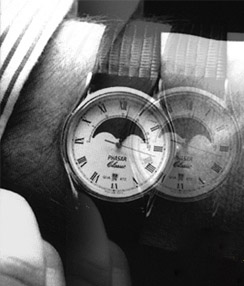
.Click to enlarge->.Photo showing how a patient experiences
")
People with double vision see two images of one object. the images are separate but often clearly focused. The disorder can be due to a number of causes and it usually disappears when one eye is closed. You should consult your ophthalmologist immediately if you start to experience double vision because it may indicate that you have a serious underlying disorder.
Click to see the pictures……………………..(1)……...(2).…(3.)....(4)
It is commonly known as Diplopia or Double vision. It is the simultaneous perception of two images of a single object. These images may be displaced horizontally, vertically, or diagonally (i.e. both vertically and horizontally) in relation to each other.
Binocular diplopia or double vision
Binocular diplopia is double vision arising as a result of the misalignment of the two eyes relative to each other, such as occurs in Esotropia or Exotropia. In such a case whilst the fovea of one eye is directed at the object of regard, the fovea of the other is directed elsewhere, and the image of the object of regard falls on an extra-foveal area of retina.
The brain calculates the ‘visual direction’ of an object based upon the position of its image relative to the fovea. Images falling on the fovea are seen as being directly ahead, whilst those falling on retina outside the fovea may be seen as above, below, right or left of straight ahead depending upon the area of retina stimulated. Thus, when the eyes are misaligned, the brain will perceive two images of one target object, as the target object simultaneously stimulates different, non-corresponding, retinal areas in either eye, thus producing double vision.
This correlation of particular areas of the retina in one eye with the same areas in the other is known as Retinal correspondence. This relationship also gives rise to an associated phenomenon of binocular diplopia, although one that is rarely noted by those experiencing diplopia: Because the fovea of one eye corresponds to the fovea of the other, images falling on the two foveas are ‘projected’ to the same point in space. Thus, when the eyes are misaligned, the brain will ‘project’ two different images in the same visual direction. This phenomenon is known as ‘Confusion’.
Double vision is dangerous to survival, therefore, the brain naturally guards against its occurrence. In an attempt to avoid double vision, the brain can sometimes ignore the image from one eye; a process known as suppression. The ability to suppress is to be found particularly in childhood when the brain is still developing. Thus, those with childhood strabismus almost never complain of diplopia whilst adults who develop strabismus almost always do. Whilst this ability to suppress might seem a wholly positive adaptation to strabismus, in the developing child this can prevent the proper development of vision in the affected eye resulting in amblyopia. Some adults are also able to suppress their diplopia, but their suppression is rarely as deep or as effective and takes longer to establish. They are not at risk of permanently damaging their vision as a result though. It can appear sometimes, therefore, that diplopia disappears without medical intervention. However, in some cases the cause of the double vision may still be present.
Monocular diplopia
More rarely, diplopia can also occur when viewing with only one eye; this is called monocular diplopia, or, where the patient perceives more than two images, monocular polyopia. In this case, the differential diagnosis of multiple image perception includes a structural defect within the eye, a lesion in the anterior visual cortex (rarely cause diplopia, more commonly polyopia or palinopsia) or non-organic conditions.
Temporary diplopia
Temporary diplopia can also be caused by intoxication from alcohol or head injuries, such as concussion. If temporary double vision does not resolve quickly, one should see an eye doctor immediately. It can also be a side effect of the anti-epileptic drugs Phenytoin and Zonisamide, and the anti-convulsant drug Lamotrigine, as well as the hypnotic drug Zolpidem and the dissociative drug Ketamine.
Voluntary diplopia
Some people are able to consciously uncouple their eyes, inducing double vision on purpose. These people do not consider their double vision dangerous or harmful, and may even consider it enjoyable. It makes viewing stereograms much easier.
Causes:
There are two possible causes. Refractive. Light from an object is split into two images by a defect in the eye’s optical system. Cataracts might, for example, cause such a defect.
Failure of both eyes to point at the object being viewed, a condition referred to as “strabismus” or “squint”. In normal vision, both eyes look at the same object. The images seen by the two eyes are fused into a single picture by the brain. If the eyes do not point at the same object, the image seen by each eye is different and cannot be fused. The result is double vision. Why might eyes not point in the same direction? Possibly because of a defect in the muscles which control the movement of the eyes or in the control of these muscles through the nerves and brain.
The most common cause of double vision is weakness or paralysis of one of more of the muscles that control the movements of one eye. The movement of the affected eye is impaired, causing crossed eyes. Two different views of the same object are received by the visual system and the brain cannot combine them. Tilting or turning the head may briefly correct the problem. however, not all types of crossed eyes cause double vision.
Many serious conditions that affect the brain and nervous system may cause impaired eye movements, leading to double vision. Potential causes include multiple sclerosis, head injuries, brain tumors and bulging of an artery inside the head due to a weakness in the vessel wall. In older people, impaired eye movement resulting in double vision may be linked with diabetes mellitus and rarely with atherosclerosis and high blood pressure.
Double vision can also occur as a result of a tumor or blood clot behind one of the eyes, causing the movement of that eye to be affected.
Implications:
Double vision can be extremely discomforting. The brain acts to alleviate the discomfort by suppressing, or blanking out, one of the images. In young children, if this suppression persists over a continued length of time, it can lead to an impairment of the development of the visual system. The suppressed eye may get to the point where it is unable to see well, no matter how good the spectacle or contact lens correction. Doctors call this condition amblyopia. Since it is a result of a defect in the interpretive mechanisms of the eye and brain, it is more difficult to treat than a refractive condition (one having to do with the eye’s ability to bend light).
Diagnosis:
Your doctor may ask you to shut one eye at a time to see whether the double vision disappears. He or she may also ask you to describe the double images, or ask if they appear side by side or one on top of the other or whether one of the images appears to be tilted. Your doctor will probably observe the movements of your eyes closely in order to establish whether any of the eye muscles are weak or paralyzed. He or she may also carry out special vision tests to identify weak eye movement.
If double vision has come on suddenly, or if no obvious cause can be found, or urgent CT scanning or MRI may be done to check for any abnormality in the eye sockets or brain that might be affecting the alignment of the eyes. You should also have a neurological examination.
Treatment:
Treatment of double vision is aimed at the underlying cause. A serious disorder such as an aneurysm may need hospital treatment. Double vision caused by diabetes mellitus will usually disappear over time. But, if it does not disappear, your doctor may advise wearing a patch cover one eye to eliminate the second image. Muscle surgery is also useful if double vision has been present for some time.
Treatment of double vision consists of eye exercises, surgical straightening of the eye or a combination of the two. Therapy is aimed at re-aligning the squinting eye where possible without surgery and re-stimulating the part of the visual pathway to the brain which is not working correctly.
Efforts must first be made to identify and treat the underlying cause of the problem. Treatment options includes prism lenses and/or vision therapy and/or surgery, and also botulinum toxin can be used. On occasions, in certain conditions such as Oculomotor nerve palsy for example, it may be necessary to occlude one eye either temporarily or permanently. Daily wear of prism lenses is a passive compensatory treatment. Vision therapy is an active treatment which retrains the visual and vestibular systems (brain, eye muscles, and body). Vision therapy may eliminate the need for daily wear of prism lenses but is only suitable for a minority of those with diplopic symptoms.
Disclaimer: This information is not meant to be a substitute for professional medical advise or help. It is always best to consult with a Physician about serious health concerns. This information is in no way intended to diagnose or prescribe remedies.This is purely for educational purpose
Resources:
http://www.charak.com/DiseasePage.asp?thx=1&id=56
http://www.broomeoptical.com/double_vision.html
http://en.wikipedia.org/wiki/Diplopia
http://concise.britannica.com/ebc/art-3425/Physiological-diplopia
Related articles


