Categories
Ailmemts & Remedies

Kidney dialysis

Introduction:
In order for blood to perform its essential functions of bringing nutrients and oxygen to the cells of the body, and carrying waste materials away from those cells, the chemical composition of the blood must be carefully controlled. Blood contains particles of many different sizes and types, including cells, proteins, dissolved ions, and organic waste products. Some of these particles, such as proteins like hemoglobin, are essential for the body. Others, such as urea (a waste product from protein metabolism), must be removed from the blood or they will accumulate and interfere with normal metabolic processes. Still other particles, including many of the simple ions dissolved in the blood, are required by the body in certain concentrations that must be tightly regulated, especially when the intake of these chemicals varies. The body has many different means of controlling the chemical composition of the blood. For instance, you learned in the “Iron Use and Storage in the Body: Ferritin and Molecular Representations” tutorial that the ferritin protein can help to control the amount of free iron in the blood. As you will discover in the tutorial entitled, “Blood, Sweat, and Buffers: pH Regulation During Exercise”, buffers dissolved in the blood can help regulate the blood’s pH. But the largest responsibility for maintaining the chemistry of the blood falls to the kidneys, a pair of organs located just behind the lining of the abdominal cavity. It is the job of the kidneys to remove the harmful particles from the blood and to regulate the blood’s ionic concentrations, while keeping the essential particles in the blood

CLICK & SEE THE PICTURES

Healthy kidneys clean the blood by removing excess fluid, salt and wastes. When they fail, harmful wastes build up, blood pressure may rise, and the body may retain excess fluid. When this happens, treatment – dialysis or a kidney transplant – is needed to replace the work of the failed kidneys, which is known as end-stage renal failure (ESRF).

There are three primary and two secondary types of dialysis: hemodialysis (primary), peritoneal dialysis (primary), hemofiltration (primary), hemodiafiltration (secondary), and intestinal dialysis (secondary).

Hemodialysis:
Haemodialysis (HD) is the most common method used to treat ESRF and has been available since the 1960s. Despite some advances in dialysis machines in recent years, HD is still a complicated and inconvenient therapy requiring a coordinated effort from a large healthcare team, including:

•GP
•Nephrologist (kidney doctor)
•Dialysis nurse
•Dialysis technician
•Dietitian
•Social worker
One important step before starting HD is a small operation to prepare a site on the body. One of the arteries in your arm is re-routed to join a vein, forming a fistula. Blood is removed from the fistula, cleaned and returned to it, allowing dialysis process to take place.

Needles are inserted into a fistula (the point of access to the bloodstream) at the start of HD. You may find this one of the hardest parts, although most people report getting used to them after a few sessions. If it’s painful, an anesthetic cream or spray can be applied to the skin.

CLICK & SEE

In HD, blood is allowed to flow, a small amount at a time, through a special filter (the ‘dialyser’ or ‘artificial kidney’) that removes wastes and extra fluids. The clean blood is then returned to your body via the fistula. This helps to keep the correct amount of water in the body, control blood pressure – and keep the proper balance of chemicals such as potassium, sodium and acid.

Most people have HD three times a week for three to five hours, with a morning, afternoon or evening ‘slot’; depending on availability and capacity at a dialysis unit, usually in a large hospital. Some receive it at a smaller satellite unit nearer home, and a few have HD in their own homes.

By learning about the treatment, and working with your healthcare team, it’s possible to have a full, active life

Peritoneal dialysis:
Peritoneal dialysis (PD) became an alternative to HD in the 1980s, with many preferring the independence it brings them.

It means you don’t have to have dialysis sessions at a unit, but can give treatments at home, at work or on holiday. Like HD, by learning about the treatment, and working with the medical team, it’s possible to have a full and active life.

CLICK & SEE

In PD, a soft tube called a catheter is used to fill the abdomen with a cleansing liquid called dialysis solution. The abdominal cavity is lined with a layer called the peritoneum. Waste products and extra fluid (and salt) then pass through the peritoneum from the blood into the dialysis solution. They then leave the body when the dialysis solution is drained. This used solution is then thrown away.

The process of draining and filling is called an ‘exchange’ and takes about 30 to 40 minutes. The period the dialysis solution is in the abdomen is called the ‘dwell time’. A typical schedule is four exchanges a day, each with a dwell time of four to eight hours.

There are many forms of PD. One doesn’t even require a machine and it’s possible to walk around with the dialysis solution in your abdomen. Talk to your specialist about what’s best for your particular situation.

Whatever form is chosen, an operation is needed to have the soft catheter placed in the abdomen, which will carry the dialysis solution in and out of the abdomen. It’s usually inserted two weeks before dialysis proceeds, to allow scar tissue to build up that will hold it in place.

Hemofiltration:
Hemofiltration is a similar treatment to hemodialysis, but it makes use of a different principle. The blood is pumped through a dialyzer or “hemofilter” as in dialysis, but no dialysate is used. A pressure gradient is applied; as a result, water moves across the very permeable membrane rapidly, “dragging” along with it many dissolved substances, importantly ones with large molecular weights, which are cleared less well by hemodialysis. Salts and water lost from the blood during this process are replaced with a “substitution fluid” that is infused into the extracorporeal circuit during the treatment. Hemodiafiltration is a term used to describe several methods of combining hemodialysis and hemofiltration in one process.

Hemodiafiltration:
Hemodialfiltration is a combination of hemodialysis and hemofiltration. In theory, this technique offers the advantages of both hemodialysis and hemofiltration.

CLICK & SEE

Intestinal dialysis:
In intestinal dialysis, the diet is supplemented with soluble fibres such as acacia fibre, which is digested by bacteria in the colon. This bacterial growth increases the amount of nitrogen that is eliminated in fecal waste.  An alternative approach utilizes the ingestion of 1 to 1.5 liters of non-absorbable solutions of polyethylene glycol or mannitol every fourth hour.

Which is better?
Neither technique ‘cures’ ESRF, as they only provide about five per cent of normal kidney function. In other words, they control kidney failure to an extent. It’s hard to state which technique is ‘better’ for which patient, as both have pros and cons. Many patients will have both in their continuing treatment.

Living with dialysis
Adjusting to the effects of ESRF and the time spent on dialysis can be difficult. Aside from the ‘lost time’ (dialysis can take six to eight hours a day) most patients feel they have less energy. Many need to make changes in their work or home life, and can feel depressed when starting the process, or after several months of treatment. It’s good to talk with a social worker, nurse or doctor as this is a common problem that can often be treated effectively.

If you’re feeling well, your kidney specialist should measure the effectiveness of the dialysis with blood tests at least once a month in HD, and once every three months in PD.

Disclaimer: This information is not meant to be a substitute for professional medical advise or help. It is always best to consult with a Physician about serious health concerns. This information is in no way intended to diagnose or prescribe remedies.This is purely for educational purpose
Resources:
http://www.bbc.co.uk/health/physical_health/conditions/in_depth/kidneys/kidneys_dialysis.shtml
http://en.wikipedia.org/wiki/Dialysis
http://www.chemistry.wustl.edu/~edudev/LabTutorials/Dialysis/Kidneys.html

Enhanced by Zemanta
Categories
Diagnonistic Test

Radionuclide Scan of the Kidneys

[amazon_link asins=’B000N3BZIA,0757004113,B003XNCHAM,B001GXQMBG’ template=’ProductCarousel’ store=’finmeacur-20′ marketplace=’US’ link_id=’81da0ccd-4fe7-11e7-b334-9fb098e8e8e6′]

Definition
A radionuclide scan of the kidneys shows a picture of your kidneys while they are at work making urine.A kidney radionuclide scan, also called a kidney scan or renal scan, is a diagnostic imaging test that involves administering a small amount of radionuclide, also called a radioactive tracer, into the body and then imaging the kidneys with a gamma camera. The images obtained can help in the diagnosis and treatment of various kidney diseases and conditions. This test can be useful to evaluate infection, blockages, injury to the kidneys, and some causes of high blood pressure.

CLICK & SEE

Precautions
A kidney scan requires the use of a radioactive material; therefore, patients who are pregnant or suspect they may be pregnant are cautioned not to have the test unless the benefits outweigh the risks. Women should inform their doctor if they are breast feeding. The doctor will recommend the woman stop breast feeding for a specified period of time, depending on the particular tracer and dose used.

Description
Kidney scans are performed either in a hospital nuclear medicine department or in an outpatient radiology or nuclear medicine facility. The patient is positioned in front of, or under, a gamma camera—a special piece of equipment that detects the radiation emitted from the body and produces an image. An intravenous injection of the radionuclide is administered. Immediately after the injection imaging begins, and, in most studies, the flow of blood to each kidney is evaluated. Serial images of the kidneys are obtained over a specified period of time, depending upon the particular radiopharmaceutical used. Kidney scans may be performed to determine the rate at which the kidneys are filtering a patient’s blood. These studies use a radiopharmaceutical called technetium DTPA (Tc99m DTPA). This radiopharmaceutical also can identify obstruction in the renal collecting system. To establish the function of the renal tubules, the radiopharmaceutical Technetium DMSA (Tc99m DMSA) is used.

A kidney scan ranges from 45 minutes to three hours in length, depending upon the goals of the test, but the test typically takes about an hour to an hour and a half. It is important to understand that kidney scans can reveal an abnormality, but they do not always identify the specific problem. They are very useful in providing information about how the various parts of the kidneys function, which, in turn, can assist in making a diagnosis.

Typically, posterior images are obtained but images are also obtained at oblique angles. If indicated, the patient may be positioned so that mobility of the kidney is demonstrated by sitting up or lying down for the images. If obstruction or renal function is being evaluated, a diuretic (drug to induce urination), such as Lasix, may be injected. If hypertension or renal artery sterosis is being evaluated, Captopril or Enalapril (ACE inhibitors) may be injected.
Preparation
No special preparation is necessary for a kidney scan. In some instances the patient may be required to drink additional liquids and to empty their bladder before the exam. If another nuclear medicine study was recently performed, the patient may have to wait for a specified period to avoid any interference from residual radioactivity in the body. The patient is instructed to remove metal items from the area to be scanned.

Let your doctor know if you could be pregnant or if you are breast-feeding a baby. The medicine used in this test would expose your baby to radiation.

What happens when the test is performed.
You have an IV (intravenous) line placed into a vein. A slightly radioactive version of a substance called sodium pertechnetate is injected through the IV. This substance helps your kidneys and urine show up on pictures.

A camera that is specially designed to detect radioactivity is placed against your back or abdomen. A number of pictures are taken over time. The camera itself does not expose you to any additional radiation, so the number of pictures is not harmful in any way. The test is usually completed within an hour.

Risk Factors:
Many people worry when they hear that the medicine used in this test is slightly radioactive. In truth, this test exposes you to a very small amount of radiation-no greater than that of routine xrays.

Moreover, Nuclear medicine procedures are very safe. Unlike some of the dyes that may be used in x-ray studies, radioactive tracers rarely cause side effects. There are no long-lasting effects of the tracers themselves, because they have no functional effects on the body’s tissues. If pharmaceuticals are injected these can temporarily raise or lower blood pressure, or cause one to urinate.

Aftercare
Patients can resume their normal daily activities immediately after the test. Most radioactive tracers are excreted through the urinary system, so drinking fluids after a kidney scan can help flush the tracer out of the body more quickly.

Results
The scan should reveal normal kidney function for the patient’s age and medical status, as well as show normal relative position, size, configuration, and location of the kidneys. Initial blood flow images should reflect that blood circulation to both kidneys is equal. Patients whose images suggest a space-occupying lesion or obstruction may require other imaging procedures, such as CT or ultrasound, to provide more information. Also, if the kidneys appear to be abnormal in size, have an unusual contour, or are unusually positioned, other imaging procedures may be required.

Resources:
https://www.health.harvard.edu/fhg/diagnostics/radionuclide-scan-of-the-kidneys.shtml
http://www.enotes.com/nursing-encyclopedia/kidney-radionuclide-scan

Reblog this post [with Zemanta]
Categories
Featured Health Problems & Solutions News on Health & Science

Kidney Donation is Safe

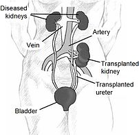
Kidney location after transplantation.
Image via Wikipedia

[amazon_link asins=’0998198404,B071FRPBCZ,1449568394,B00BKNAOL6′ template=’ProductCarousel’ store=’finmeacur-20′ marketplace=’US’ link_id=’85dda5cb-4fe5-11e7-bf56-dd96d881794a’]

People who decide to donate a kidney to their near and dear ones need not fear any long term implications as donation has no negative impact on donor’s general health, say experts. Donors can lead a perfectly normal life and, on the contrary, are benefitted psychologically, having the satisfaction of saving a life, they add. Similar observations have been made by a study done in puerto rico which has been reported in the transplantion proceedings . People with one normal kidney can lead a perfectly nomal life and in fact one in 1,000 people are born with single kidney, Dr. S.   C. Tiwari, professor at the department of nephrology at aiims, said. Hence, mortality for donors is same as for normal people with there being hardly any chance of death because of complications arising out of donation, he said. Tiwari said the institute, where on an average two kidney transplants are performed in a week, did not have a policy of keeping a track of kidney donors.

………………………..CLICK & SEE

However, recipients, who usually come for health follow up regularly, are enquired about the health of donors, generaly related to them. The only difference a donation makes for donors is that their remaining kidney is under more pressure as it has to work for the whole body. thus, donors are asked to live a regulated life and control their diet, blood pressure and physical activity so that work load on kidney does not exceed, Tiwari said. till three months after donation, donors’ single kidney remains hypertrophy, which reflects that it is coping with entire body’s load, and then it adjusts, Tiwari said.

During these three months, the donors are supposed to take special care. One significant outcome of kidney donaiton is that donors have a sense of “eternal satisfaction” for contributing to the life of a relative, he added. The opinion on kidney donation is strengthened by the study, carried out under the puerto rico renal transplant programme, an academic programme based in an affiliated community hospital, in puerto rico.

The study, based on a documentation of the long-term health of live kindney donors, said that in general the health after many years of donation reflects more or less the health of the general population, stressing that kidney donation is a relatively safe procedure with little morbidity and no mortality in the majority of cases. Risk of mortality is estimated to be 0.03 per cent while acute complication rates vary and are relatively low at eight per cent in places with vast experience in living donation, it said. the puerto rico study involved follow up of the health of 20 donors who had donated their kidney 20 years ago or more.

The donors were interviewed and subjected to a complete history and physical examination, including blood pressure and urine analysis, the report said. of the 20 donors, 12 were females and eight males. the donors were in the mean age of 61 years. Significantly all the donors expressed happiness over donation, the report said. in terms of health parameters, the donors had normal urine analyses, excluding one, a 73-year-old woman who had donated kidney to her daughter, who had persence of protein in urine (proteinuria), the report said adding the woman had developed de novo diabetes. Five of the 20 donors developed de novo hypertension at least 10 years after the donation.However, all of them had a strong family history of hypertension, it said. however, donors had elevated levels of creatinin, a product of muscle breakdown, in their serum and lower creatinin clearance by kidneys.

The report said that though it indicated reduced kidney function, the increase in serum creatinin was not significant. Tiwari said that at aiims he had not seen any donor having a creatinin level, which has caused any problem. Dr. D. S. Rana, a nephrologist at the ganga ram hospital, said that creatinin level sometimes increases in marginal donors – donor who are aged (above 65), or have mild hypertension, or have slightly abnormal kidney function. All these are contraindicaitons for kidney donation, but such people are sometimes accepted as donors when no other suitable donor is available, rana said, adding even such donors do not carry any major risk. If raised levels of creatinin are observed, patients are asked to avoid high protein diet, rana said. during the donation surgery also, donors are not at an additional risk. The risks are same as in any other surgery.

Sources: The Times Of India

Reblog this post [with Zemanta]
css.php