Categories
Herbs & Plants

Duchman’s Breeches

[amazon_link asins=’B01FTSN3F6,B01EXGACBI,B077K26L47,B000H4HQXS,B075598QV3,B074NZQQ6S’ template=’ProductCarousel’ store=’finmeacur-20′ marketplace=’US’ link_id=’ac555002-d44a-11e7-96b2-5dff51938678′]

Botanical Name:Dicentra cucullaria
Family : Fumariaceae
Other Name: Dicentra cucullaria
Kingdom: Plantae
Division: Magnoliophyta
Class: Magnoliopsida
Order: Ranunculales
Genus: Dicentra
Species: D. cucullaria

Syninyms:Bicuculla cucullaria[B,P] Corydalis cuccularia[H] D. cuccularia[H] D. cucullaria var. occidentalis[B,P] D. occidentalis[B,P] Fumaria cucullaria[G]

Common Names: Dicentra cucullaria , Dutchman’s breeches (derives from their white flowers that look like white breeches.)

Habitat :Woodland, Dappled Shade, Shady Edge, Deep Shade. Native to North America.It occurs mainly in the eastern half of the continent, from Nova Scotia and southern Quebec west to eastern North Dakota, and south to northern Georgia and eastern Oklahoma; there is also a disjunct population in Idaho, Oregon and Washington. It typically grows in rich woods. The common name Dutchman’s breeches derives from their white flowers that look like white breeches.

Description: It is a perennial herbaceous plant, reaching a height of 15-40 cm. The leaves are 10-36 cm long and 4-18 cm broad, with a petiole up to 15 cm long; they are trifoliate, with finely divided leaflets. The flowers appear during spring; they are white, 1-2 cm long, and are born on flower stalks 12-25 cm long. Both the leaf stalks and the flower stalks rise from an underground, scaly bulb.

CLICK & SEE THE PICTURES

Dutchman’s breeches is one of many plants whose seeds are spread by ants, a process called myrmecochory. The seeds have a fleshy organ called an elaiosome that attracts ants. The ants take the seeds to their nest, where they eat the elaiosomes, and put the seeds in their nest debris, where they are protected until they germinate. They also get the added bonus of growing in a medium made richer by the ant nest debris.

This delicate spring flower is well know to those who visit the southern mountains in the early spring. It is sometimes found in abundance on a northern slope or shaded blank that has remained undisturbed for a very long time. It is very rare in the south except for the mountains and in bloom for a very short time.
Lore: Among some northern tribes it may have been used as a love charm or for seduction. Imagine a young man chewing the root and circling the intended female breathing out the fragrance in the belief that once she smells it she will follow him even against her will.(Erichsen-Brown)Flowering time: April to May

Medical Uses: Alterative; Diaphoretic; Diuretic; Poultice; Tonic; VD.

Alterative, tonic.

The dried tubers were used as a tonic and were recommended in the treatment of VD.

A tea made from the roots is diaphoretic and diuretic.

A poultice made from the leaves has been used in the treatment of skin ailments and as a muscle rub to make them more limber.

The plant contains an alkaloid that depresses the central nervous system – it is used in the treatment of paralysis and tremors.

Native Americans and early white practitioners considered this plant useful for several conditions including syphilis, skin conditions and as a blood purifier. There are several alkaloids that may have effects on the brain and heart. Warning: May be toxic and may cause contact dermatitis in some people.

Native Americans and early white practitioners considered this plant useful for syphilis, skin conditions and as a blood purifier. Dutchman’s breeches contains several alkaloids that may have effects on the brain and heart.

However, D. cucullaria may be toxic and may cause contact dermatitis in some people.The plant is potentially poisonous and can also cause skin rashes.

Similar Species: Squirrel Corn (Dicentra canadensis) has more rounded flowers. Turkey Corn or Wild Bleeding heart (Dicentra eximia) has pink rounded flowers.

Disclaimer:The information presented herein is intended for educational purposes only. Individual results may vary, and before using any supplements, it is always advisable to consult with your own health care provider

Resources:
http://en.wikipedia.org/wiki/Dicentra_cucullaria
http://2bnthewild.com/plants/H289.htm
http://www.ibiblio.org/pfaf/cgi-bin/arr_html?Dicentra+cucullaria

Categories
Healthy Tips

Mind Your Mouth

[amazon_link asins=’1455542512,B004R1Q86I,0975415905,B01AYA38I6,B077S1ZMV7,B00I4VTUQW’ template=’ProductCarousel’ store=’finmeacur-20′ marketplace=’US’ link_id=’190a40d3-d44a-11e7-aa7b-d3cd50f70cb0′]

There are strong links between poor oral health and systemic ailments like diabetes, Alzheimer’s, heart disease and certain types of cancer.

The way to a person’s heart is through his stomach, the adage goes. Researchers now think the way to a healthy heart might be through your gums and teeth.

Evidence suggests that the healthier these are, the stronger and less disease-prone the heart is. If you don’t floss or brush, you might be setting yourself up not just for gum disease but also for heart disease. The link between what’s happening in your mouth and in the rest of your body goes further still: gum disease might be a kind of early warning system, with poor oral health linked to diabetes, kidney disease, preterm labour, osteoporosis, Alzheimer’s disease and even certain types of cancer.

“A lot of studies are coming out that suggest some possible link or associations” between oral infection and systemic disorders, says Sally Cram, a periodontist in the District of Columbia, in the US.

There’s a certain logic to the idea that your mouth — your body’s key opening to the outside — is a harbinger of bodily health. Yet the connection is one that many people, even medical professionals, often overlook. Patients tend to minimise oral health, treating mouth issues as merely “dental”. Professionals echo this artificial dichotomy: dentists and doctors don’t really talk to one another; they don’t attend the same conferences; they don’t read the same journals.

But recent research shows a startling correlation between gum health and atherosclerosis, a condition underlying much heart disease: the worse a person’s gum disease, the narrower that person’s arteries owing to a build-up of plaque. This holds even for young, healthy adults who have no other symptoms of heart disease.

Many questions remain about the nature of the body-mouth connection. In gum disease (called gingivitis in the early stages, before it develops into full-blown periodontal disease), the tissue that surrounds the bones supporting the teeth becomes infected. Often this results from the accumulation of bacteria in the plaque under the tissue holding the teeth. The bacteria release toxins and other chemicals that begin to destroy the bone. Scientists believe they circulate and cause damage elsewhere in the body; exactly how remains unclear.

When Maurizio Tonetti of the University of Connecticut in the US conducted a study into whether reversing the production of bacteria and toxins in the mouth would benefit patients who had atherosclerosis, the results were encouraging. He reported in the New England Journal of Medicine last year that patients who underwent an intensive, six-month programme of treatment for gum disease emerged not only with healthier gums but also with improved endothelial function — that is, better function of the lining of the blood vessels.

These findings merit further study of a possible link between gum infections and preterm labour. Observational studies in the US show that rates of preterm birth are higher for women with severe gum disease than those with milder or no such disease.

Gum disease may also be implicated in a “small, but significant” increase in overall cancer risk for men, according to a recent study in The Lancet Oncology. The authors linked gum disease with a higher chance of lung, kidney, pancreatic and blood cancers. A similar pattern is emerging for kidney disease and Alzheimer’s. But as with heart disease and preterm labour, what causes what is not known.

In diabetes, however, the body-mouth connection is clear. Diabetics who have uncontrolled gum disease, Cram said, “have a much harder time (than other diabetics) controlling their blood sugar levels.” The reverse, she noted, is also true: people with uncontrolled diabetes are about “three to four times at greater risk of developing periodontal disease.”

You would think physicians would be telling their diabetic patients to make regular dental visits to head off gum disease and that dentists would be advising patients who develop persistent gum disease to be tested for diabetes. But neither group of practitioners has been especially good at making the connection.

Dental patients need to get past thinking of their dentist primarily as a cosmetic practitioner — a whitener and straightener of teeth — a “fireman” to be called only when there’s extreme tooth pain.

Regular visits to a dentist can make the mouth an early warning system for a variety of problems. Sores or fungus in the mouth, for example, are often the very first indications of infection by HIV or of cancer. Osteoporosis, a disease of the bones, could show up in a routine dental X-ray before you notice its impact on your hips or spine.

“There’s a desperate need for more collaboration between the two specialities,” says Alan Douglass, associate professor of family medicine at the University of Connecticut School of Medicine, the US.

One thing both groups agree on is the power of prevention, which means brushing, flossing and having regular professional cleaning.

Doctors can also help identify patients at risk. The good news is that if you catch gum disease in its early stages, Cram says, “you can reverse it” with procedures such as scaling and root cleaning.

So take care of your mouth. Your heart, your brain and your kidneys may thank you for it. As another US dentist puts it: “Oral health is not an out-of-body experience.”

Sources: The Telegraph (Kolkata, India)

Reblog this post [with Zemanta]
Categories
Featured

Farm Pregnancy ‘Cuts Asthma Risk’

A picture of my wifeImage via Wikipedia

[amazon_link asins=’B01LWBYWO1,B074GFGW96,B071Z4HGGZ,B077KK4YCV,B073RNVRW2,B01MXE7SG6,B071VTR652,B01C3TZ9N8,B071VRXVGR’ template=’ProductCarousel’ store=’finmeacur-20′ marketplace=’US’ link_id=’f04fef7d-da3d-11e7-b353-05e373a53f9d’]

Living on a farm during pregnancy may help reduce the chance of the child developing asthma, eczema and even hayfever, say scientists.

..
Living on a farm while pregnant may benefit the baby

The New Zealand researchers suggest that exposure to animals and the bacteria they carry may affect the foetus’s immune system.

Writing in the European Respiratory Journal, they said exposure before and after birth halved the risk.

But experts warn some animals carry infections which may harm the baby.

The research, carried out at Massey University, adds to other studies which have suggested that living on a farm, with regular contact with animals, during the early years of life, could cut the risk of asthma and other allergic diseases.

But the study of more than 1,300 farmers’ children goes further, suggesting that this protection could start building even before birth.

It found that the greatest apparent protection – a 50% reduction in asthma, and an even greater reduction in eczema and hay fever – was gained by children whose mothers had been exposed to farm life during pregnancy, and who currently lived on a farm.

The reasons why this might happen are unclear, although they are likely to be related to the way that the child begins to develop its immune system.

Milk bacteria

Living on a farm means frequent contact with animal bacteria, perhaps through the consumption of unpasteurised milk, or contact with the animals directly.

The researchers suggested that this might suppress the production of particular immune cells linked to the development of asthma.

However, they suggested that while exposure during pregnancy might be useful, it might only persist if the child was exposed after its birth as well.

The findings are unlikely to lead to any change in current advice to pregnant women, which urges caution about contact with certain farm animals.

In particular, an infection which can cause miscarriage in pregnant ewes can lead to the same result in humans.

The faeces of other animals can also carry infections which can affect a pregnancy.

Dr Elaine Vickers, research manager at Asthma UK, said: “This study adds to existing evidence supporting the hygiene hypothesis, which states that early exposure to potential allergens results in a reduced risk of asthma development.

“However, the causes of asthma are still largely unknown and the processes involved in asthma development are incredibly complicated, including family history, environment and lifestyle.”

Sources: BBC NEWS:30Th. Aug.’08

Reblog this post [with Zemanta]
Categories
Ailmemts & Remedies

Keloids

Definition : A keloid is a type of scar which results in an overgrowth of tissue at the site of a healed skin injury. Keloids are firm, rubbery lesions or shiny, fibrous nodules, and can vary from pink to flesh-colored or red to dark brown in color. A keloid scar is benign, non-contagious, and usually accompanied by severe itchiness, sharp pains, and changes in texture. In severe cases, it can affect movement of skin. Keloids should not be confused with hypertrophic scars, which are raised scars that do not grow beyond the boundaries of the original wound and may reduce over time.

CLICK & SEE THE PICTURES

History in medicine
Keloids were described by Egyptian surgeons around 1700 BC. Baron Jean-Louis Alibert (1768-1837) identified the keloid as an entity in 1806. He called them cancroide, later changing the name to cheloide to avoid confusion with cancer. The word is derived from the Greek chele, meaning crab’s claw, and the suffix -oid, meaning like. For many years Alibert’s clinic at the L’Hôpital Saint-Louis was the world’s center for dermatology.Earlobe keloid as a result of piercing

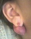
Locations of keloids
Keloids can develop in any place that an abrasion has occurred. They can be the result of pimples, insect bites, scratching, burns, or other skin trauma. Keloid scars can develop after surgery. Lack of proper precautions (e.g., too much movement and/or heavy lifting after an abdominal surgery) can cause keloid scars to develop.

Incidence
People of all ages can develop a keloid. Children under 11 are less likely to develop keloids, even when they get their ears pierced. Keloids may also develop from pseudofoliculitis barbae, continued shaving when one has razor bumps will cause irritation to the bumps, infection and over time keloids will form. It would thus be wise for a man with razor bumps to stop shaving for a while and have the skin repair itself first before undertaking any form of hair removal. It is also speculated that the tendency to form keloids is hereditary and may be passed down from generation to generation.

Causes : Keloids occur from such skin injuries as surgical incisions, traumatic wounds, vaccination sites, burns, chickenpox, acne, or even minor scratches. They are fairly common in young women and African Americans. Keloids often run in families. Keloidosis is a term used when multiple or repeated keloids occur.

Most keloids will flatten and become less noticeable over a period of several years. They may become irritated from rubbing on clothing or other forms of friction. Extensive keloids may become binding, limiting mobility. They may cause cosmetic changes and affect the appearance.

Exposure to the sun during the first year of the keloid’s formation will cause the keloid to tan darker than surrounding skin. This dark coloration may become permanent.

Keloids expand in claw-like growths over normal skin. They have the capability to hurt with a needle-like pain or to itch without warning, although the degree of sensation varies from patient to patient.

If the keloid becomes infected, it may ulcerate. The only treatment is to remove the scar completely. However, the probability that the resulting surgery scar will also become a keloid is high, usually greater than 50%.Keloids form within scar tissue. Collagen, used in wound repair, tends to overgrow in this area, sometimes producing a lump many times larger than that of the original scar. Although they usually occur at the site of an injury, keloids can also arise spontaneously. They can occur at the site of a piercing and even from something as simple as a pimple or scratch. They can occur as a result of severe acne or chickenpox scarring, infection at a wound site, repeated trauma to an area, excessive skin tension during wound closure or a foreign body in a wound. Keloids can sometimes be sensitive to chlorine.

Biologically, keloids are fibrotic tumors characterized by a collection of atypical fibroblasts with excessive deposition of extracellular matrix components, especially collagen, fibronectin, elastin, and proteoglycans. Generally, keloids contain relatively acellular centers and thick, abundant collagen bundles that form nodules in the deep dermal portion of the lesion.

Keloids present a therapeutic challenge that must be addressed, as these lesions can cause significant pain, pruritus (itching), and physical disfigurement. They may not improve in appearance over time and can limit mobility if located over a joint.

Keloids affect both sexes equally, although the incidence in young female patients has been reported to be higher than in young males, probably reflecting the greater frequency of earlobe piercing among women. There is a fifteen times higher frequency of occurrence in highly pigmented people. It is speculated that people who possess any degree of African descent, regardless of skin color, may be especially susceptible to keloid occurrences.

Intentional keloids
The Olmec of Mexico in pre-Columbian times used keloid scarification as a means of decoration. In the modern era, women of the Nubia-Kush in Sudan are intentionally scarified with facial keloids as a means of decoration. The Nuer and Nuba use lip plugs, keloid tattoos along the forehead, keloid tattoos along the chin and above the lip, and cornrows. As a part of a ritual, the people of Papua, New Guinea cut their skin and insert clay or ash into the wounds so as to develop permanent bumps (known as keloids or weals). This painful ritual honors members of their tribe who are celebrated for their courage and endurance.

Symptoms:

A skin lesion that is:

*Flesh-colored, red, or pink
*Located over the site of a wound, injury, or other lesion
*Nodular or ridged

The lesion may itch during formation and growth

Diagnosis:
Diagnosis is made on the basis of the appearance of the skin or scar. A skin biopsy may be needed to rule out other skin growths (tumors).

Treatment:

Keloids usually are not medically dangerous, but they may affect the cosmetic appearance. In some cases, they may spontaneously reduce in size over time. Removal or reduction may not be permanent, and surgical removal may result in a larger keloid scar.

No treatment for keloids is considered to be 100% effective. Some of the treatments that are currently available are described below. These treatments have varying degrees of effectiveness. All the invasive methods of treatment like surgery carry a serious risk of the keloid recurring and becoming bigger than it previously was.

*Contractubex Gel / Hexilak Gel — These gels contain allium cepa extract, heparin and allantoin. Developed for the treatment of post-thyroidectomy scars, these gels are now indicated for the treatment of all post traumatic (burns, acne, piercings) or post surgery scars and keloids. Treatment is simple but requires perseverance. They have shown exceptional results, especially in newer scars.[citation needed] The earlier the initiation of treatment, the better the prognosis. This is now the first line of approach in conservative treatment of keloids.

*Natural treatments :— Some scar treatments contain mucin from the snail helix aspersa müller. The secretion from the snail regulates the skin healing and scar formation process. Topical application of treatments with this ingredient on keloid scars regulates and/or decreases dermal fibroblast proliferation and excess collagen production, and thus prevents and reduces keloid scars and hyperthropic scars.

*Tea tree oil — Keloids that result from piercing can be treated with frequent (1-3 times daily) application of pure tea tree oil, which is most effective on newly formed keloids.

*Crushed aspirin paste — Keloids resulting from piercing can be treated with a crushed aspirin paste applied directly to the scar formation once a day. This is most effective on newly formed keloids.

*Surgery :— Surgery requires great care during and after the operation. Keloids that return after being excised may be larger than the original. There is a 50% chance of recurrence after surgical removal. However, keloids are less likely to return if surgical removal is combined with other treatments. Surgical or laser excision may be followed by intralesional injections of a corticosteroid. Plastic closure of the skin including techniques such as v-plasty or w-plasty to reduce skin tension are known to reduce recurrence of keloids following excision.

*Dressings
— Moistened wound coverings made of silicone gel (such as Dermatix) or silastic have been shown in studies to reduce keloid prominence over time. This treatment is safe and painless, although some patients may experience increased itchiness from wearing the dressing for an extended period of time.

*Steroid injections — Steroid injections are best used as the scar begins to thicken or if the person is a known keloid former. A series of injections with triamcinolone acetonide or another corticosteroid may reduce keloid size and irritation. However, injections are often uncomfortable and in large and/or hard scars can be difficult to perform, requiring local anesthetic for people over 16, and full anesthetic for people under. The treatment area can become very painful as the anesthetic wears off.

*Compression — Compression bandages applied to the site over several months, sometimes for as long as six to twelve months, may lead to a reduction in the size of the keloid. This is the best treatment for preventing new scars.

*Cryosurgery — Cryosurgery is an excellent treatment for keloids which are small and occur on lightly pigmented skin. It is often combined with monthly cortisone injections. The use of cryotherapy is limited since it causes skin blanching. It freezes the skin and causes sludging of the circulation beneath, effectively creating an area of localized frostbite. There is a slough of skin and keloid with re-epithelization.

*Radiation therapy — Electron beam radiation can be used at levels which do not penetrate the body deeply enough to affect internal organs. Orthovoltage radiation is more penetrating and slightly more effective. Radiation treatments reduce scar formation if they are used soon after a surgery while the surgical wound is healing. This is one of the most effective procedures.

*Laser therapy — This is an alternative to conventional surgery for keloid removal. Lasers produce a superficial peel but often do not reduce the bulk of the keloid. The use of dye-tuned lasers has not shown better results than that of cold lasers. A relatively new approach is to combine laser therapy with steroid injections. It is said[who?] that the laser helps in softening the scar tissue, allowing the steroid to work more effectively.

*Newer treatments — Drugs that are used to treat autoimmune diseases or cancer have shown promise. These include alpha-interferon, 5-fluorouracil and bleomycin. However, there is a need for further study and evaluation of this treatment technique.

Possible Complications:
*Psychological distress if keloid is large or disfiguring
*Recurrence of keloid
*Discomfort, tenderness, irritation of the keloid

Disclaimer: This information is not meant to be a substitute for professional medical advise or help. It is always best to consult with a Physician about serious health concerns. This information is in no way intended to diagnose or prescribe remedies.This is purely for educational purpose.

Resources:
http://en.wikipedia.org/wiki/Keloid
http://www.nlm.nih.gov/MEDLINEPLUS/ency/article/000849.htm

Reblog this post [with Zemanta]
Categories
News on Health & Science

Antipsychotic Drug ‘Stroke Risk’

More people than previously thought could be at higher risk of having a stroke caused by their antipsychotic drugs, say UK scientists.

……………………………..
Antipsychotic drugs are given to people with schizophrenia and dementia


Previous research suggested only some types of the drug increased the risk, particularly for people with dementia.

However a study published in the British Medical Journal says all forms of antipsychotics boost the risk, in all patients.

A mental health charity said patients on the drugs must be closely monitored.

“This is another warning that all antipsychotics should be prescribed with great thought and care”…says Marjorie Wallace Sane

Antipsychotic drugs are generally used to control psychotic symptoms in patients with disorders such as schizophrenia, and some severe forms of depression.

They are also thought to be widely used to control symptoms of dementia such as aggression, leading to accusations they were being used unnecessarily as a “chemical cosh” in some circumstances.

They fall into two types – newer “atypical” and older “typical” antipsychotics.

When the first concerns were raised in 2002, these focused on the “atypical” drugs.

These worries led to a recommendation from drug safety watchdogs in the UK that they not be given to people with dementia, and the government has been urged to strengthen this in England in its forthcoming dementia strategy.

The latest findings, from researchers at the London School of Hygiene and Tropical Medicine, confirm the fears over dementia patients, but raise wider concerns.

They identified 6,700 patients from a GP database, all with an average age of 80, and concluded that there was more than a tripling of risk for dementia patients taking any sort of anti-psychotic drug.

Patients without dementia taking any sort of antipsychotic had a 40% increase in risk.

The researchers repeated the recommendation that patients with dementia should not be prescribed these drugs.

‘Last resort’

Neil Hunt, from the Alzheimer’s Society, said that doctors now needed to heed these warnings.

“The over-prescription of antipsychotics is a serious breach of human rights, these drugs should only be a last resort.

“The forthcoming National Dementia Strategy is a crucial opportunity to stop this dangerous over-prescribing and we look forward to its launch in the autumn.”

Marjorie Wallace, the chief executive of the mental health charity Sane, said that while the drugs were capable of transforming lives, different patients reacted differently to their side-effects.

“This study should remind us all that antipsychotics are powerful drugs which can both be essential for some people, while carrying other risks.

“This is another warning that all antipsychotics should be prescribed with great thought and care and be subject to rigorous follow-up.”

Sources: BBC NEWS:30Th. AUG.’08

Reblog this post [with Zemanta]
css.php