Categories
Human Organ Transplantation

Kidney Transplantation

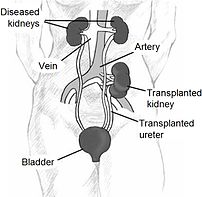
Kidney location after transplantation.
Image via Wikipedia

[amazon_link asins=’1496326156,1455740969,0781753228,1493903411,1416033432,144191689X,1841843164,0062418173,0967038448′ template=’ProductCarousel’ store=’finmeacur-20′ marketplace=’US’ link_id=’11a01150-0970-11e8-81ed-39a60e701c07′]

Alternative Names:Renal transplant; Transplant – kidney

Definition:

A kidney transplant is surgery to place a healthy kidney into a person with kidney failure. Kidney transplantation or renal transplantation is the organ transplant of a kidney in a patient with end-stage renal disease. Kidney transplantation is typically classified as deceased-donor (formerly known as cadaveric) or living-donor transplantation depending on the source of the recipient organ. Living-donor renal transplants are further characterized as genetically related (living-related) or non-related (living-unrelated) transplants, depending on whether a biological relationship exists between the donor and recipient.

..CLICK & SEE THE PICTURES

Description :
Kidney transplants are one of the most common transplant operations in the United States.

A donated kidney is needed to perform a kidney transplant.

The donated kidney may be from:

*Living related donor — related to the recipient, such as a parent, sibling, or child
*Living unrelated donor — such as a friend or spouse

Indications:
The indication for kidney transplantation is end-stage renal disease (ESRD), regardless of the primary cause. This is defined as a drop in the glomerular filtration rate (GFR) to 20-25% of normal. Common diseases leading to ESRD include malignant hypertension, infections, diabetes mellitus and glomerulonephritis; genetic causes include polycystic kidney disease as well as a number of inborn errors of metabolism as well as autoimmune conditions including lupus and Goodpasture’s syndrome. Diabetes is the most common cause of kidney transplant, accounting for approximately 25% of those in the US. The majority of renal transplant recipients are on some form of dialysis – hemodialysis, peritoneal dialysis, or the similar process of hemofiltration – at the time of transplantation. However, individuals with chronic renal failure who have a living donor available often elect to undergo transplantation before dialysis is needed.

Sources of kidneys:
Since medication to prevent rejection is so effective, donors need not be genetically similar to their recipient. Most donated kidneys come from deceased donors, with some coming from living donors. However, the utilization of living donors in the United States is on the rise. In the year 2006, 47% of donated kidneys were actually from living donors (Organ Procurement and Transplantation Network, 2007). It is important to note that this varies by country: for example, only 3% of transplanted kidneys during 2006 in Spain came from living donors (Organización Nacional de Transplantes (ONT), 2007).

Living donors:
Potential donors are carefully evaluated on medical and psychological grounds. This ensures that the donor is fit for surgery and has no kidney disease whilst confirming that the donor is purely altruistic. Traditionally, the donor procedure has been through a single, 4-7 inch incision but live donation is being increasingly performed by laparoscopic surgery. This reduces pain and accelerates recovery for the donor. Excellent results have been demonstrated with laparoscopic donor nephrectomy, for both donor and recipient outcomes. Overall, recipients of kidneys from live donors do extremely well, in comparison to deceased donor recipients.

In 2004 the FDA approved the Cedars-Sinai High Dose IVIG therapy which reduces the need for the living donor to be the same blood type (ABO compatible) or even a tissue match. The therapy reduced the incidence of the recipient’s immune system rejecting the donated kidney in highly-sensitized patients

PROCEDURE FOR A LIVING KIDNEY DONOR:-
If you are donating a kidney, you will be placed under general anesthesia before surgery. This means you will be asleep and pain-free. The surgeon makes a cut in the side of your abdomen, removes the proper kidney, and then closes the wound. The procedure used to require a long surgical cut. However, today surgeons can use a short surgical cut (mini-nephrectomy) or laparoscopic techniques.

Deceased donors:-
Deceased donors can be divided in two groups:

Brain-dead (BD) donors
Donation after Cardiac Death (DCD) donors
Although brain-dead (or “heart-beating”) donors are considered dead, the donor’s heart continues to pump and maintain the circulation. This makes it possible for surgeons to start operating while the organs are still being perfused. During the operation, the aorta will be cannulated, after which the donor’s blood will be replaced by an ice-cold storage solution, such as UW (Viaspan), HTK, or Perfadex. [Depending on which organs are transplanted, more than one solution may be used simultaneously.] Due to the temperature of the solution (and since large amounts of cold NaCl-solution are poured over the organs for a rapid cooling of the organs), the heart will stop pumping.

Donation after Cardiac Death”
donors are patients who do not meet the brain-dead criteria, but due to the small chance of recovery have elected, via a living will or through family, to withdraw support. In this procedure, treatment is discontinued (mechanical ventilation is shut off). Usually, a certain amount of minutes after death has been pronounced, the patient is rushed to the operating theatre, where the organs are recovered, after which the storage solution is flushed through the organs itself. Since the blood is no longer being circulated, coagulation must be prevented with relatively large amounts of anti-coagulation agents, such as heparin. It is important to note that several ethical and procedural guidelines must be followed, chief of which is that the organ recovery team should not participate in the patient’s care in any manner until after death has been declared.

Kidneys from brain-dead donors are generally of a superior quality, since they have not been exposed to warm ischemia (the time between the heart stopping and the kidney being cooled).

Compatibility:
If plasmapheresis or IVIG is not performed, the donor and recipient have to be ABO blood group compatible. Also, they should ideally share as many HLA and “minor antigens” as possible. This decreases the risk of transplant rejection and the need for another transplant. The risk of rejection may be further reduced if the recipient is not already sensitized to potential donor HLA antigens, and if immunosuppressant levels are kept in an appropriate range. In the United States, up to 17% of all deceased donor kidney transplants have no HLA mismatch. However, it is important to note that HLA matching is a relatively minor predictor of transplant outcomes. In fact, living non-related donors are now almost as common as living (genetically)-related donors.

In the 1980s, experimental protocols were developed for ABO-incompatible transplants using increased immunosuppression and plasmapheresis. Through the 1990s these techniques were improved and an important study of long-term outcomes in Japan was published. . Now, a number of programs around the world are routinely performing ABO-incompatible transplants.

In 2004 the FDA approved the Cedars-Sinai High Dose IVIG protocol which eliminates the need for the donor to be the same blood type.

Procedure:
Since in most cases the barely functioning existing kidneys are not removed because this has been shown to increase the rates of surgical morbidities, the kidney is usually placed in a location different from the original kidney (often in the iliac fossa), and as a result it is often necessary to use a different blood supply:

*The renal artery of the kidney, previously branching from the abdominal aorta in the donor, is often connected to the external iliac artery in the recipient.

*The renal vein of the new kidney, previously draining to the inferior vena cava in the donor, is often connected to the external iliac vein in the recipient.

Why the Procedure is Performed :

A kidney transplant may be recommended if you have kidney failure caused by:

*Diabetes
*Glomerulonephritis
*Severe, uncontrollable high blood pressure
*Certain infections

A kidney transplant alone may NOT be recommended if you have:

*Certain infections, such as TB or osteomyelitis
*Difficulty taking medications several times each day for the rest of your life
*Heart, lung, or liver disease
*Other life-threatening diseases

Risks  Factor:

The risks for any anesthesia are:

*Problems breathing
*Reactions to medications

The risks for any surgery are:
*Bleeding
*Infection

Other risks include:
Infection due to medications that suppress the immune response that must be taken to prevent transplant rejections

Post operation:
The transplant surgery lasts about three hours. The donor kidney will be placed in the lower abdomen and its blood vessels connected to arteries and veins in the recipient’s body. When this is complete, blood will be allowed to flow through the kidney again, so the ischemia time is minimized. In most cases, the kidney will soon start producing urine. Since urine is sterile, this has no effect on the operation. The final step is connecting the ureter from the donor kidney to the bladder.

Depending on its quality, the new kidney usually begins functioning immediately. Living donor kidneys normally require 3-5 days to reach normal functioning levels, while cadaveric donations stretch that interval to 7-15 days. Hospital stay is typically for four to seven days. If complications arise, additional medicines may be administered to help the kidney produce urine.

Medicines are used to suppress the immune system from rejecting the donor kidney. These medicines must be taken for the rest of the patient’s life. The most common medication regimen today is : tacrolimus, mycophenolate, and prednisone. Some patients may instead take cyclosporine, rapamycin, or azathioprine. Cyclosporine, considered a breakthrough immunosuppressive when first discovered in the 1980’s, ironically causes nephrotoxicity and can result in iatrogenic damage to the newly transplanted kidney. Blood levels must be monitored closely and if the patient seems to have a declining renal function, a biopsy may be necessary to determine if this is due to rejection or cyclosporine intoxication.

Acute rejection occurs in 10% to 25% of people after transplant during the first sixty days. Rejection does not necessarily mean loss of the organ, but may require additional treatment.

Complications:
Problems after a transplant may include:

*Transplant rejection (hyperacute, acute or chronic)

*Infections and sepsis due to the immunosuppressant drugs that are required to decrease risk of rejection

*Post-transplant lymphoproliferative disorder (a form of lymphoma due to the immune suppressants)

*Imbalances in electrolytes including calcium and phosphate which can lead to bone problems amongst other things

*Other side effects of medications including gastrointestinal inflammation and ulceration of the stomach and esophagus, hirsutism (excessive hair growth in a male-pattern distribution), hair loss, obesity, acne, diabetes mellitus (type 2), hypercholesterolemia, and others.

*The average lifetime for a donor kidney is ten to fifteen years. When a transplant fails a patient may opt for a second transplant, and may have to return to dialysis for some intermediary time.

Prognosis:
Kidney transplantation is a life-extending procedure. The typical patient will live ten to fifteen years longer with a kidney transplant than if kept on dialysis. The years of life gained is greater for younger patients, but even 75 year-old recipients (the oldest group for which there is data) gain an average four more years’ life. People generally have more energy, a less restricted diet, and fewer complications with a kidney transplant than if they stay on conventional dialysis.

Some studies seem to suggest that the longer a patient is on dialysis before the transplant, the less time the kidney will last. It is not clear why this occurs, but it underscores the need for rapid referral to a transplant program. Ideally, a kidney transplant should be pre-emptive, i.e. take place before the patient starts on dialysis.

At least three professional athletes have made a comeback to their sport after receiving a transplant: NBA players Sean Elliott and Alonzo Mourning; and New Zealand rugby union player Jonah Lomu as well as the German-Croatian Soccer Player Ivan Klasni?.

Recovery
The recovery period is 4 – 6 weeks for people who donate a kidney. If you’ve done so, you should avoid heavy activity during this time. Your doctor removes the stitches after a week or so.

If you received a donated kidney, you will need to stay in the hospital for about a week. Afterwards, you will need close follow-up by a doctor and regular blood tests.

Resources:
http://en.wikipedia.org/wiki/Kidney_transplantation
http://www.nlm.nih.gov/medlineplus/ency/article/003005.htm

Reblog this post [with Zemanta]
Categories
News on Health & Science

Just what is it about moobs?

[amazon_link asins=’B00AH0NSKM,B074DTRRCH,B0078IPR64,B01E50SCZU’ template=’ProductCarousel’ store=’finmeacur-20′ marketplace=’US’ link_id=’670c8e24-f81f-11e7-84ce-554e03e2a38a’]

The number of men having breast reduction operations in the UK is rising dramatically, but is this really the result of the media spotlighting the physical flaws of male celebrities?

click & see

This is an era when glossy magazines and tabloids delight in the most minor flaw of the female celebrity.

The actress with bags under her eyes, the singer with an untrimmed armpit, the model with a sweat patch, all are presented blinking in the paparazzo’s flashbulb as their imperfections are chronicled.

All are highlighted with red circles and magnification. And the same process has been applied to male celebrities in recent years

When both the then Prime Minister Tony Blair and leader of the opposition David Cameron were pictured enjoying the sun in the summer of 2006, newspapers from tabloid to broadsheet passed comment on their “moobs”.

Every man has breast tissue, but some have excessive breasts. This ranges from classical cases of gynaecomastia, prompted by a range of causes, to breasts enlarged entirely by deposits of fat over the pectoral muscles. But whatever the cause British men seem to be increasingly concerned over the state of their chests.

The latest figures from the British Association of Aesthetic Plastic Surgeons (Baaps) seem to bear out this obsession.

Surgeons carried out 323 male breast reduction procedures in 2008, up a staggering 44% from 2007.

EXCESSIVE MALE BREASTS
*Pubertal gynaecomastia, common in boys, sees breast tissue grow due to hormonal imbalance

*In most boys it disappears by end of puberty

*Breast growth can be side effect of drugs used to suppress prostate cancer

*Can be caused by genetic condition like Klinefelter’s Syndrome

Other causes include:
*Obesity

*Anabolic steroid use

It would be easy to assume that the UK is a nation where men are rapidly becoming more obese, and they are taking a surgical shortcut to get rid of male breasts that are merely deposits of fat on top of their pectoral muscles.

But this is not the full picture says consultant plastic surgeon and Baaps member Dalia Nield.

She concedes that anything up to a third of the men seeking breast reductions are simply obese. But she says the rest of the rising numbers of operations are people who are suffering gynaecomastia – excessive breasts – caused by other factors, such as a hormonal imbalance.

Among these, a common type is pubertal gynaecomastia, where boys develop the excessive breast tissue during adolescence.

Many of those young men if they don’t have a very marked gynaecomastia they don’t necessarily seek help,” says Ms Nield. “But I see many of these pubertal cases later in life when they put on weight and it becomes more obvious.”

Genetic disorders like Klinefelter’s Syndrome – having an extra “X” chromosome – also account for some cases, and there are a rising number of men suffering from excessive breast tissue as a side effect of drugs prescribed for prostate cancer. Treatment of this type of cancer has improved in recent years, says Mrs Nield, leading to more cases.

But how can one explain the dramatic upwards trajectory for male breast reduction procedures? In 2005, only 22 were performed.

‘Tremendous distress’

Mrs Nield suggests that much of the increase may be due to the media publicising the surgery option.

Many of those pieces mocking the imperfections of the middle-aged celebrity also contain a factbox that talks about non-obesity gynaecomastia and explains that surgery is an option.

MOOBS: THE ETYMOLOGY
*Portmanteau word of “man” and “boobs”

*First reference in UK newspaper in June 2004

*Satirical website manboobs.co.uk domain name registered in January 2003

*Term assumed to be of US origin

The effect, Mrs Nield suggests, is that men who might have been suffering in silence for years, realise they are not alone and are spurred on to seek out surgery.

“It is a cause of tremendous distress,” says the surgeon.

And there is no doubting that the last few years have seen an increasing attention to this particular physical flaw.

A search of the LexisNexis newspaper databases suggests the word made its debut in a British newspaper in June 2004. Since then it has been used 161 times. There have been more than 350 references to “man boobs” over the same period. “Moobs” clocks up 281,000 hits on Google.

Kerri McPherson, a chartered health psychologist at Glasgow Caledonian University and a member of the men’s health group, Scotland, is an expert on male body image.

“I would argue that what the media is really discussing is just representing the growing concerns of everyday men. This concern has always been there but they have not been able to articulate it.”

And it could be argued that media mockery reinforces the negative body image of the excessive male breast sufferer, it also might free some from isolation and paranoia that they could have been burdened with a decade ago.

The presentation of “moobs” as something suffered by a slew of male celebrities might make life easier for the ordinary bloke sitting in a pub discussing his problem with his mates.

“More and more people are being given a language to talk about concerns about their body,” says Dr McPherson.

“Particularly with what is a very feminine [characteristic] if a man was talking about [having] breasts [decades ago] they would have been a source of ridicule.”
Paula Singleton, a researcher in the health faculty at Leeds Metropolitan University, is doing a PhD on the attitudes shown by men planning to have breast reduction surgery, entitled “Bruises heal but moobs last forever – men’s account of cosmetic surgery for gynaecomastia.”

“It seems like you can hardly turn on the telly and open a newspaper without it being mentioned,” she says.

“[Those planning surgery] described feelings of shame, anxiety and embarrassment. They had suffered everything from being shouted at from a bus to teasing from work colleagues… doctors smirking and laughing at them and saying ‘get down the gym’.”

Of course, it would be wrong to group men with excessive breasts into justifiable “moobs” – ie a hormonal, chemical or genetic cause – and unjustifiable “moobs” – those caused primarily by obesity.

Both sets of men may be suffering psychologically at a time when the male body is under increasing scrutiny.

In the academic world, most of the theorising about body image has traditionally been about women, but now researchers are starting to look at changing attitudes among men.

“Men are starting to feel those appearance pressures more and more,” says Ms Singleton.

And this growing body consciousness could lead to more men making their way through the surgeon’s doors.

Sources:BBC News: 28th.Jan.2009

Reblog this post [with Zemanta]
Categories
Herbs & Plants

Astralagus membranaceus

[amazon_link asins=’B01MSWTMXV,B001100OVI,B00GBK9NUQ,B00GPDLJ92,B01MXDZ58U,B00AAXDFI6,B01N8PJ8IK,B01N2I03NP,B01MXIKUCN’ template=’ProductCarousel’ store=’finmeacur-20′ marketplace=’US’ link_id=’0a87a856-8949-11e7-9236-35c93ab47b84′]

Botanical Name:Astragalus membranaceus
Family:Leguminosae (pea family)
Common Names:Tragacanth, Gum Dragon, Milk Vetch, Canada Milk Vetch, Membranous Milk Vetch, Slender Milk Vetch, Standing Milk Vetch, Astragali, Huang Qi (Chinese), Beg Kei, Bei Qi, Hwanggi.
Part Used : Root.
Other Names : Milk-vetch root, huang qi

Different Species:A. membranaceus ,A. gummifer ,A. gracilis ,A. adsurgens var. robustior

Habitat:Native to Mongolia and northern and eastern China.

Description:Astralagus is a low-growing, perennial shrub that reaches sixteen inches. It thrives in sandy, well-drained soil, with plenty of sun. It produces hairy stems and leaves divided into twelve to eighteen pairs of leaflets.A. gummifer is now found growing in Turkey, Syria, Lebanon, northwest Iraq, and the border area between Iran and Iraq.
.CLICK TO SEE THE PICTURES.>.....(01).....(1).……..(2)..…………………….

There are now more than 2000 species worldwide, including some 400 in North America. A. australis is an endemic plant of the Olympic Mountains in the US state of Washington. However, the medicinal varieties are found only in central and western Asia, where it has been extensively tested, both chemically and pharmacologically.

The root readily pulls apart and shreds into a million smaller pieces rather like tissue paper. A yellow core in the center of the sweet-tasting black root is the medicinal substance. The roots are harvested in autumn from four-year-old plants in several Chinese provinces and shipped worldwide. The latex is extracted by making an incision in the trunk and branches of trees growing in the wild.

History:-
The plant is one of the oldest used medicinally, dating to about 200 BCE. It was known even then to balance the body systems and especially good for the lungs and spleen.
The yellow colour of the root contributes to the Chinese name, huang qi, meaning “yellow leader”. It has been used in China for thousands of years to strengthen qi (pronounced “chee”), the body’s life force and protective energy. In Western terminology, this means to strengthen the immune system.

Folk medicine in Europe and Arabia have used the herb for treating tumors of the eyes, liver, and throat.

Tragacanth is the latex that exudes from under the bark and is extracted by making an incision in the trunk and branches. When it dries, it forms flakes that swell in water to form a gelatinous mass used in various treatments, including that of constipation.

European botanists first wrote about its medicinal qualities in the 1700s.

Some of the poisonous species are referred to as Poison Milk Vetch or Loco Weed.

Some of the Native American names came about as a reference to its seeds which rattle in the pods when dried.

A tea of the root was used by the Dakota tribes as a febrifuge for children. The Lakotas pulverized the roots and chewed it for chest and back pains and to relieve coughing. Also, a vapour was inhaled to treat a child’s aching chest. The roots were chewed and applied to cuts before they were bandaged. When combined with the roots of wild licorice, it arrested the spitting of blood. Lakota women who had little or no breast milk, chewed the roots to promote milk production. The Cheyenne used one species for cases of poison ivy or dermatitis. They also ground the leaves and stems and sprinkled the powder on weepy, inflamed, skin conditions.

When the explorer John Bradbury visited the Arikara village along the Missouri River in 1809, he was shown two new species of Astralagus, that were unknown to him, by the local medicine man.

Medicinal Uses: This herb has a variety of benefits as a convalescent and rejuvenating tonic and is also useful in the treatment of Chronic Fatigue Syndrome. Astragalus have been shown to intensify phagocytosis of reticulo-endothelial systems, stimulate pituitary-adrenal cortical activity and restore depleted red blood cell formation in bone marrow. Astragalus is also one of the herbs known to stimulate the bodies natural production of interferon. Astragalus is an ideal remedy for any one who might be immuno-compromized in any way. This can range from someone who easily catches colds to someone with cancer.

Astragalus help maintain normal functions of the liver. Astragalus strengthens immunity to disease. It has certain inhibiting effects on molecular pathological changes caused by viruses, increases growth of plasma cells, stimulates synthesis of antibodies, and builds up body defense.  It enhances body energy. It promotes metabolism of serum and liver proteins, stimulates growth of antibodies, increases white blood cells, and thus increases resistance to viruses. Studies in the West confirm that astragalus enhances immune function by increasing activity of several kinds of white blood cells and boosting production of antibodies and interferon, the body’s own antiviral agent. It is diuretic, detoxifying and reduces proteinuria and cures kidney disease. It inhibits gastric secretions, reduces gastric acid, and thus helps cure stomach ulcers. It is cardiotonic. It has even more remarkable effects on heart failure due to poisoning or exhaustion. It protects the liver and alleviates liver injury.

Key Components: asparagine ,calcyosin ,formononetin ,astragalosides ,kumatakenin ,sterols

Key medical  Actions:
*adaptogenic
*antiviral
*antioxidant
*cardiovascular toner
*diuretic
*immune stimulant
*laxative
*liver protector
*strengthens gastrointestinal tract
*tonic
*vasodilator

Medicinal Parts used: Root, gum-like exudate

*It contains numerous active compounds which bolster immunity.

*The polysaccharides seem to stimulate white blood cell production and spurs the activity of killer T cells, increasing the number of cells and the aggressiveness of their activity. Increased macrophage activity has been measured as lasting up to seventy-two hours.

*It also increases production of interferon, a natural protein that stimulates production of other proteins that help prevent and fight viral infections.

*It increases the number of stem cells in the marrow and lymph tissues, stimulates their maturation into active immune cells, increases spleen activity, increases the release of antibodies, and boosts the production of hormonal messenger molecules that signal for virus destruction.

*Studies at the University of Texas Medical Center found that astragalus was able to restore completely the function of cancer patients compromised immune cells.

*It protects the liver from a variety of liver toxins, including carbon tetrachloride and the anticancer compound stilbenemide.

*Gamma-aminobutyric acid extracts have been found to kill bacteria and lower blood sugar and blood pressure levels

*Chinese experiments indicated that the herb was able to protect against the absorption of toxic chemicals into the liver.

*Studies have shown that patients given the herb suffered less angina and had a greater improvement in the EKGs and other measurements than patients given such standard heart drugs as nifedipine.

Chinese researchers report that the herb improves funtion of the heart’s left ventricle after a heart attack, which they theorize may derive from the herb’s antioxidant effects. Other Chinese researchers found heart-protective effects in people with Coxsackie B virus which can cause viral myocarditis. Staphylococcus aureus, Salmonella spp., and Proteus mirabilis.

Strengthens digestion, raises metabolism, strengthens the immune system, and promotes the healing of wounds and injuries.  It treats chronic weakness of the lungs with shortness of breath, collapse of energy, prolapse of internal organs, spontaneous sweating, chronic lesions, and deficiency edema.  It is very effective in cases of nephritis that do not respond to diuretics.

In China astragalus enjoyed a long history of use in traditional medicine to strengthen the Wei Ch’i or “defensive energy” or as we call it, the immune system. Regarded as a potent tonic for increasing energy levels and stimulating the immune system, astragalus has also been employed effectively as a diuretic, a vasodilator and as a treatment for respiratory infections.

Antibacterial; used with the ginsengs; helpful for young adults for energy production and respiratory endurance; warming energy; helpful for hypoglycemia; used for “outer energy” as ginseng is used for “inner energy”; American Cancer Society publication reports it restored immune functions in 90% of the cancer patients studied; use to bolster the white blood cell count; strengthens the body’s resistance; use for debilitating conditions; helps to promote the effects of other herbs; helps to improve digestion. Astragalus is of the most popular herbs used in the Orient; the Chinese name for astragalus is Huang Ch’i. It is a tonic producing warm energy and specifically tonifying for the lungs, spleen, and triple warmer via meridians.

In studies performed at the Nation Cancer Institute and 5 other leading American Cancer Institutes over the past 10 years, it has been positively shown that astragalus strengthens a cancer patient’s immune system. Researchers believed on the basis of cell studies that astragalus augments those white blood cells that fight disease and removes some to those that make the body more vulnerable to it. There is clinical evidence that cancer patients given astragalus during chemotherapy and radiation, both of which reduce the body’s natural immunity while attacking the cancer, recover significantly faster and live longer. It is evident that astragalus does not directly attack cancers themselves, but instead strengthens the body’s immune system. In these same studies, both in the laboratory and with 572 patients, it also has been found that Astragalus promotes adrenal cortical function, which also is critically diminished in cancer patients.

Astragalus also ameliorates bone marrow pression and gastointestinal toxicity caused by chemotherapy and radiation. Astragalus is presently being looked upon as a possible treatment for people living with AIDS and for its potentials to prolong life.

Scientists have isolated a number of active ingredients contained in astragalus, including bioflavanoids, choline, and a polysaccharide called astragalan B. Animal studies have shown that astragalan B is effective at controlling bacterial infections, stimulating the immune system, and protecting the body against a number of toxins.

Astragalan B seems to work by binding to cholesterol on the outer membranes of viruses, destabilizing their defenses and allowing for the body’s immune system to attack the weakened invader. Astragalus also increases interferon production and enhances NK and T cell function, increasing resistance to viral conditions such as hepatitis, AIDS and cancer. Astragalus shows support for peripheral vascular diseases and peripheral circulation.

Traditional Uses
In China, it has long been used as a classic energy tonic and is considered to be superior to ginseng for young people. It is believed to warm and tone wei qi (a protective energy that circulates just beneath the skin), helping the body to adapt to external influences, especially to the cold. It raises immune resistance, improves physical endurance, and encourages the body systems to function correctly.
By encouraging blood flow to the surface, the herb is effective in controlling night sweats, relieving fluid retention, and reducing thirstiness.

It is used to treat prolapsed organs and is beneficial in uterine bleeding.

In Chinese medicine, the herb has been used alone, or in combination with other herbs, to treat liver fibrosis, acute viral myocarditis and other viral infections, heart failure, and small cell lung cancer, liver and kidney diseases, and amenorrhea.

Taken internally, it is commonly used to strengthen the immune system, especially in such immuno-compromised individuals as those with HIV or during chemotherapy.

Infusions are used to ward off or help treat colds and other infections, to improve heart function especially after a heart attack, to improve memory and learning, to temporarily increase urinary output, and to promote the healing of burns and skin sores.

A decoction of the root in combination with Chinese angelica is used to treat anemia but when combined with cinnamon, it is used to treat cold and numbness.

When the root is dry-fried alone or with honey added, it is used as a stimulating tonic and eaten with meals.

Asragalus boosts the spleen when symptoms indicate that it is not functioning as it should. These symptoms include chronic fatigue, diarrhea, and a loss of appetite.

The herb is also used to treat anorexia, arthritis, diabetes, hypertension, malaria, kidney inflammations, painful urination, prolapsed uterus, uterine bleeding or weakness, edema, water retention, skin ulcers that will not heal, fever, lack of stamina, and generalized weakness.

Tinctures are often used for night sweats.

Disclaimer:The information presented herein is intended for educational purposes only. Individual results may vary, and before using any supplements, it is always advisable to consult with your own health care provider.

Resources:
http://www.innvista.com/health/herbs/astralag.htm
http://www.herbs-herbal-remedies.com/list_of_herbs.htm

http://www.neerlandstuin.nl/plantenc/astralagus.html

http://www.godsremedy.com/hepatitis/prodadd.htm

http://www.herbnet.com/Herb%20Uses_AB.htm

Categories
Diagnonistic Test

Bronchoscopy

[amazon_link asins=’034096832X,1455703206,1107449529,3805593104,0781700957,0521711096,9350903407,1517270898,B015UWKSPE’ template=’ProductCarousel’ store=’finmeacur-20′ marketplace=’US’ link_id=’164a9041-894a-11e7-aff1-f166518e5c19′]

Definition;
Bronchoscopy is a technique of visualizing the inside of the airways for diagnostic and therapeutic purposes. An instrument (bronchoscope) is inserted into the airways, usually through the nose or mouth, or occasionally through a tracheostomy. This allows the practitioner to examine the patient’s airways for abnormalities such as foreign bodies, bleeding, tumors, or inflammation. Specimens may be taken from inside the lungs: biopsies, fluid (bronchoalveolar lavage), or endobronchial brushing. The construction of bronchoscopes ranges from rigid metal tubes with attached lighting devices to flexible fibreoptic instruments with realtime video equipment.
CLICK & SEE THE PICTURES
A bronchoscope is a long snakelike instrument with a tiny video camera and biopsy instruments on one end. It can be maneuvered through your mouth and directly into the airways of your lungs. Bronchoscopy is usually done to obtain a sample of deep lung mucus or lung tissue to help diagnose cancer, pneumonia, or other lung disease.

Why it is done?
Bronchoscopy is usually done to find the cause of a lung problem. Samples of mucus or tissue may be taken from the patient’s lungs during the procedure to test in a lab.

Bronchoscopy may show a tumor, signs of an infection, excess mucus in the airways, the site of bleeding, or something blocking the airway, like a piece of food.

Sometimes bronchoscopy is used to treat lung problems. It may be done to insert a stent in an airway. An airway stent is a small tube that holds the airway open. It is used when a tumor or other condition blocks an airway.

In children, the procedure is most often used to remove something blocking the airway. In some cases, it is used to find out what’s causing a cough that has lasted for at least a few weeks.

How do you prepare for the test?
You will need to sign a consent form giving your doctor permission to perform this test. Some patients have this test done in a clinic procedure area, while others are admitted to the hospital for it. Generally your doctor will decide whether you need to be in the hospital based on your medical condition. If you are not staying in the hospital afterward, you should arrange for a ride home.

Talk with your doctor ahead of time if you are taking insulin, or if you take aspirin, nonsteroidal antiinflammatory drugs, or other medicines that affect blood clotting. It may be necessary to stop or adjust the dose of these medicines before your test. Most people need to have a blood test done some time before the procedure to make sure they are not at high risk for bleeding complications. Also tell your doctor if you have ever had an allergic reaction to the medicine lidocaine or the numbing medicine used at the dentist’s office.

Usually you will be told not to eat anything after midnight on the night before the test. This is so you will have an empty stomach in case you experience nausea from anti-anxiety medicines (sedatives) or have a choking sensation or nausea when the camera is first lowered past your throat.

What happens when the test is performed?
You wear a hospital gown during the procedure. You have an IV (intravenous) line inserted into a vein in case you need medicines or fluid during the procedure.

Bronchoscopy can be performed in a special room designated for such procedures, operating room, intensive care unit, or other location with resources for the management of airway emergencies. The patient will often be given antianxiety and antisecretory medications (to prevent oral secretions from obstructing the view), generally atropine, and sometimes an analgesic such as morphine. During the procedure, sedatives such as midazolam or propofol may be used. A local anesthetic is often given to anesthetise the mucous membranes of the pharynx, larynx, and trachea. The patient is monitored during the procedure with periodic blood pressure checks, continuous ECG monitoring of the heart, and pulse oximetry.

During the procedure, a thin, flexible tube called a bronchoscope is passed through the patient’s nose (or sometimes the mouth), down the throat, and into the airways. If the patient has a breathing tube, the bronchoscope can be passed through it to the airways.

At the bronchoscope’s tip are a light and a mini-camera, so the doctor can see your windpipe and airways. The patient will be given medicine to make them relaxed and sleepy during the procedure.

In some cases, your doctor decides that this procedure would be safer or easier if you were intubated before the test and for a short time afterward. This means having a plastic tube placed through your mouth into your main airway. If you are intubated, you are able to breathe, but you cannot speak while the tube is in place, as it passes between your vocal cords in your voice box. Intubation is always done with the assistance of an anesthesiologist, who gives you medicines to relax your throat muscles and make you unconscious for a minute or two while the tube is placed. Most patients do not require intubation.

If you are not intubated, your doctor or nurse sprays a numbing medicine onto the back of your throat just before the procedure. This medicine makes it easier for you to have the bronchoscope placed. Most patients are also given some medicine through the IV to relax them.

You lie on a hospital bed for the procedure. Your doctor (usually a pulmonary specialist) moves one end of the bronchoscope through your mouth and throat and into your trachea (windpipe). Some patients cough or gag briefly when this is done. The bronchoscope is much narrower than your trachea, so you are able to breathe easily during the procedure.

The doctor can see into your lungs by watching a TV screen that shows the view from the camera on the end of the bronchoscope. Your doctor can control a miniature vacuum at the end of the scope that allows him or her to take a sample of mucus from inside the lung. It is also possible for the doctor to take a biopsy sample of the lung tissue using a needle that can be moved through the scope. At the end of the test, the bronchoscope is pulled out, and you might cough forcefully a few times, possibly coughing out some phlegm.

Bronchoscopy usually takes 30 minutes to an hour, including setup time. The camera is usually in place for less than 20 minutes.

What risks are there from the test?
Besides the risks associated with the drug used, there are also specific risks of the procedure. Although the rigid bronchoscope can scratch or tear airway or damage the vocal cords, the risk of bronchoscopy is limited. Complications from fiberoptic bronchoscopy remain extremely low. Common complications include excessive bleeding following biopsy. A lung biopsy also may cause leakage of air called pneumothorax. Pneumothorax occurs in less than 1% of cases requiring lung biopsy. Laryngospasm is a rare complication but may sometimes require intubation. Patients with tumors or significant bleeding may experience increased difficulty breathing after a bronchoscopic procedure, sometimes due to swelling of the mucous membranes of the airways.

The risks of bronchoscopy are primarily associated with the needle biopsy procedure that is sometimes done through the bronchoscope. If a biopsy is done, the risks include bleeding in the lung or the formation of an air leak. If a patient vomits during the procedure and stomach contents leak down around the bronchoscope, this can irritate the lung and cause a type of pneumonia called aspiration pneumonia. Some patients have a hoarse voice or a sore throat for a day or two after bronchoscopy. Most people have no side effects from the procedure.

The other risks include:

*A drop in a patient’s oxygen level during the procedure. Oxygen will be administered if this happens.
*A slight risk of minor bleeding and developing a fever or pneumonia.

A rare but more serious side effect is a pneumothorax. A pneumothorax is a condition in which air or gas collects in the space around the lungs. This can cause the lung(s) to collapse.

This condition is easily treated and may go away on its own. If it interferes with breathing, a tube may need to be placed in the space around the lungs to remove the air.

A chest X-ray may be done after bronchoscopy to check for problems

Must you do anything special after the test is over?
You will probably feel sleepy after the procedure for a few hours, due to the anti-anxiety medicines. Generally, patients either spend a few hours in a recovery room or stay overnight in the hospital after bronchoscopy. If you do go home the same day, you should not drive or drink alcohol.

What does bronchoscopy show?
Bronchoscopy may show a tumor, signs of an infection, excess mucus in the airways, the site of bleeding, or something blocking the airway.

The doctor will use the procedure results to decide how to treat any lung problems that were found. Other tests may be needed.
Recovery and recuperation :
Patients will be advised by their doctors about when they can return to their normal activities, such as driving, working, and physical activity. For the first few days, a sore throat, cough, and hoarseness are common. The doctor should be called right away if the patient:

*Develops a fever
*Has chest pain
*Has trouble breathing
*Coughs up more than a few tablespoons of blood

How long is it before the result of the test is known?
Your doctor can tell you what the airways in your lungs look like as soon as the test is over. If a sample of mucus or lung tissue was obtained, analysis will require anywhere from a few hours to a few days.

Resources:
https://www.health.harvard.edu/diagnostic-tests/bronchoscopy.htm
http://www.daviddarling.info/encyclopedia/B/bronchoscopy.html
http://en.wikipedia.org/wiki/Bronchoscopy

Reblog this post [with Zemanta]
Categories
Herbs & Plants

Aspalathus Linearis (Rooibos)

[amazon_link asins=’B01DDMWLCG,B001A1PXNC,B00GWQK29U,B003D4IY26,3869558725,3838108949′ template=’ProductCarousel’ store=’finmeacur-20′ marketplace=’US’ link_id=’343ca8b3-fef2-11e6-bce1-a3f7e7879ea6′]

Botanical Name : Aspalathus linearis
Family:    Fabaceae
Subfamily:Faboideae
Tribe:    Crotalarieae
Genus:    Aspalathus
Species:linearis
Kingdom:Plantae
Order:    Fabales

Synonyms: Aspalathus contaminatus – auct. Borbonia pinifolia – Marloth.
Common names : rooibos tea ( Eng. ), rooibostee, bossietee (Afr.)
Habitat : Aspalathus linearis is naturally distributed in the winter rainfall area from about Vanrhynsdorp in the north to the Cape Peninsula and the Betty’s Bay area in the south. The area experiences cold wet winters and hot dry summers with about 300-350 mm of rain per annum. Rooibos tea is made from selected forms of the species found mainly on the Cederberg Mountains. It is cultivated on sandy soils in the valleys of the Olifants, Breede and Hex Rivers (Dahlgren 1988).

Derivation of name and historical aspects :
The genus name Aspalathus is derived from the Greek aspalathos, which was the name of a scented bush that grew in Greece. The epithet linearis is derived from the Latin word for linear, which in this case refers to the shape of the leaves.

Description
Aspalathus linearis is an erect to spreading, highly variable shrub or shrublet up to 2 m high. Its young branches are often reddish. The leaves are green and needle-like, 15-60 mm long and up to about 1 mm thick. They are without stalks and stipules and may be densely clustered. The yellow flowers, which appear in spring to early summer, are solitary or arranged in dense groups at the tips of branches. The fruit is a small lance-shaped pod usually containing one or two hard seeds.

click to see the pictures…>…..(01)…....(1).…....(2)……..…(3).….

Although many of the plants in the genus Aspalanthus are attractive, they have apparently seldom been grown in gardens. This is thought to be due to the difficulty in propagation by seed or root cuttings and in providing the optimal growing conditions for the plants. In order to grow Aspalathus linearis successfully, seeds must first be scarified and then planted in acid, sandy soils.

There are commercial plants of rooibos at the Cape. According to Mr S de Beer of Lambertshoek farm, Clanwillian, seeds which are obtained from the local rooibos tea management board have been treated and germinate easily. They are planted in seedbeds in March to a depth of 5-10cm and are ready for planting out by July. Plants are generally rainfall dependent and the plants prefer not to be too wet. No fertilizing is required and the plants grow quite well in nutrient poor conditions. The most common pest is Loopers or “Landmeter wurmpies” (the larvae of the family Geometridae and of the order Lepidoptera)

Cultivation:Aspalathus  Linearis  grows in sandy hills and on the sides of mountains. Well-drained, sandy but moisture-retaining, non-acidic soils. Generally farmers plant seeds in February and March and then transfer the seedlings to plantations. It takes 12- 18 months before the shrubs are ready to be harvested. The plants are harvested once each year, from December through April. They are harvested up to period of five years and then pulled out and new plants are planted.

Propagation:
Seed – sow late spring in a greenhouse covering the seed with about 10mm of soil. It will probably be beneficial to pre-soak the seed for 12 hours in warm water prior to sowing. Prick out the seedlings into individual pots of well-drained sandy soil as soon as they are large enough to handle. Grow them on in the greenhouse for at least their first winter and plant them out in late spring or early summer after the last expected frosts. It will probably be wise to give the plants protection from the cold and from excessive rain for at least their first winter outdoors. Cuttings of half-ripe wood in a closed frame in early summer

Hervesting:

The basic method of rooibos harvesting has remained largely the same as the process used centuries ago. An environmentally friendly way of harvesting tea is used that involves cutting only the young branches. Once they are cut, they are neatly bound and transported to the process yards. The older branches are left on the tree and the bushes get slightly taller every year. The tea cuttings are chopped very fine and then bruised to ensure that the important chemical reaction which develops the characteristic colour and flavour of the tea can take place. After watering and airing, the tea is left to “sweat” in heaps and it at this point that the tea acquires its typical reddish brown colour and develops its sweet flavour. After the sweating process has been completed, it is spread out in a large drying yard to dry in the sun.
The rest of the process involves sorting and grading the tea according to length, colour and flavour. The finished Rooibos is finally weighed, bagged, and sold to companies who pack the product in either teabags or in loose leaf form under their own brand names.

Main constituents:Rooibos contains Magnesium, zinc and iron which are all essential to a healthy nervous system. Zinc and iron in particular are important for brain functioning and concentration. It also contains Vitamin C, Alphahydroxy Acid, potassium, copper, calcium, iron, manganese and fluoride.

Uses:
Rooibos tea is a most popular drink for health-conscious people, as it contains no colourants, additives or preservatives and is free of caffeine.Aspalathus linearis is of great economic value. It was first used by the indigenous people of the Cederberg area and is currently a very popular tea.
A tea made from the dried fermented leaves tastes similar to oriental tea made from Camellia sinensis. It is less astringent, however, due to the lower tannin content. It is caffeine-free, but has a higher content of fluoride which might help to protect against tooth decay. Recent research has shown that this tea contains a substance similar to superoxide dismutase, an antioxidant compound that is thought to retard the ageing process. The leaves and stems are harvested in the summer, fermented and sun dried for later use. The leaves are sometimes used as a flavouring in foods and in baking.

Medicinal Uses:
It is considered healthy as it is caffeine-free, low in tannins and rich in anti-oxidants. It is not only enjoyed as a herbal tea, but is also used as an ingredient in cosmetics, in slimming products, as a flavouring agent in baking, cooking and cocktails and even as a treatment for infants who are prone to colic.

it has been used in the treatment of vomiting, diarrhoea and other mild gastric complaints. It has also been shown to be of benefit when used internally and externally in the treatment of a wide range of allergies especially milk allergy, eczema, hay fever and asthma in infants.

Many children with ADHD symptoms also suffer from various allergies and food intolerances. Rooibos is of great benefit in the management of allergies and to build up the immune system. Rooibos improves overall liver functioning, which helps the body to eliminate toxins and improves overall body functioning thereby increasing the efficiency of all organs of the body. Rooibos is endemic to the Cedarberg Mountains of the Cape and is not found anywhere else in the world.
(Treatment for Acne)  (Ingredient in ClearSkin FaceWash)
(Treatment for ADHD)  ( Ingredient in Focus ADHD)
(Treatment for High Cholesterol)  Ingredient in Cholesto-Rite)

Now known worldwide for its anti-oxidant and healing properties, the soothing and healing effect of Rooibos on the skin is remarkable. It is an extremely nutritious herb. Rooibos can help to control blood sugar levels, lower blood pressure and enhance immune functioning.

Disclaimer:The information presented herein is intended for educational purposes only. Individual results may vary, and before using any supplements, it is always advisable to consult with your own health care provider.

Resources:
http://www.herbs-herbal-remedies.com/list_of_herbs.htm
http://www.plantzafrica.com/plantab/aspallinearis.htm

http://www.pfaf.org/database/plants.php?Aspalathus+linearis


css.php