Categories
Ailmemts & Remedies

Laryngomalacia

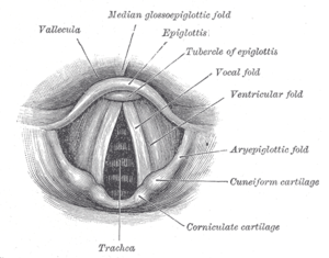
A labeled anatomical diagram of the vocal fold...
A labeled anatomical diagram of the vocal folds or cords. (Photo credit: Wikipedia)

Definition:
Laryngomalacia is a softening of the tissues of the larynx (voice box) above the vocal cords. This softening causes the tissues to become floppy, and they may fall over the airway opening and partially block it.

CLICK TO SEE THE PICTURES

Laryngomalacia (literally, “soft larynx”) is a very common condition of infancy, in which the soft, immature cartilage of the upper larynx collapses inward during inhalation, causing airway obstruction. It can also be seen in older patients, especially those with neuromuscular conditions resulting in weakness of the muscles of the throat. However, the infantile form is much more common.

There are several types – the mildest may cause no problems, while the most severe can be associated with other abnormalities of the respiratory tract, and with neuromuscular and gastroenterological problems.

Symptoms:
Until the larynx becomes stronger, problems can arise for several reasons:

•The soft limp tissues of the larynx can collapse as the baby breathes in. This interrupts the flow of air and causes noisy breathing, with a sound called stridor, which is a sign of obstructed air flow – in fact laryngomalacia is the commonest cause of stridor in babies. It may be worse if the baby has a respiratory infection.

•In some children, laryngomalacia can interfere with feeding. The effort required to draw air in through the obstructed airway can cause reflux of food from the stomach back up into the oesophagus or gullet.

•There may be other ear, nose and throat problems, and rarely problems with the lungs. Low oxygen levels may disrupt normal growth.

Common symptoms are :-
*Nosy breathing (stridor) – An audible wheeze when your baby breathes in. It is often worse when the baby is agitated, feeding, *crying or sleeping on the back
*High pitched sound
*Difficulty feeding
*Poor weight gain
*Choking while feeding
*Apnea — Breathing stoppage
*Pulling in neck and check with each breath
*Cyanosis — Turning blue
*Gastroesophageal reflux — Spitting, vomiting and regurgitation
*Aspiration – Inhalation of food into the lungs

Causes:
Laryngomalacia is thought to be the result of abnormally slow maturation of the tissues of the larynx, possibly because of genetic factors. This simply means that at birth the baby’s respiratory tract isn’t developed and string enough to cope with the mechanical demands of drawing breath.

Although doctors believe there’s a link between laryngomalacia and gastro-oesophageal reflu, there isn’t a single common mechanism to link these two problems, so several theories exist. In some patients with laryngomalacia, reflux may be the primary cause of their airway problems. In others, it’s an additional factor on top of neurological or anatomical abnormalities.

Reflux is common in babies less than one year old, because the muscular valve at the entrance to the stomach (which holds food in the stomach) may be weak in small infants.

Research suggests that a very large number, if not all, of babies with laryngomalacia also have reflux of gastric acid and digestive enzymes up to the pharynx (back of the throat). This may have detrimental effects on the larynx and tracheobronchial tree (air passages into the lungs). This may cause persistent swelling (oedema) of the larynx lining, which is common in children with laryngomalacia.

There’s no consensus yet about managing this link, but it makes sense to think simple treatments to control reflux could help resolve the laryngomalacia more quickly, too. More interventional treatments such as surgery, with all their inherent risks, are best avoided if possible.

Although laryngomalacia is not associated with a specific gene, there is evidence that some cases may be inherited.

Diagnosis:
Your doctor will ask you some questions about your baby’s health problems and may recommend a test called a flexible laryngoscopy (lar ring os co pee) to further evaluate your baby’s condition.

During this test, done in your doctor’s office, a tiny camera that looks like a strand of spaghetti with a light on the end is passed through your baby’s nostril and into the lower part of the throat where the larynx is. This allows your doctor to see your baby’s voicebox.

After the diagnosis — additional tests:
If laryngomalacia is diagnosed, the doctor may want to do other diagnostic tests to evaluate the extent of your child’s problems and to see whether the lower airway is affected. These tests may include:

X-ray of the neck;
A neck X-ray is done to make sure that your baby does not have other problems below the voice box (in the subglottis, trachea or chest). These are areas that the doctor cannot see during the flexible laryngoscopy.

Airway fluoroscopy;
The doctor may also order a motion picture X-ray of the trachea to make sure that there are no other problems.

Microlaryngoscopy (my crow lar ring os co pee)and bronchoscopy (brawn cos co pee), also known as MLB
This test is done when a neck X-ray shows additional problems in the lower airway. Your child is taken to the operating room and given anesthesia. Then the doctor passes a tiny camera through your child’s mouth and down past the vocal cords (larynx) to look at the area below the vocal folds that may be contributing to the stridor (noisy breathing). The surgeon will take some pictures and will review the results with you afterward.

EGD or esophagogastroduodenoscopy pH probe
This test will be done if your child’s doctor suspects that there may be a more severe problem.

Treatment :
In almost all cases (99 percent), laryngomalacia resolves without treatment by the time your child is 18 to 20 months of age. However, if the laryngomalacia is severe, your child’s treatment may include medication or surgery.

Medication:
Your child’s GI doctor may prescribe an anti-reflux medication to help manage the gastroesophageal reflux (GERD). This is important because your child’s chronic neck and chest retractions from the laryngomalacia can worsen GERD. Also, the acid reflux can cause swelling above the vocal cords and worsen the noisy breathing.

Surgery:
Surgery is the treatment of choice if your child’s condition is severe. Symptoms that signal the need for surgery include:

*Life-threatening apneas (stoppages of breathing)

*Significant blue spells

*Failure to gain weight with feeding

*Significant chest and neck retractions

*Need for extra oxygen to breathe

*Heart or lung issues related to your child’s inability to get enough oxygen

Supraglottoplasty:
In this surgery, extra tissue above the vocal cords is trimmed in the operating room. Your child will be under general anesthesia while the surgeon does a thorough evaluation of the airway and removes the tissue. After surgery, your child will be taken to the pediatric intensive care unit (PICU) and will spend one night with a breathing tube in the nose. If there is not much swelling in this area, and if the surgeon feels it will be safe, the breathing tube will be removed the next day in the PICU. Your child will then be observed for another day to ensure that the airway is safe, and that your child is getting enough oxygen and is drinking normally.

This surgery may not completely eliminate the noisy breathing but it should help to:

*Reduce the severity of the symptoms

*Lessen the apneas (breathing stoppages)

*Reduce the extra oxygen requirements

*Improve swallowing

*Help your child gain weight.

Disclaimer: This information is not meant to be a substitute for professional medical advise or help. It is always best to consult with a Physician about serious health concerns. This information is in no way intended to diagnose or prescribe remedies.This is purely for educational purpose

Resources:
http://en.wikipedia.org/wiki/Laryngomalacia
http://www.chop.edu/service/airway-disorders/conditions-we-treat/laryngomalacia.html
http://www.bbc.co.uk/health/physical_health/conditions/laryngomalacia.shtml

Enhanced by Zemanta
Categories
Ailmemts & Remedies

Laryngeal cancer

Alternative Name:Cancer of the larynx or Laryngeal carcinoma,Vocal cord cancer; Throat cancer; Laryngeal cancer; Cancer of the glottis

Definition:
The larynx is located in the neck at the top of the windpipe (trachea) and is used when we talk, swallow and breathe. It’s also called the voice box and is made up of cartilage – the large cartilage in the front is often called the Adam’s apple. Inside the larynx are the vocal cords.

The larynx is made up of three main parts:
•Supraglottis – the tissue at the top of the larynx
•Glottis – the middle part of the larynx where the vocal cords are located
•Subglottis – the tissue at the bottom of the larynx that connects the larynx to the windpipe
Laryngeal cancer can develop in any of these parts, but most commonly develops on the vocal cords.
CLICK  &  SEE  THE  PICTURES

Most laryngeal cancers are squamous cell carcinomas, reflecting their origin from the squamous cells which form the majority of the laryngeal epithelium. Cancer can develop in any part of the larynx, but the cure rate is affected by the location of the tumor. For the purposes of tumour staging, the larynx is divided into three anatomical regions: the glottis (true vocal cords, anterior and posterior commissures); the supraglottis (epiglottis, arytenoids and aryepiglottic folds, and false cords); and the subglottis.

Most laryngeal cancers originate in the glottis. Supraglottic cancers are less common, and subglottic tumours are least frequent.

Laryngeal cancer may spread by direct extension to adjacent structures, by metastasis to regional cervical lymph nodes, or more distantly, through the blood stream. Distant metastates to the lung are most common.
Incidence:

Two in 20,000 (12,500 new cases per year) in the USA. The American Cancer Society estimates that 9,510 men and women (7,700 men and 1,810 women) will be diagnosed with and 3,740 men and women will die of laryngeal cancer in 2006.

Laryngeal cancer is listed as a “rare disease” by the Office of Rare Diseases (ORD) of the National Institutes of Health (NIH). This means that laryngeal cancer affects fewer than 200,000 people in the U.S.

Each year, about 2,200 people in the U.K. are diagnosed with laryngeal cancer

Symptoms:
Symptoms of laryngeal cancer depend on where the cancer develops. Since it most often develops on the vocal cords, hoarseness or other changes in the voice are common.

Other symptoms that may occur when cancer develops above or below the vocal cords, or if it spreads from the vocal cords, include:

*A persistent sore throat  or feeling that something is stuck in the throat
*Ear pain or Ear ache (“referred”)
*Noisy breathing
*Difficulty swallowing
*Difficulty breathing
*A lump in the neck
*Painful swallowing
*A feeling of a lump in the throat
*A persistent cough
*Hoarseness or other voice changes
*Stridor
*Bad breath

If the cancer spreads outside the larynx, the lymph glands in the neck may become enlarged.

Causes:
The precise causes of laryngeal cancer aren’t known. However, it’s more common:

*On the vocal cords
*In men
*Between the ages of 55 and 65
*In smokers
*In those who drink alcohol heavily

Risk Factors:
Smoking is the most important risk factor for laryngeal cancer. Death from laryngeal cancer is 20 times more likely for heaviest smokers than for nonsmokers.  Heavy chronic consumption of alcohol, particularly alcoholic spirits, is also significant. When combined, these two factors appear to have a synergistic effect. Some other quoted risk factors are likely, in part, to be related to prolonged alcohol and tobacco consumption. These include low socioeconomic status, male sex, and age greater than 55 years.

People with a history of head and neck cancer are known to be at higher risk (about 25%) of developing a second cancer of the head, neck, or lung. This is mainly because in a significant proportion of these patients, the aerodigestive tract and lung epithelium have been exposed chronically to the carcinogenic effects of alcohol and tobacco. In this situation, a field change effect may occur, where the epithelial tissues start to become diffusely dysplastic with a reduced threshold for malignant change. This risk may be reduced by quitting alcohol and tobacco.

Diagnosis:
Diagnosis is made by the doctor on the basis of a medical history, physical examination, and special investigations which may include a chest x-ray, CT or MRI scans, and tissue biopsy. The examination of the larynx requires some expertise, which may require specialist referral.

The physical exam includes a systematic examination of the whole patient to assess general health and to look for signs of associated conditions and metastatic disease. The neck and supraclavicular fossa are palpated to feel for cervical adenopathy, other masses, and laryngeal crepitus. The oral cavity and oropharynx are examined under direct vision. The larynx may be examined by indirect laryngoscopy using a small angled mirror with a long handle (akin to a dentist’s mirror) and a strong light. Indirect laryngoscopy can be highly effective, but requires skill and practice for consistent results. For this reason, many specialist clinics now use fibre-optic nasal endoscopy where a thin and flexible endoscope, inserted through the nostril, is used to clearly visualise the entire pharynx and larynx. Nasal endoscopy is a quick and easy procedure performed in clinic. Local anaesthetic spray may be used.

If there is a suspicion of cancer, biopsy is performed, usually under general anaesthetic. This provides histological proof of cancer type and grade. If the lesion appears to be small and well localised, the surgeon may undertake excision biopsy, where an attempt is made to completely remove the tumour at the time of first biopsy. In this situation, the pathologist will not only be able to confirm the diagnosis, but can also comment on the completeness of excision, i.e., whether the tumour has been completely removed. A full endoscopic examination of the larynx, trachea, and esophagus is often performed at the time of biopsy.

For small glottic tumours further imaging may be unnecessary. In most cases, tumour staging is completed by scanning the head and neck region to assess the local extent of the tumour and any pathologically enlarged cervical lymph nodes.

The final management plan will depend on the site, stage (tumour size, nodal spread, distant metastasis), and histological type. The overall health and wishes of the patient must also be taken into account.

Treatment :
Treatment of laryngeal cancer may involve:

*Radiotherapy – when high-energy x-rays are used to kill cancer cells.

*Surgery – this may involve the removal of a vocal cord (cordectomy), part of the larynx (partial laryngectomy) or the entire larynx (total laryngectomy) and lymph glands may also be removed (neck dissection). For early laryngeal cancers, laser therapy may be used.

*Chemotherapy – when drugs are used to kill cancer cells.
A team of experts is involved in caring for a person with laryngeal cancer. This team may include an ear, nose and throat (ENT) surgeon, a clinical oncologist, a medical oncologist, a specialist cancer nurse, a dietician, a dentist and a speech therapist.

Following treatment, a person may need specialist help and advice with talking and breathing, especially if the whole larynx has been removed. Helping someone speak involves using a speaking valve inserted at time of surgery, using a special electronic device that generates sound or using the oesophagus (gullet) to speak.

Emotional help and support is also often needed following diagnosis and treatment.

Prognosis:
Throat cancers can be cured in 90% of patients if detected early. If the cancer has spread to surrounding tissues or lymph nodes in the neck, 50 – 60% of patients can be cured. If the cancer has spread (metastasized) to parts of the body outside the head and neck, the cancer is not curable and treatment is aimed at prolonging and improving quality of life.

After treatment, patients generally need therapy to help with speech and swallowing. A small percentage of patients (5%) will not be able to swallow and will need to be fed through a feeding tube.

Complications:
•Airway obstruction
•Difficulty swallowing
•Disfigurement of the neck or face
•Hardening of the skin of the neck
•Loss of voice and speaking ability
•Spread of the cancer to other body areas (metastasis)

Prevention:
Avoid smoking and other tobacco exposure. Limit or avoid alcohol use.

Disclaimer: This information is not meant to be a substitute for professional medical advise or help. It is always best to consult with a Physician about serious health concerns. This information is in no way intended to diagnose or prescribe remedies.This is purely for educational purpose

Resources:
http://www.bbc.co.uk/health/physical_health/conditions/in_depth/cancer/typescancer_larynx.shtml
http://en.wikipedia.org/wiki/Laryngeal_cancer
http://www.nlm.nih.gov/medlineplus/ency/article/001042.htm
http://www.dwp.gov.uk/publications/specialist-guides/medical-conditions/a-z-of-medical-conditions/laryngeal-cancer/
https://www.aarphealthcare.com/galecontent/laryngeal-cancer-1
http://www.robertsreview.com/cancer_pictures_larynx.html

Enhanced by Zemanta
css.php