Categories
Health Quaries

Some Medical Questions and Answers by Dr.Gita Mathai

Dealing with motion sickness:-

Q: My son vomits every time we travel, whether it is by car, bus, train or in a plane. It is exhausting to us and irritating for other passengers.

[amazon_link asins=’B001F731N0,B00S9FSPOG,B00SD9IE9O,B004FJFF2A,B00J6P9U2Y,B004S30AAK,B00DMBTZQO,B007HBLKNG,B001P4C8N2′ template=’ProductCarousel’ store=’finmeacur-20′ marketplace=’US’ link_id=’54c8cf68-3ef7-11e7-9156-6ba0b1aad181′]

.A: Your son has the classic symptoms of motion sickness. In some people like him, movement by all the modes of transportation you have mentioned causes a dissociation in the information that the brain receives. The person is immobile, seated in a chair, but is actually moving. The balance centre in the ear becomes affected, causing dizziness, nausea and eventually vomiting.
…………..CLICK & SEE THE PICTURES

Simple methods like facing forwards or smelling a lemon may ease motion sickness

Simple methods like facing forwards, or sitting in the centre of the vehicle may help. Smelling a lemon or sucking on ginger-flavoured sweets helps some people overcome the nausea. Medications like Dramamine or Avomine taken half an hour before the journey usually stop the vomiting. Consult your paediatrician, who will be able to prescribe appropriate medication if required.

Fortunately, some children outgrow motion sickness as they grow older and travel more frequently.

Blocked nose :-

Q: One side of my nose is permanently blocked and if I get a cold I cannot breathe at all.

A: If your nose has been blocked from birth, there may be a congenital absence of the opening, a condition called chonal atresia. This requires surgical correction. If the block is recent, you need to consult an ear, nose and throat surgeon to evaluate the nasal passages. He will be able to tell you if the obstruction is due to a mechanical cause like a deviated nasal septum or nasal polyps or a reactive intermittent block caused by a local response to allergens. Just using nosal drops and sprays is not the answer. Many of the chemical drops cause rebound congestion. The saline drops are safer but they are milder and short acting.

Insect stings :-

Q: I got stung by a wasp and the sting remained in my flesh for a long time. Please advise.

[amazon_link asins=’B00D8DGX9S,B00D8DEYO4,B003WS0C28,B003DKOYHY,B00OYJDJCE,B004K690LA,B01N0ZPR6C,B008SIEL1U,B007YJQJTG’ template=’ProductCarousel’ store=’finmeacur-20′ marketplace=’US’ link_id=’c754d8f4-3ef7-11e7-b749-65a01c7dede2′]

A: An insect sting can be very painful and may cause allergic reactions. The proboscis (stinging apparatus) should be quickly removed. The easiest way to do this is to apply ice to the site of the injury. The swelling subsides and enough of the sting is usually exposed to facilitate removal. If there is redness and itching, calamine lotion can be applied. If the allergy is severe, antihistamines many need to be taken.

Some people can develop life-threatening allergic reactions to insect bites or stings, with swelling in the lips, tongue and throat and breathing obstructed. They need immediate medical attention.

Varicose veins :-

Q: I have ugly blue veins on my legs which swell up when I stand. What can I do?

[amazon_link asins=’B0016J0NP6,B00JW1XCXK,B00GU1GHXW,B01N7FS6KH,B00SSBE6JY,B0169PFLFI,B01KB2D6A2,B01FF1MURY,B01KIINISO’ template=’ProductCarousel’ store=’finmeacur-20′ marketplace=’US’ link_id=’02622f24-3ef8-11e7-a600-b144163e2a2d’]

A: The swellings you describe are varicose veins. This condition is commoner in women. It tends to get aggravated during pregnancy. It is due to weak and faulty valves in the veins of the leg. Many patients can manage this with weight reduction, exercises and elastic stockings. If there is constant pain and repeated ulcer formation, it is better to opt for surgery.

Pregnancy after a caesarean :-

Q: I delivered my first baby by caesarean and was advised to wait for three years before the second baby. As I did not menstruate for seven months, I thought I did not need contraception. Now I find I am pregnant. Can I have a medical termination of the pregnancy?

A: Unfortunately, after vague post natal instructions stating   “come for a check up after six weeks   or  use contraception  (details unspecified) for three years, most couples are left to their own devices. Here, unfortunately, old wives   tales   You cannot get pregnant as long as you breast feed the baby.” “I did not become pregnant for three years and neither did your grand mother.” “If you have not menstruated, you are safe.” “If you have intercourse infrequently, you will not get pregnant.”

None of these theories has any scientific basis. Even a single act of intercourse can result in pregnancy. In your case, options are limited. Return to the obstetrician who performed the first caesarean and follow her advice.

Sources: The Telegraph (Kolkata, India)

Categories
Ailmemts & Remedies

Chalazion

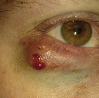
A chalazion immediately after excision
Image via Wikipedia

Alternative Names:Meibomian gland lipogranuloma

Definition:
A chalazion is a small bump in the eyelid caused by a blockage of a tiny oil gland.
It is a cyst in the eyelid that is caused by inflammation of a blocked meibomian gland, usually on the upper eyelid.

..click to see the pictures >....…(01)....`(1)..…....(2)………….(3)..………………

..
…………………………………………...Eyelid affected by Chalazion
Chalazions differ from styes (hordeolums) in that they are usually painless apart from the tenderness caused when they swell up, and in size (chalazia tend to be larger than styes). A chalazion may eventually disappear on its own after a few months, though more often than not, some treatment is necessary.
A large chalazion ca. 20 minutes upon excision. This bipartite chalazion was removed via two separate incisions. Further along the lower eyelid, signs of chronic inflammation are visible.

Causes:

A chalazion develops within the glands that produce the fluid that lubricate the eye. These are called Meibomian glands. The eyelid has approximately 100 of these glands, which are located near the eyelashes.

A chalazion is caused by blockage of the duct that drains one of these glands. A chalazion begins as swelling and tenderness, and later forms a cyst-like growth. Many chalazia disappear without treatment after a few months

Signs and symptoms:

*Painful swelling on the eyelid

*Eyelid tenderness

*Sensitivity to light

*Increased tearing

*Swelling on the eyelid

*Heaviness of the eyelid

Diagnosis:
Exams and Tests

Examination of eyelid confirms the diagnosis.
Rarely, the Meibomian gland duct may be blocked by a skin cancer. If this is suspected, a biopsy may be needed to diagnose the disorder.

Treatment:
A chalazion will often disappear without treatment within a month or so.

The primary treatment is application of warm compresses for 10-15 minutes at least 4 times a day. This may soften the hardened oils blocking the duct and promote drainage and healing. If the chalazion continues to get bigger, it may need to be surgically removed. This is usually done from underneath the eyelid to avoid a scar on the skin.

Topical antibiotic eye drops or ointment (eg chloramphenicol or fusidic acid) are sometimes used for the initial acute infection, but are otherwise of little value in treating a chalazion. Chalazia will often disappear without further treatment within a few months and virtually all will resorb within two years.

If they continue to enlarge or fail to settle within a few months, then smaller lesions may be injected with a corticosteroid or larger ones may be surgically removed using local anesthesia. . If the chalazion is located directly under the eyelid’s outer tissue, however, an excision from above may be more advisable so as not to inflict any unnecessary damage on the lid itself. Eyelid epidermis usually mends well, without leaving any visible traces of cicatrisation. Depending on the chalazion’s texture, the excision procedure varies: while fluid matter can easily be removed under minimal invasivion, by merely puncturing the chalazion and exerting pressure upon the surrounding tissue, hardened matter usually necessitates a larger incision, through which it can be scraped out. Any residual matter should be metabolized in the course of the subsequent healing process, generally aided by regular appliance of dry heat. The excision of larger chalazia may result in visible hematoma around the lid, which will wear off within three or four days, whereas the swelling may persist for longer. Chalazion excision is an ambulant treatment and normally does not take longer than fifteen minutes. Nevertheless, owing to the risks of infection and severe damage to the eyelid, such procedures should only be performed by a doctor.

Rarely chalazia may reoccur and these will be biopsied to help rule out tumors.

Antibiotic eye drops are usually used several days before and after removal of the cyst, but are otherwise of little value in treating a chalazion.

A chalazion that keeps coming back should be biopsied to rule out tumor.

Click for :->Chalazion and its online homeopathic treatment

Prognosis: Chalazia usually heal on their own. The outcome with treatment is usually excellent.

Complications:
A large chalazion can cause astigmatism due to pressure on the cornea. This will resolve with resolution of the chalazion.Complications including, but not limitedtohypopigmentation may occur with corticosteroid injection.The presence of recurring chalazion in the same area sometimes leads to a consideration of sebaceous cell carcinoma.The minor operation is quite painless, the eyelid is injected with a local anesthetic a clamp is put on the eyelid, then the eyelid is turned over and the chalazion is scraped out.

When to contact your Healthcare Provider:
Apply warm compresses and call your health care provider if the swelling progresses or persists longer than 1 month.
Call for an appointment with your health care provider if lumps on the eyelid continue to enlarge despite treatment or are associated with an area of eyelash loss.

Prevention:
Proper cleansing of the eyelid may prevent recurrences in people prone to chalazia. Cleaning the eyelash area with baby shampoo will help reduce clogging of the ducts.

Click to see also:-> Stye
Disclaimer: This information is not meant to be a substitute for professional medical advise or help. It is always best to consult with a Physician about serious health concerns. This information is in no way intended to diagnose or prescribe remedies.This is purely for educational purpose.
Resources:
http://www.nlm.nih.gov/medlineplus/ency/article/001006.htm
http://en.wikipedia.org/wiki/Chalazion

Enhanced by Zemanta
Categories
Featured

Is Your Full Figure an Increased Risk for Diabetes?

A new study suggest that women who have smaller breasts in their late teens and early 20s may enjoy a lower risk of developing type 2 diabetes later in life. However, many doctors have cautioned that the results may have more to do with obesity than they do with breast size alone.

……………...click & see

Researchers surveyed more than 92,000 women with an average age of 38, asking each of the participants to recall her bra size at the age of 20.

Women who recalled having a D cup or larger had about three times the risk of developing type 2 diabetes. Women who reported wearing B cup and C cup bras also experienced a higher risk than women who wore an A cup, even after figuring in age, body mass index (BMI), waist circumference, eating habits, family history of diabetes, physical activity level and pregnancies.

The study lead investigator believes that the correlation has something to do with how breasts develop during puberty. Puberty is a period marked by raised insulin resistance. Just as breast development is both accelerated and more pronounced in obese girls, their levels of insulin resistance may be as well.

However, if that is the case, many experts question why they should abandon the tried-and-true methods of evaluating type 2 diabetes risk by calculating their BMIs and evaluating lifestyles.

Sources:
ABC News January 28, 2008
Canadian Medical Association Journal January 29, 2008; 178(3): 313–315 (Free Full Text Article)

Categories
Ailmemts & Remedies

Cleft Lip And Plate

What is cleft lip and palate?
At around six weeks of pregnancy, your baby’s upper lip and palate develop from tissue lying on either side of the tongue. Normally these tissues grow towards each other and join up in the middle.

When the tissues that form the upper lip fail to join up in the middle, a gap forms in the lip. Usually, there will be a single gap below a nostril (unilateral cleft lip). Sometimes there are two gaps in the upper lip, each below a nostril (bilateral cleft lip). When the palate fails to join up, a gap is left in the roof of the mouth, going up into the nose.

About half of all clefts involve both the lip and palate. About 2 in 10 are of the lip alone and 3 in 10 are of the palate alone. Of clefts that involve the lip, 8 in 10 are unilateral and 2 in 10 are bilateral.

A clear upper lip and palate are among the most common defects in babies and affect about 1 in 700 babies in the us. These conditions may occur singly or together and are present at birth. both conditions are very upsetting for parents, but plastic surgery usually produces excellent results.

………….CLICK & SEE

The defects occur when the upp & SEEer lip or roof of the mouth does not fuse completely in the fetus. In many cases, the cause is unknown, but the risk if higher if certain anticonvulsant drugs, such as phenytoin, are taken during pregnancy or if the mother is a heavy drinker. cleft lip and /or palate sometimes run in families.

If a baby is severely affected, he or she may find it difficult to feed at first, and, if the condition is not treated early, speech may be delayed. Children with a cleft lip and/or palate are also susceptible to persistent buildup of fluid in the middle ear that impairs hearing and may delay speech.

Cleft lip and cleft palate, which can also occur together as cleft lip and palate are variations of a type of clefting congenital deformity caused by abnormal facial development during gestation. This type of deformity is sometimes referred to as a cleft. A cleft is a sub-division in the body’s natural structure, regularly formed before birth. A cleft lip or palate can be successfully treated with surgery soon after birth. Cleft lips or palates occur in somewhere between one in 600-800 births. The term hare lip is sometimes used colloquially to describe the condition because of the resemblance of a hare’s lip. The Chinese word for cleft lip is tuchun , literally “harelip.”

Cleft lip
If only skin tissue is affected one speaks of cleft lip. Cleft lip is formed in the top of the lip as either a small gap or an indentation in the lip (partial or incomplete cleft) or continues into the nose (complete cleft). Lip cleft can occur as one sided (unilateral) or two sided (bilateral). It is due to the failure of fusion of the maxillary and medial nasal processes (formation of the primary palate).


.
.Unilateral incomplete..….Unilateral complete..…..Bilateral complete

A mild form of a cleft lip is a microform cleft. A microform cleft can appear as small as a little dent in the red part of the lip or look like a scar from the lip up to the nostril. In some cases muscle tissue in the lip underneath the scar is affected and might require reconstructive surgery. It is advised to have newborn infants with a microform cleft checked with a craniofacial team as soon as possible to determine the severeness of the cleft. The actor Joaquin Phoenix is an example of a person with a microform cleft that did not require surgry.

Cleft palate
Cleft palate is a condition in which the two plates of the skull that form the hard palate (roof of the mouth) are not completely joined. The soft palate is in these cases cleft as well. In most cases, cleft lip is also present. Cleft palate occurs in about one in 700 live births worldwide.

Palate cleft can occur as complete (soft and hard palate, possibly including a gap in the jaw) or incomplete (a ‘hole’ in the roof of the mouth, usually as a cleft soft palate). When cleft palate occurs, the uvula is usually split. It occurs due to the failure of fusion of the lateral palatine processes, the nasal septum, and/or the median palatine processes (formation of the secondary palate).

The hole in the roof of the mouth caused by a cleft connects the mouth directly to the nasal cavity.

A direct result of an open connection between the oral cavity and nasal cavity is velopharyngeal insufficiency (VPI). Because of the gap, air leaks into the nasal cavity resulting in a hypernasal voice resonance and nasal emissions. Secondary effects of VPI include speech articulation errors (e.g., distortions, substitutions, and omissions) and compensatory misarticulations (e.g., glottal stops and posterior nasal fricatives). Possible treatment options include speech therapy, prosthetics, augmentation of the posterior pharyngeal wall, lengthening of the palate, and surgical procedures.
………………..Pictures showing unilateral and bilateral cleft lip and palate

.Symptoms:
Feeding
Most babies with a cleft lip can be breastfed. However, some babies have difficulty creating a seal around the nipple and may not be able to breastfeed. A special squeezy bottle can be used for feeding and can help if the baby can’t suck hard enough. These bottles are provided by specialist cleft nurses and are also available from the support charity CLAPA (see Further Information).

Babies who find it difficult to feed may gain weight slowly at first. A specialist cleft nurse can give advice about changing the type of formula milk and other feeding issues.

Speech
Cleft palate can cause problems with speech. The size of the cleft is not an indicator of how serious speech problems are likely to be – even a small cleft can affect speech. Most children go on to speak normally after some speech therapy, although sometimes further surgery will be needed to improve palate function. Children with clefts can sometimes have nasal sounding speech.

Hearing

Children with clefts sometimes have hearing problems. This is because the tube that connects the ear to the palate (the Eustachian tube) can be affected. Having a cleft can increase the chance of developing a condition known as glue ear. This is quite a common condition in all children and occurs when thick, sticky fluid builds up behind the eardrum. It can cause temporary hearing loss. As part of surgery to repair a cleft palate, surgeons often put a tiny plastic tube (a grommet) into the eardrum so that the fluid can drain out.

Teeth
Occasionally, a cleft palate may also affect the growth of you child’s jaw and the development of the teeth. Looking after teeth well and having regular care from a dentist or orthodontist can minimise problems.

Your child may need to have extensive orthodontic treatment to make sure the teeth come through straight and in the right place. This may involve wearing braces, especially around the time the second teeth are coming through and during the early teens. Your child may also need to have some teeth removed to prevent overcrowding.

Causes:
There are many factors that hinder the joining up process of the lip or palate during a baby’s development. If you have had a child with a cleft lip or palate, your chance of future children being affected is increased.

However, doctors can’t reliably predict which pregnancies will be affected because cleft lip and palate is usually caused by a combination of genetic and other unknown factors. The unknown factors may include an illness during pregnancy or being exposed to certain substances such as tobacco smoke or certain medicines.

Treatment:
Specialist centres
Ideally, children with cleft lip and palate should be treated by a multidisciplinary specialist “cleft team” that may include surgeons, speech and language therapists, audiologists (hearing experts), dentists, orthodontists, psychologists, geneticists and specialist cleft nurses. Care and support of your child and the family should last from birth until your child stops growing at about age 18.

If you have a baby born with a cleft lip or palate, your maternity hospital should refer you to one specialist centre. Often they have specialist nurses who can visit you to provide immediate support and advice. This can be invaluable in the early days.

Surgery
The timing of surgery varies, but usually an operation to close the gap in the lip will be done about three months after the baby is born. Surgery to close the gap in the palate is usually done at about six months.

Both operations are done while your baby asleep under general anaesthetic and involve a hospital stay of 3 to 5 days.

As your child grows older, further surgery may be needed to improve the appearance of the lip and nose and the function of the palate. If there is a gap in the gum, a bone graft will normally be done when your child is between 9 and 12 years old. This will help their second teeth to anchor properly into the gum. Bone is usually taken from the hip or shin and grafted into the gap in the gum.

Prevention:
If you have had a child with a cleft lip or palate, you may be offered genetic counselling to find out the chances of your next child being affected. However, in most cases the most sensible approach is simply to aim to have a healthy pregnancy. Smoking and drinking alcohol have been shown to increase the risk of babies being affected, and can cause other birth defects.

Research has shown that taking a daily supplement of 400 micrograms of folic acid in the month before conception and in the first two months of pregnancy can help prevent cleft lip. This is the same amount of supplement recommended to reduce the risk of neural tube defects such as spina bifida.

It’s thought that certain medicines may slightly increase the risk of cleft lip and palate. These include anti-epilepsy medicines such as phenytoin (eg Epanutin) and sodium valproate (eg Epilim). Steroid tablets and a medicine called methotrexate (eg Metoject) that is used to treat some cancers and inflammatory conditions, such as rheumatoid arthritis, may also increase the risk. If you are on these medicines, you should discuss the benefits and possible risks with your doctor before trying for a baby.

Help and support:
If you are a new parent of a child who has a cleft lip or palate, or a child who was born with a cleft, a specialist psychologist working in the cleft team can help you cope with some of the challenges you may have to deal with. It can also help to get support from other people who have had have had similar experiences, either as parents, or as someone who has grown up with a cleft leaf.

Click for more knowledge & information:

www.clapa.com
www.changingfaces.org.uk
Cleft Plate Foundation
Best Way to Manage Cleft Lip and Palate

Disclaimer: This information is not meant to be a substitute for professional medical advise or help. It is always best to consult with a Physician about serious health concerns. This information is in no way intended to diagnose or prescribe remedies.This is purely for educational purpose.

.Resources:
http://hcd2.bupa.co.uk/fact_sheets/Mosby_factsheets/cleft_lip.html
http://www.charak.com/DiseasePage.asp?thx=1&id=341
http://en.wikipedia.org/wiki/Cleft_lip_and_palate

Categories
Ailmemts & Remedies

Hydrocele

A hydrocele is an accumulation of clear fluid in the tunica vaginalis, the most internal of
membranes containing a testicle. A primary hydrocele causes a painless enlargement in the scrotum on the affected side and is thought to be due to the defective absorption of fluid secreted between the two layers of the tunica vaginalis (investing membrane). A secondary hydrocele is secondary to either inflammation or a neoplasm in the testis.

Click to see the picture

A hydrocele usually occurs on one side. The accumulation can be a marker of physical trauma, infection or tumor, but the cause is generally unknown.

.Click to see the picture.

A hydrocele is a fluid-filled sac surrounding a testicle that results in swelling of the
scrotum, the loose bag of skin underneath the penis. About one in 10 male infants has a
hydrocele at birth, but most hydroceles disappear without treatment within the first year of life. Additionally, men  usually older than 40 can develop a hydrocele due to
inflammation or injury within the scrotum.

click to see…..(01)…...(1).…....(2).…….(3)...…….

Hydroceles usually aren’t painful. Typically not harmful, hydroceles may require no
treatment. However, if you have scrotal swelling, see your doctor to rule out other causes, such as testicular cancer or other conditions

Symptoms
A hydrocele feels like a small fluid filled balloon inside the scrotum. It is smooth, and is
mainly in front of one of the testes. Hydroceles vary greatly in size. Hydroceles are
normally painless and harmless. Large hydroceles cause discomfort because of their size. As the fluid of a hydrocele is transparent, light shone through a hydrocelic region will be
visible from the other side.

Symptoms of a hydrocele can easily be distinguished from testicular cancer, as a hydrocele is soft and fluidy, where a testicular cancer feels hard and rough.
Through diagnostic ultrasound the accumulation of fluids can be diagnosed correctly.

Causes:
For baby boys, a hydrocele can develop in the womb. At about 28 weeks of gestation, the
testicles descend from the developing baby’s abdominal cavity into the scrotum. A sac
(processus vaginalis) accompanies each testicle, allowing fluid to surround them.

In most cases, the sac closes and the fluid is absorbed. However, if the fluid remains after
the sac closes, the condition is known as a noncommunicating hydrocele. Because the sac is closed, fluid can’t flow back into the abdomen. Usually the fluid gets absorbed within a
year.

In some cases, however, the sac remains open. With this condition, known as communicating hydrocele, the sac can change size or, if the scrotal sac is compressed, fluid can flow back into the abdomen.

In older males, a hydrocele can develop as a result of inflammation or injury within the
scrotum. Inflammation may be the result of infection of the small coiled tube at the back of each testicle (epididymitis) or of the testicle (orchitis).

Risk factors
Most hydroceles are present at birth (congenital). Otherwise, the condition generally
affects men 40 or older. Risk factors include:

*Scrotal injury
*Infection
*Radiation therapy

Possible Complications

Complications may occur from hydrocele treatment.

Risks related to hydrocele surgery may include:

  • Blood clots
  • Infection
  • Injury to the scrotal tissue or structures

Risks related to aspiration and sclerosing may include:

  • Infection
  • Fibrosis
  • Mild-to-moderate pain in the scrotal area
  • Return of the hydrocele

When to seek medical advice
For yourself
See your doctor if you experience scrotal swelling. It’s important to rule out other
possible causes for the swelling, such as a tumor. Sometimes a hydrocele is associated with an inguinal hernia, in which a weak point in the abdominal wall allows a loop of intestine to extend into the scrotum and which may require treatment.

For your child
In infants, a hydrocele typically disappears on its own. However, if your baby’s hydrocele
doesn’t disappear after a year or if it enlarges, you may need to have it evaluated.

Diagnosis
Screening and diagnosis entails a physical exam, which may reveal an enlarged scrotum that isn’t tender to the touch. Pressure to the abdomen or scrotum may enlarge or shrink the
fluid-filled sac, which may indicate an associated inguinal hernia.

Because the fluid in a hydrocele usually is clear, your doctor may shine a light through the scrotum (transillumination). With a hydrocele, the light will outline the testicle,
indicating that clear fluid surrounds it. If your doctor suspects your hydrocele is caused by inflammation, blood and urine tests may help determine whether you have an infection, such as epididymitis.

The fluid surrounding the testicle may keep the testicle from being felt. In that case,
further study may be needed. Possible tests include:

Ultrasound imaging. This test, which uses high-frequency sound waves to create images of structures inside your body, may be used to rule out a testicular tumor or other cause of
scrotal swelling.
Abdominal X-ray. A basic X-ray uses electromagnetic radiation to make images of your bones, teeth and internal organs. An X-ray may distinguish a hydrocele from an inguinal hernia.

Other Complications
A hydrocele typically isn’t dangerous and usually doesn’t affect fertility. However, it may be associated with an underlying testicular condition that may cause serious complications:

*Infection or tumor. Either may impair sperm production or function.

*Inguinal hernia. A loop of intestine could become trapped in the weak point in the

abdominal wall (strangulated), a life-threatening condition.

Treatment
For baby boys, hydroceles typically disappear on their own within a year. If a hydrocele
doesn’t disappear after a year or if it continues to enlarge, it may need to be surgically
removed.

For adult males as well, hydroceles often go away on their own. A hydrocele requires
treatment only if it gets large enough to cause discomfort or disfigurement. Then it may
need to be removed.

Surgical excision (hydrocelectomy). . The procedure is called hydrocelectomy, the tunica vaginalis is excised, the fluid drained, and the edges of the tunica are sutured to prevent the reaccumulation of fluid.

If the hydrocele is not surgically removed, it may continue to grow. The hydrocele fluid can be aspirated, the procedure is less invasive but recurrence rates are high. Sclerotherapy, the injection of a solution following aspiration of the hydrocele fluid may increase success rates. In many patients, the procedure of aspiration and sclerotherapy is repeated as the hydrocele recurrs.

Needle aspiration. Another option is to remove the fluid in the scrotum with a needle. This treatment isn’t widely used because it’s common for the fluid to return. The injection of a
thickening or hardening (sclerosing) drug after the aspiration may help prevent the fluid
from reaccumulating. Aspiration and injection may be an option for men who have risk factors that make surgery more dangerous. Risks of this procedure include infection and scrotal pain.

Sometimes, a hydrocele may recur after treatment.

Outlook (Prognosis)

Generally, a simple hydrocele goes away without surgery. If surgery is necessary, it is a simple procedure for a skilled surgeon, and usually has an excellent outcome.

Homeopathic Treatment & Medicines of Hydrocoele –
Following homeopathic medicines are known to help in cases of hydrocoele – Apis, Aurum-met, Calc-c, Con, Flour-acid, Graph, Iod, Kali-iod, Puls, Rhod, Selen, Sil, Spong.

Hydrocele treatment: alternative therapy

Herbal Remedies of Hydrocele

Disclaimer: This information is not meant to be a substitute for professional medical advise or help. It is always best to consult with a Physician about serious health concerns. This information is in no way intended to diagnose or prescribe remedies.This is purely for educational purpose.

Resources:
http://www.mayoclinic.com/health/hydrocele/DS00617/DSECTION=1
http://en.wikipedia.org/wiki/Hydrocele_testis
http://health.nytimes.com/health/guides/disease/hydrocele/overview.html

Enhanced by Zemanta
css.php