Categories
Ailmemts & Remedies

Brusists

Definition:Whether you’re at work or at play, if you overuse or repetitively stress your body’s joints, you may eventually develop a painful inflammation called bursitis.

Click to see the picture…….>.(01)....(1)…..(2).…...(3)……...(4)…….(5)…..     (6)

You have more than 150 bursae in your body. These small, fluid-filled sacs lubricate and cushion pressure points between your bones and the tendons and muscles near your joints. They help your joints move with ease. Bursitis occurs when a bursa becomes inflamed. When inflammation occurs, movement or pressure is painful.

Bursitis often affects the joints in your shoulders, elbows or hips. But you can also have bursitis by your knee, heel and the base of your big toe. Bursitis pain usually goes away within a few weeks or so with proper treatment, but recurrent flare-ups of bursitis are common.

Symptoms:
If you have bursitis, you may notice:

A dull ache or stiffness in the area around your elbow, hip, knee, shoulder, big toe or other joints:-

*A worsening of pain with movement or pressure

*An area that feels swollen or warm to the touch

*Occasional skin redness in the area of the inflamed bursa

Bursitis of the hip doesn’t cause any visible swelling or skin redness because the bursae are located beneath some of your body’s bulkiest muscles. In this type of bursitis, pain is primarily over the greater trochanter, a portion of your thighbone (femur) that juts out just below where the bone joins the hip.

Causes:
Common causes of bursitis are overuse, stress and direct trauma to a joint, such as with repeated bumping or prolonged pressure from kneeling. Bursitis may also result from an infection, arthritis or gout. Many times, the cause is unknown.

Bursitis in certain locations of your body is caused by repetitive motion related to certain activities:

Shoulder. Bursitis of the shoulder often results from injury to the rotator cuff, the muscles and tendons that connect your upper arm bone to your shoulder blade. Causes of the injury may include falling, lifting and repetitive overhead arm activities. Sometimes it’s hard to distinguish between the pain caused by bursitis and that caused by a rotator cuff injury.

Elbow. This type of bursitis is associated with actions requiring you to repeatedly bend and extend your Elbow. You may get such an inflammation by pushing a vacuum cleaner back and forth. Throwing a baseball and swinging a tennis racket or a golf club are other examples of repeated physical activities that may lead to bursitis or tendinitis of the elbow or shoulder. Simple repeated leaning on your elbows could lead to bursitis over the tip of your elbow

Buttocks. This type of bursitis describes an inflamed bursa over the bone in your buttocks. It may result from sitting on a hard surface for long periods, such as on a bike.

Hip. Bursitis of the hip is frequently associated with arthritis or a hip injury. The pressure from standing or sitting for a prolonged time also may lead to bursitis of the hip.

Knee. In this form of bursitis, a soft, egg-shaped bump occurs on the front of your knee, the result of repetitive kneeling while installing tiles, scrubbing a floor, gardening or doing other activities that place pressure on your knees. A sharp blow to the knee can cause inflammation of the bursae around the kneecap. People with arthritis who are overweight often develop bursitis of the knee.

Ankle.
Inflammation of the bursa in the ankle commonly occurs as a result of improper footwear or prolonged walking or in sports, such as ice-skating.

You may not be able to pinpoint a specific incident or activity that led to your bursitis. In some cases, the inflammation may stem from a staphylococcal infection.

Diagnosis:
Your doctor may have you undergo a physical examination and ask you about your recent activities. By feeling the painful joint and surrounding area, your doctor may be able to identify a specific area of tenderness.

If it appears that something else may be causing the discomfort, your physician may request an X-ray of the affected area. If bursitis is the cause, X-ray images can’t positively establish the diagnosis, but they can help to exclude other causes of your discomfort.

Although you usually can trace bursitis to events of overuse or pressure, there may be no obvious cause. In the latter case, your doctor may want to perform additional screening to rule out other causes of joint inflammation and pain. This may include blood tests or an analysis of fluid from the inflamed bursa.

Treatments :
Bursitis treatment is usually simple and includes:

*Resting and immobilizing the affected area

*Applying ice to reduce swelling

*Taking nonsteroidal anti-inflammatory drugs (NSAIDs) to relieve pain and reduce inflammation

*With simple self-care and home treatment, bursitis usually disappears within a couple of weeks.

Sometimes, your doctor may recommend physical therapy or exercises to strengthen the muscles in the area. Additionally, your doctor may inject a corticosteroid drug into the bursa to relieve inflammation. This treatment generally brings immediate relief and, in many cases, one injection is all you’ll need.

If your bursitis is caused by an infection, you’ll need to take antibiotics. Sometimes the bursa must be surgically drained, but only rarely is surgical removal of the affected bursa necessary.

Lifestyle and home remedies:
To take care of your bursitis at home:

*Take nonsteroidal anti-inflammatory drugs (NSAIDs). NSAIDs such as aspirin, ibuprofen (Advil, Motrin, others) or naproxen sodium (Aleve) can provide relief. Use as directed.

* Consult your doctor if you need NSAIDs for an extended period of time.

*Apply ice packs. Use them for 20 minutes several times a day during the first few days, or for as long as the joint area is warm to the touch.

*Apply heat. Use heat after the affected joint is no longer warm or red to help relieve muscle and joint pain and stiffness. But don’t overdo it. Don’t apply heat for more than 20 minutes at a time. Sometimes moist heat seems to penetrate deeper and give you more relief than does dry heat.

*Perform stretching exercises. Stretching can help restore full range of motion.

*Elevate the affected joint. Raising your knee or elbow can help reduce swelling.

Keep pressure off your joint. If possible, use an elastic bandage, sling or soft foam pad to protect a joint until the swelling goes down.

Herbal Remedy:

YOU can promote the healing of inflamed fluid sacs between tendons and bones, and fight the pain and tenderness of “tennis elbow” and “frozen shoulder” with these herbs from Mother Nature’s medicine chest:

Coral calcium with trace minerals, glucosamine sulfate, shavegrass.

Prevention:
To help prevent bursitis or reduce the severity of flare-ups:

*Stretch your muscles. Warm up or stretch before physical activity.

*Strengthen your muscles. Strengthening can help protect your joints. Wait until the pain and inflammation are gone before starting to exercise a joint that has bursitis.

*Take frequent breaks from repetitive tasks. Alternate repetitive tasks with rest or other activities.

*Cushion your joint. Use cushioned chairs, foam for kneeling or elbow pads. Avoid resting your elbows on hard surfaces. Avoid shoes that don’t fit properly or that have worn-down heels.

*Don’t sit still for long periods. Get up and move about frequently.

*Practice good posture. For example, avoid leaning on your elbows.

If your bursitis is caused by a chronic underlying condition, such as arthritis, it may recur despite these preventive measures.

Disclaimer: This information is not meant to be a substitute for professional medical advise or help. It is always best to consult with a Physician about serious health concerns. This information is in no way intended to diagnose or prescribe remedies.This is purely for educational purpose.

Resources:
http://www.mayoclinic.com/health/bursitis
http://www.herbnews.org/bursitisdone.htm

Enhanced by Zemanta
Categories
Ailmemts & Remedies

Injuries in Toe, Foot, and Ankle

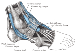
The mucous sheaths of the tendons around the a...
The mucous sheaths of the tendons around the ankle. Lateral aspect. (Photo credit: Wikipedia)

At one time or another, everyone has had a minor toe, foot, or ankle injury that caused pain or swelling. Most of the time our body movements do not cause problems, but it’s not surprising that symptoms develop from everyday wear and tear, overuse, or an injury. Toe, foot, or ankle injuries most commonlyoccur during:

You may click to see the picture

*Sports or recreational activities.
*Work-related tasks.
*Work or projects around the home.

In children, most toe, foot, or ankle injuries occur during sports or play or accidental falls. The risk for injury is higher in sports with jumping, such as basketball, or sports with quick direction change, such as soccer or football. Any bone injury near a joint may injure the growth plate (physis) in a child and needs to be evaluated.

Certain athletes, such as dancers, gymnasts, or soccer or basketball players, have an increased risk of toe, foot, or ankle injuries.

Older adults are at higher risk for injuries and fractures because they lose muscle mass and bone strength (osteopenia) as they age. They also have more problems with vision and balance, which increases their risk for accidental injury.

Most minor injuries will heal on their own, and home treatment is usually all that is needed to relieve your symptoms and promote healing.

Sudden (acute) injury

An acute injury may occur from a direct blow, a penetrating injury, a fall, or from twisting, jerking, jamming, or bending a limb abnormally. Your pain may be sudden and severe. Bruising and swelling may develop soon after your injury. Acute injuries include:

*Bruises. After an ankle injury, bruising may extend to your toes from the effects of gravity.

See a illustration of a bruise (contusion)->

*Injuries to ligaments that support your joints. See an illustration of a ligament tear……

*Injuries to tendons, such as ruptured tendons in your heel (Achilles tendon). Young boys between 8 and 14 years old may have a condition known as Sever’s disease, which causes injury to the growing bone where the Achilles tendon is attached. This usually occurs during activity and is relieved with home treatment……

*Injuries to your joints (sprains). If a sprain does not appear to be healing, a condition known as osteochondritis dissecans may be present, causing persistent symptoms. See an illustration of a sprained ankle…….

*Pulled muscles (strains). Muscles of the foot and ankle can be strained and can also rupture.

*Broken bones (fractures), such as a broken toe.

*A bone moving out of place (dislocation).

*A crushing injury, which can lead to compartment syndrome.

Overuse injuries:

Overuse injuries occur when too much stress is placed on your joint or other tissue, often by “overdoing” an activity or repeating the same activity over and over. Overuse injuries include:

*Retrocalcaneal bursitis, which is inflammation of the bursa. This condition causes swelling and tenderness of the heel. Pain usually worsens while wearing shoes and during activity and improves during rest. See an illustration of the back of the heel and ankle.

*Achilles tendinitis or tendinosis (tendinopathy), which is the breakdown of soft tissues in and around the Achilles tendon that connects the calf muscles to the heel bone.

*Stress fracture, which is a hairline crack in a bone. See an illustration of stress fractures of the foot.
Plantar fasciitis, which is an inflammation of the plantar fascia, a broad, flat ligament on the bottom of the foot that extends from the front of the heel to the base of the toes and helps maintain the arch of the foot. See an illustration of the plantar fascia.

*Metatarsalgia, which is pain in the front (ball) of the foot. See an illustration of metatarsalgia.

Treatment:
Treatment for your toe, foot, or ankle injury may include first aid measures (such as the application of a brace, splint, or cast), a special shoe (orthotic device), physical therapy, medicine, and in some cases surgery. Treatment depends on:

*The location, type, and severity of your injury.

*When the injury occurred.

*Your age, your overall health condition, and your activities (such as work, sports, or hobbies)

Review the Emergencies and Check Your Symptoms sections to determine if and when you need to see a doctor.

Click for :->Emergency

>Prevention

>Home Treatment

Sources: MSN Health.

Enhanced by Zemanta
Categories
Featured Therapies

Osteopathy

[amazon_link asins=’1517173671,1479297518,1451193416,B00H3NOFQM,1909141240,B00ODNYGLO,B01J4AUQ3W,142320316X’ template=’ProductCarousel’ store=’finmeacur-20′ marketplace=’US’ link_id=’f0fdbf33-1ba3-11e7-8900-91dc58aaa52e’]

Definition
Osteopathy is a system and philosophy of health care that separated from traditional (allopathic) medical practice about a century ago. It places emphasis on the musculoskeletal system, hence the name—osteo refers to bone and path refers to disease. Osteopaths also believe strongly in the healing power of the body and do their best to facilitate that strength. During this century, the disciplines of osteopathy and allopathic medicine have been converging.

It is a system of therapy founded in the 19th century based on the concept that the body can formulate its own remedies against diseases when the body is in a normal structural relationship, has a normal environment and enjoys good nutrition.

While osteopathy takes a “holistic” approach to medical care, it also embraces modern medical knowledge, including medication, surgery, radiation, and chemotherapy when warranted. Osteopathy is particularly concerned with maintaining correct relationships between bones, muscles, and connective tissues. The practice of osteopathy often includes chiropractic-like adjustments of skeletal structures. Craniosacral therapy, a practice in which the bones and tissues of the head and neck are manipulated, also arose in osteopathy.
CLICK & SEE THE PICTURES

Purpose
Osteopathy shares many of the same goals as traditional medicine, but places greater emphasis on the relationship between the organs and the musculoskeletal system as well as on treating the whole individual rather than just the disease.

Precautions
Pain is the chief reason patients seek musculoskeletal treatment. Pain is a symptom, not a disease by itself. Of critical importance is first to determine the cause of the pain. Cancers, brain or spinal cord disease, and many other causes may be lying beneath this symptom. Once it is clear that the pain is originating in the musculoskeletal system, treatment that includes manipulation is appropriate.

History

Osteopathy was founded in the 1890s by Dr. Andrew Taylor, who believed that the musculoskeletal system was central to health. The primacy of the musculoskeletal system is also fundamental to chiropractic, a related health discipline. The original theory behind both approaches presumed that energy flowing through the nervous system is influenced by the supporting structure that encase and protect it—the skull and vertebral column. A defect in the musculoskeletal system was believed to alter the flow of this energy and cause disease. Correcting the defect cured the disease. Defects were thought to be misalignments—parts out of place by tiny distances. Treating misalignments became a matter of restoring the parts to their natural arrangement by adjusting them.

As medical science advanced, defining causes of disease and discovering cures, schools of osteopathy adopted modern science, incorporated it into their curriculum, and redefined their original theory of disease in light of these discoveries. Near the middle of the 20th century the equivalance of medical education between osteopathy and allopathic medicine was recognized, and the D.O. degree (Doctor of Osteopathy) was granted official parity with the M.D. (Doctor of Medicine) degree. Physicians could adopt either set of initials.

However, osteopaths have continued their emphasis on the musculoskeletal system and their traditional focus on “whole person” medicine. As of 1998, osteopaths constitute 5.5% of American physicians, approximately 45,000. They provide 100 million patient visits a year. From its origins in the United States, osteopathy has spread to countries all over the world.

Practice

Osteopaths, chiropractors, and physical therapists are the experts in manipulations (adjustments). The place of manipulation in medical care is far from settled, but millions of patients find relief from it. Particularly backs, but also necks, command most of the attention of the musculoskeletal community. This community includes orthopedic surgeons, osteopaths, general and family physicians, orthopedic physicians, chiropractors, physical therapists, massage therapists, specialists in orthotics and prosthetics, and even some dentists and podiatrists. Many types of headaches also originate in the musculoskeletal system. Studies comparing different methods of treating musculoskeletal back, head, and neck pain have not reached a consensus, in spite of the huge numbers of people that suffer from it.

The theory behind manipulation focuses on joints, mostly those of the vertebrae and ribs. Some believe there is a very slight offset of the joint members—a subluxation. Others believe there is a vacuum lock of the joint surfaces, similar to two suction cups stuck together. Such a condition would squeeze joint lubricant out and produce abrasion of the joint surfaces with movement. Another theory focuses on weakness of the ligaments that support the joint, allowing it freedom to get into trouble. Everyone agrees that the result produces pain, that pain produces muscle spasms and cramps, which further aggravates the pain.

Some, but not all, practitioners in this field believe that the skull bones can also be manipulated. The skull is, in fact, several bones that are all moveable in infants. Whether they can be moved in adults is controversial. Other practitioners manipulate peripheral joints to relieve arthritis and similar afflictions.

Manipulation returns the joint to its normal configuration. There are several approaches. Techniques vary among practitioners more than between disciplines. Muscle relaxation of some degree is often required for the manipulation to be successful. This can be done with heat or medication. Muscles can also be induced to relax by gentle but persistent stretching. The manipulation is most often done by a short, fast motion called a thrust, precisely in the right direction. A satisfying “pop” is evidence of success. Others prefer steady force until relaxation permits movement.

Return of the joint to its normal status may be only the first step in treating these disorders. There is a reason for the initial event. It may be a fall, a stumble, or a mild impact, in which case the manipulation is a cure. On the other hand, there may be a postural misalignment (such as a short leg), a limp, or a stretched ligament that permits the joint to slip back into dysfunction. Tension, as well as pain, for emotional reasons causes muscles to tighten. If the pain has been present for any length of time, there will also be muscle deterioration. The osteopathic approach to the whole person takes all these factors into account in returning the patient to a state of health.

Other repairs may be needed. A short leg is thought by some to be a subluxation in the pelvis that may be manipulated back into position. Other short legs may require a lift in one shoe. Long-standing pain requires additional methods of physical therapy to rehabilitate muscles, correct posture, and extinguish habits that arose to compensate for the pain. Medications that relieve muscle spasm and pain are usually part of the treatment. Psychological problems may need attention and medication.

Risks
Manipulation has rarely caused problems. Once in a while too forceful a thrust has damaged structures in the neck and caused serious problems. The most common adverse event, though, is misdiagnosis. Cancers have been missed; surgical back disease has been ignored until spinal nerves have been permanently damaged.

Normal results
Many patients find that one or a series of manipulations cures long-standing pain. Other patients need repeated treatments. Some do not respond at all. It is always a good idea to reassess any treatment that is not producing the expected results.

Sources:http://www.healthline.com/galecontent/osteopathy-1 and http://www.medterms.com/script/main/art.asp?articlekey=4684

Categories
Ailmemts & Remedies

Frozen Shoulder

[amazon_link asins=’B017AD9B9K,1612436439,B071149H43,1452510725,1569757380,157224447X,B01MQUF0O4,B000BF70RA,B06XY58F5T’ template=’ProductCarousel’ store=’finmeacur-20′ marketplace=’US’ link_id=’8492b58a-e549-11e7-868d-157bdd34ede3′]

Frozen shoulder, medically referred to as adhesive capsulitis, is a disorder in which the shoulder capsule, the connective tissue surrounding the glenohumeral joint of the shoulder, becomes inflamed and stiff. Movement of the shoulder is severely restricted. The condition is sometimes caused by injury that leads to lack of use due to pain but also often arises spontaneously with no obvious preceding trigger factor. These seemingly spontaneous cases are usually referred to as Idiopathic Frozen Shoulder. Rheumatic disease progression and recent shoulder surgery can also cause a pattern of pain and limititation similar to frozen shoulder. Intermittent periods of use may cause inflammation.

You may click  to see the pictures


Abnormal bands of tissue (adhesions) grow between the joint surfaces, restricting motion. There is also a lack of synovial fluid, which normally helps the shoulder joint move by lubricating the gap between the humerus (upper arm bone) and the socket in the scapula (shoulder blade). It is this restricted space between the capsule and ball of the humerus that distinguishes adhesive capsulitis from a less complicated, painful, stiff shoulder. People with diabetes, stroke, lung disease, rheumatoid arthritis, and heart disease, or who have been in an accident, are at a higher risk for frozen shoulder. Adhesive capsulitis has been indicated as a possible adverse effect of some forms of highly active antiretroviral therapy (HAART). The condition rarely appears in people under 40 years old and (at least in its idiopathic form) is much more common in women than in men. Frozen shoulder in diabetic patients is generally thought to be a more troublesome condition than in the non-diabetic population. If a diabetic patient develops frozen shoulder then the time to full recovery is often prolonged.

Causes:
There are several different causes of a frozen shoulder. Some are obvious, whereas the others are difficult to find. A history of a fracture, a previous dislocated shoulder, or other trauma to the shoulder, can often aggravate the process of scar tissue formation. This is often made much worse by a period of prolonged immobilization in which the arm is held in a sling — a measure that is often necessary as a fracture heals or because pain from the original trauma limits motion. Loss of motion can also commonly occur as the result of a prior shoulder surgery for the treatment of other conditions — such as fractures or a torn rotator cuff.

The term “idiopathic adhesive capsulitis” is used to describe the gradual loss of shoulder motion which has no obvious cause or explanation. The reasons why this process occurs are unknown and are still the subject of debate among orthopedic surgeons. It is known that people with diabetes, neurologic illnesses, and other forms of inflammatory arthritis are at increased risk of developing a frozen shoulder. In general, this is a self-limiting disease, which means that over the course of several years it should run its course and then resolve itself. When there is no objective evidence of an obvious cause of a frozen shoulder (such as a prior fracture), then the first step in restoring motion is a program of supervised physical therapy in combination with a home program of maintenance exercises.

Signs and diagnosis:
With a frozen shoulder, one sign is that the joint becomes so tight and stiff that it is nearly impossible to carry out simple movements, such as raising the arm. People complain that the stiffness and pain worsen at night. Pain due to frozen shoulder is usually dull or aching. It can be worsened with attempted motion. A doctor, or therapist (occupational, massage or physical), may suspect the patient has a frozen shoulder if a physical examination reveals limited shoulder movement. Frozen shoulder can also be diagnosed if limits to the active range of motion (range of motion from active use of muscles) are the same or almost the same as the limits to the passive range of motion (range of motion from a person manipulating the arm and shoulder). An arthrogram or an MRI scan may confirm the diagnosis – although in practice this is rarely required. Most orthopaedic specialists make the diagnosis of frozen shoulder by recognising the typical pattern of signs and symptoms.

Physicians have described the normal course of a frozen shoulder as having three stages:

Stage one: In the “freezing” or painful stage, which may last from six weeks to nine months, the patient has a slow onset of pain. As the pain worsens, the shoulder loses motion.

Stage two: The “frozen” or adhesive stage is marked by a slow improvement in pain, but the stiffness remains. This stage generally lasts four months to nine months.

Stage three: The “thawing” or recovery, during which shoulder motion slowly returns toward normal. This generally lasts five months to 26 months.

Modern Treatment:
The natural course of a frozen shoulder can be separated into a few different phases. The first phase can be considered the “inflammatory” phase, during which the shoulder is painful and becomes less mobile. In the second phase, the shoulder is stiff, but the pain gradually decreases. In the third phase of “resolution”, the motion gradually improves in the shoulder. The natural course of this disorder can be very long, and the goals of treatment are to speed you towards the “resolution” phase faster.

A supervised physical therapy program can be successful in helping improve the shoulder pain and limited motion of the frozen shoulder. Physical therapy is more often successful in people who suffer from “idiopathic” adhesive capsulitis, and it allows them to gain a lot more use and function from their shoulder. Unfortunately, this approach is less successful in the treatment of shoulder problems after a previous surgery or a bad injury.

Because there is some scientific evidence that inflammation of the shoulder joint is one of the causes of a frozen shoulder, many doctors will inject the shoulder joint in order to calm the inflammation down. This injection uses a long acting local anesthetic like the Xylocaine® that the dentist uses and a powerful anti-inflammatory steroid as well. This is a very safe procedure, it is not terribly painful, and sometimes a single shot can make a very significant difference in the amount of shoulder pain and motion. The reason that an injection is more effective than oral medications is that it allows doctors to deliver a higher concentration of a more powerful anti-inflammatory medication to the inflamed tissues. If these treatments prove to be unsuccessful after a period of several months, then there are several options. The first option after these measures have failed is for a doctor to perform manipulation under general anesthesia.

Manipulation under anesthesia and What happens if this doesn’t work?

Shoulder Surgery Explained

Alternative Medication:

Zenotin: Simple time tested natural medicine as remedy for Frozen Shoulder

Treatment of Frozen Shoulder Using Chinese Medicine

Exercise program by physical therapist to treat frozen shoulder

Self-Treatment of Frozen Shoulder

Prevention:
To prevent the problem, a common recommendation is to keep the shoulder joint fully moving to prevent a frozen shoulder. Often a shoulder will hurt when it begins to freeze. Because pain discourages movement, further development of adhesions that restrict movement will occur unless the joint continues to move full range in all directions (adduction, abduction, flexion, rotation, and extension). Therapy will help one continue movement to discourage freezing and warm it. A medical doctor referral is needed before occupational or physical therapy can begin under law in most US states. Medical referral is not required for physical or occupational therapy in most Canadian provinces.

Management:
Management of this disorder focuses on restoring joint movement and reducing shoulder pain. Usually, it begins with nonsteroidal anti-inflammatory drugs (NSAIDs) and the application of heat, followed by gentle stretching exercises. These stretching exercises, which may be performed in the home with the help of a physical, massage or occupational therapist, are the treatment of choice. In some cases, transcutaneous electrical nerve stimulation (TENS) with a small battery-operated unit may be used to reduce pain by blocking nerve impulses.

One of the most successful treatments for frozen shoulder has been shown to be The Bowen Technique with average range of motion improvement of 23° during controlled trials.

If these measures are unsuccessful, the doctor may recommend manipulation of the shoulder under general anesthesia to break up the adhesions. Surgery to cut the adhesions is only necessary in some cases.

Disclaimer: This information is not meant to be a substitute for professional medical advise or help. It is always best to consult with a Physician about serious health concerns. This information is in no way intended to diagnose or prescribe remedies.This is purely for educational purpose.

Resources:
http://en.wikipedia.org/wiki/Frozen_shoulder
http://www.shouldersolutions.com/frozen_2.php


Enhanced by Zemanta
Categories
Ailmemts & Remedies

Vertigo

Vertigo, a specific type of dizziness, is a major symptom of a balance disorder. It is the sensation of spinning or swaying while the body is stationary with respect to the earth or surroundings. There are two types of vertigo: subjective and objective. A person experiencing subjective vertigo feels a false sensation of movement. When a person experiences objective vertigo, the surroundings will appear to move past his or her field of vision.

The effects of vertigo may be slight. It can cause nausea and vomiting and, if severe, may give rise to difficulty with standing and walking.

The word “vertigo” comes from the Latin “vertere”, to turn + the suffix “-igo”, a condition = a condition of turning about.

CLICK & SEE THE PIICTURES

Causes of vertigo
Vertigo is usually associated with a problem in the inner ear balance mechanisms (vestibular system), in the brain, or with the nerve connections between these two organs.

The most common cause of vertigo is benign paroxysmal positional vertigo, or BPPV. Vertigo can be a symptom of an underlying harmless cause, such as in BPPV or it can suggest more serious problems. These include drug toxicities (specifically gentamicin), strokes or tumors (though these are much less common than BPPV).

Vertigo can also be brought on suddenly through various actions or incidents, such as skull fractures or brain trauma, sudden changes of blood pressure, or as a symptom of motion sickness while sailing, riding amusement rides, airplanes or in a vehicle.

Vertigo-like symptoms may also appear as paraneoplastic syndrome (PNS) in the form of opsoclonus myoclonus syndrome. A multi-faceted neurological disorder associated with many forms of incipient cancer lesions or virus. If conventional therapies fail, consult with a neuro-oncologist familiar with PNS.

Vertigo is typically classified into one of two categories depending on the location of the damaged vestibular pathway. These are peripheral or central vertigo. Each category has a distinct set of characteristics and associated findings.

Peripheral vertigo
The lesions, or the damaged areas, affect the inner ear or the vestibular division of the auditory nerve or (Cranial VIII nerve). Vertigo that is peripheral in origin tends to be felt as more severe than central vertigo, intermittent in timing, always associated with nystagmus in the horizontal plane and occasionally hearing loss or tinnitus (ringing of the ears).

Peripheral vertigo can be caused by BPPV, labyrinthitis, Ménière’s disease, perilymphatic fistula or acute vestibular neuronitis. Peripheral vertigo, compared to the central type, though subjectively felt as more severe, is usually from a less serious cause.

Central vertigo
The lesions in central vertigo involve the brainstem vestibulocochlear nerve nuclei. Central vertigo is typically described as constant in timing, less severe in nature and occasionally with nystagmus that can be multi-directional. Associated symptoms include motor or sensory deficits, dysarthria (slurred speech) or ataxia.

Causes include things such as migraines, multiple sclerosis, posterior fossa tumors, and Arnold-Chiari malformation. Less commonly, strokes (specifically posterior circulation stroke), seizures, trauma (such as concussion) or infections can also cause central vertigo.

Vertigo in context with the cervical spine
According to chiropractors, ligamental injuries of the upper cervical spine can result in head-neck-joint instabilities which can cause vertigo.[citation needed] In this view, instabilities of the head neck joint are affected by rupture or overstretching of the alar ligaments and/or capsule structures mostly caused by whiplash or similar biomechanical movements.

Symptoms during damaged alar ligaments besides vertigo often are

dizziness

reduced vigilance, such as somnolence

seeing problems, such as seeing “stars”, tunnel views or double contures.

Some patients tell about unreal feelings that stands in correlation with:

depersonalization and attentual alterations

Medical doctors (MDs) do not endorse this explanation to vertigo due to a lack of any data to support it, from an anatomical or physiological standpoint. Often the patients are having an odyssey of medical consultations without any clear diagnosis and are then sent to psychiatrist because doctors think about depression or hypochondria. Standard imaging technologies such as CT Scan or MRI are not capable of finding instabilities without taking functional poses.

Neurochemistry of vertigo
The neurochemistry of vertigo includes 6 primary neurotransmitters that have been identified between the 3-neuron arc that drives the vestibulo-ocular reflex (VOR). Many others play more minor roles.

Three neurotransmitters that work peripherally and centrally include glutamate, acetylcholine, and GABA.

Glutamate maintains the resting discharge of the central vestibular neurons, and may modulate synaptic transmission in all 3 neurons of the VOR arc. Acetylcholine appears to function as an excitatory neurotransmitter in both the peripheral and central synapses. GABA is thought to be inhibitory for the commissures of the medial vestibular nucleus, the connections between the cerebellar Purkinje cells and the lateral vestibular nucleus, and the vertical VOR.

Three other neurotransmitters work centrally. Dopamine may accelerate vestibular compensation. Norepinephrine modulates the intensity of central reactions to vestibular stimulation and facilitates compensation. Histamine is present only centrally, but its role is unclear. It is known that centrally acting antihistamines modulate the symptoms of motion sickness.

The neurochemistry of emesis overlaps with the neurochemistry of motion sickness and vertigo. Acetylcholinc, histamine, and dopamine are excitatory neurotransmitters, working centrally on the control of emesis. GABA inhibits central emesis reflexes. Serotonin is involved in central and peripheral control of emesis but has little influence on vertigo and motion sickness.

Diagnostic testing
Tests of vestibular system (balance) function include electronystagmography (ENG), rotation tests, Caloric reflex test,[2] and Computerized Dynamic Posturography (CDP).

Tests of auditory system (hearing) function include pure-tone audiometry, speech audiometry, acoustic-reflex, electrocochleography (ECoG), otoacoustic emissions (OAE), and auditory brainstem response test (ABR; also known as BER, BSER, or BAER).

Other diagnostic tests include magnetic resonance imaging (MRI) and computerized axial tomography (CAT, or CT).

Treatment:

Treatment is specific for underlying disorder of vertigo.
Vestibular rehabilitation
anticholinergics
antihistamines
benzodiazepines
calcium channel antagonists, Specific Verapamil and Nimodipine
GABA modulators, specifically gabapentin and baclofen
Neurotransmitter reuptake inhibitors such as SSRI’s, SNRI’s and Tricyclics

Click to read : Benign paroxysmal positional vertigo (BPPV)

Vertigo: Its Causes and Treatment

Herbal Treatment:

THE HERBS listed below can help ease impaired sense of balance often described as “light-headedness” or “dizziness,” either of which can be symptoms of serious conditions, such as heart attack or stroke.

Butcher’s broom, cayenne 40,000 Scoville heat units, ginkgo biloba, coral calcium with trace minerals, kelp.

Quik Tip: Diminished blood flow to the brain can cause dizziness and lightheadedness, making circulatory stimulants like cayenne good choices for relief.

EXERCISE  TO  CURE VERTIGO

YOGA EXERCISES  FOR VERTIGO

Disclaimer: This information is not meant to be a substitute for professional medical advise or help. It is always best to consult with a Physician about serious health concerns. This information is in no way intended to diagnose or prescribe remedies.This is purely for educational purpose.

Resources:http://en.wikipedia.org/wiki/Vertigo_%28medical%29    http://www.herbnews.org/vertigodone.htm

Enhanced by Zemanta
css.php