Categories
Ailmemts & Remedies Pediatric

Epispadias

Definition:
An epispadias is a rare type of malformation of the penis in which the urethra ends in an opening on the upper aspect (the dorsum) of the penis. It can also develop in females when the urethra develops too far anteriorly. It occurs in around 1 in 120,000 male and 1 in 500,000 female births.

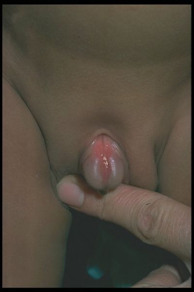
An epispadia occurs when the urethra opening is abnormally placed. In a male infant with epispadias, the urethra will be generally open on the top or side of the penis.

Click to see the picture…>…...(01).…..(2).…..(1)

Boys will suffer from a short, wide penis and widened pubic bone. In a female infant with epispadias, the urethra will generally be located between the clitoris and the labia or in the abdominal area. Girls will suffer from a widened pubic bone and an abnormal clitoris and labia. In both males and females, urine will flow into the kidney and urinary tract infections are common. It is also common for the child to have urinary incontinence, kidney damage and often infertility issues as an adult.

A doctor will perform a series of tests to diagnose epispadias, which may include blood tests, x-rays and ultrasounds. Treatment involves surgery to help with urine control and appearance.

It is also called bladder exstrophy

Symptoms:

In males:
*Abnormal opening from the joint between the pubic bones to the area above the tip of the penis
*Backward flow of urine into the kidney (reflux nephropathy)
*Short, widened penis with an abnormal curvature
*Urinary tract infections
*Widened pubic bone

In females:……..Picture
*Abnormal clitoris and labia
*Abnormal opening where the from the bladder neck to the area above the normal urethral opening
*Backward flow of urine into the kidney (reflux nephropathy)
*Widened pubic bone
*Urinary incontinence
*Urinary tract infections

Causes:
The causes of epispadias are unknown at this time. It may be related to improper development of the pubic bone.

In boys with epispadias, the urethra generally opens on the top or side of the penis rather than the tip. However, it is possible for the urethra to be open along the entire length of the penis.

In girls, the opening is usually between the clitoris and the labia, but may be in the belly area.

Epispadias can be associated with bladder exstrophy, an uncommon birth defect in which the bladder is inside out, and sticks through the abdominal wall. However, epispadias can also occur with other defects.

Epispadias is an uncommon and partial form of a spectrum of failures of abdominal and pelvic fusion in the first months of embryogenesis known as the exstrophy – epispadias complex. While epispadias is inherent in all cases of exstrophy it can also, much less frequently, appear in isolation as the least severe form of the complex spectrum. It occurs as a result of defective migration of the genital tubercle primordii to the cloacal membrane, and so malformation of the genital tubercle, at about the 5th week of gestation.

Presentation:
Most cases involve a small and bifid penis, which requires surgical closure soon after birth, often including a reconstruction of the urethra. Where it is part of a larger Exstrophy, not only the urethra but also the bladder (bladder exstrophy) or the entire perineum (cloacal exstrophy) are open and exposed on birth, requiring closure.

Relationship to other conditions:
Despite the similarity of name, an epispadias is not a type of hypospadias, and involves a problem with a different set of embryologic processes.

In women:
Women can also have this type of congenital malformation. Epispadias of the female may occur when the urethra develops too far anteriorly, exiting in the clitoris or even more forward. For females, this may not cause difficulty in urination but may cause problems with sexual satisfaction. Frequently, the clitoris is bifurcated at the site of urethral exit, and therefore clitoral sensation is less intense during sexual intercourse due to frequent stimulation during urination. However, with proper stimulation, using either manual or positional techniques, clitoral orgasm is definitely possible

Diagnosis:
•Blood test to check electrolyte levels
•Intravenous pyelogram (IVP), a special x-ray of the kidneys, bladder, and ureters
•MRI and CT scans, depending on the condition
•Pelvic x-ray
•Ultrasound of the urogenital system

Treatment:
The main treatment for isolated epispadias is a comprehensive surgical repair of the genito-urinary area usually during the first 7 years of life, including reconstruction of the urethra, closure of the penile shaft and mobilisation of the corpora. The most popular and successful technique is known as the modified Cantwell-Ransley approach. In recent decades however increasing success has been achieved with the complete penile disassembly technique despite its association with greater and more serious risk of damage

Prognosis:
Even with successful surgery, patients may have long-term problems with:
*incontinence, where serious usually treated with some form of continent urinary diversion such as the Mitrofanoff
*depression and psycho-social complications
*sexual dysfunction

Disclaimer: This information is not meant to be a substitute for professional medical advise or help. It is always best to consult with a Physician about serious health concerns. This information is in no way intended to diagnose or prescribe remedies.This is purely for educational purpose

Resources:
http://www.nlm.nih.gov/medlineplus/ency/article/001285.htm
http://en.wikipedia.org/wiki/Epispadias
http://health.stateuniversity.com/pages/794/Hypospadias-Epispadias.html
http://www.wikidoc.org/index.php/Epispadias
http://www.eclips.consult.com/eclips/article/Pediatrics/S0084-3954(07)70134-3

Enhanced by Zemanta
Categories
Ailmemts & Remedies Pediatric

Hypospadias

Definition:
Hypospadias is a birth defect found in boys in which the penile meatus is not at the tip of the penis. The meatus is the term for the opening of the penis through which urine normally exits the bladder. The incidence is reported to be 1 in 300 live male births. There is some family risk of hypospadias, as familial tendencies have been noted. Up to 14% of male siblings are affected.

Hypospadias is usually classified according to the location of the opening. As the defect increases in severity, the opening to the penis will be found further back on the penis. The most severe types can have openings at the region of the scrotum and even in the perineum (the region between the anus and scrotum).

Click for picture

In some men with hypospadias, there’s another abnormality called chordee, in which the penis curves downwards and the foreskin only covers the front of it.In the most severe forms of hypospadias, the urethral opening is so far back it’s almost in the scrotum. The scrotum itself may be small and the testes may not have descended (that is, they’re still deep in the abdomen). When babies are born like this, it can be difficult to work out which sex they are without further tests.

click tom see the picture

Both hypospadias and chordee must be repaired so that a child can have normal urinary and reproductive health.

Symptoms:
Hypospadias is a structural abnormality that doesn’t progress or put the man at risk of any other serious illness. However, as with any abnormalities of the urinary system, there may be an increased risk of urinary infection in more severe cases.

Hypospadias may cause emotional turmoil when a boy realises he’s different from his friends. It can also cause practical problems with passing urine (those with the condition usually have to sit down to pee) and later with sexual intercourse, which may be embarrassing or difficult to cope with. Hypospadias may cause general worries about sexuality and fertility.

Signs and symptoms of hypospadias may include:

*Opening of the urethra at a location other than the tip of the penis
*Downward curve of the penis (chordee)
*Hooded appearance of the penis because only the top half of the penis is covered by foreskin
*Abnormal spraying during urination

Causes:
Hypospadias is present at birth (congenital). The exact reason this defect occurs is unknown. Sometimes hypospadias is inherited.

As the penis develops in a male fetus, certain hormones stimulate the formation of the urethra and foreskin. Hypospadias results when a malfunction occurs in the action of these hormones, causing the urethra to develop abnormally.

As a boy is developing in utero, the penis begins to form in the sixth week of fetal life. Two folds of tissue join each other in the middle and a hollow tube is formed in the middle of the future penis. This tube is the urethra and its opening is called the penile meatus. As the skin folds develop to form the penis, any interruption in this process leads to the meatus being located in a location further from the end of the penis. The exact etiology for this premature cessation of urethral formation is poorly understood. In addition, the etiology of the often-associated abnormal downward curvature (chordee) is also poorly understood.

Risk Factors:
This condition is more common in infants with a family history of hypospadias.

Some research suggests that there may be an increased risk of hypospadias in infant males born to women of an advanced age or those who used in vitro fertilization (IVF) to conceive. The connection to IVF may be due to the mother’s exposure to progesterone, a natural hormone, or to progestin, a synthetic form of progesterone, administered during the IVF process. Other research, however, hasn’t confirmed a link between IVF and hypospadias, but did find an association between a mother’s exposure to pesticides and hypospadias.

Diagnosis:
A physical examination can diagnose this condition. Imaging tests may be needed to look for other congenital defects.

Treatment
The treatment of hypospadias is always surgical. Initially when the child is born and hypospadias is identified, it is important to delay any thoughts of circumcision until seen by a urologist. This is because the foreskin can provide essential additional skin needed to reconstruct the urethra.

Hypospadias is often repaired  before a child is one year of age. This way, the boy is in diapers and management of dressings are made easier. However, the exact age of repair can vary according to the size of the penis and severity of the defect. It can be repaired in most of the  cases with a single operation, but on occasion, a second operation may be needed. The operation is performed under general anesthesia with the child completely asleep. Most of the boys will have a small tube exiting the tip of their new meatus. This “stent” will protect the new urethra and allow for adequate healing. Most patients leave the hospital the same day or the following day. However, more complex repairs for the more severe types of hypospadias can require longer hospital stays due to the need for bedrest and immobilization in the immediate post-operative setting.


Click for the picture

The exact type of operation employed varies according to the severity of the defect. For the more distal defects that have openings closer to the normal position at the end of the penis, a new tube can be created from the surrounding skin. This creation of a tube is known as a Thiersch-Duplay repair. For more severe defects, the options range. Additional hairless skin is often needed to recreate the urethral tube when longer defects are seen. Here, the subdermal skin of the foreskin can be used. For the most severe defects, we can remove mucosal skin from the inside of the cheek or use subdermal skin from other hairless parts of the body. It is important to use hairless skin as future hair growth in the neourethra can present multiple problems.

Complications:
The usual risks of surgery are present at the time of performing  hypospadias repairs. Risk of infection is controlled with use of antibiotics with the surgery and in the post-operative setting. Bleeding is well controlled by using a penile tourniquet during the operation. This limits the blood loss to a very minimal amount, while allowing for good visualization of the tissues for the surgeon.

By using good surgical techniques   the longer-term complications of the surgery are minimised. The most common problems that present are fistula and stricture. A fistula occurs if a hole develops along the pathway of the repair proximal to the tip of the penis. In other words, a hole can develop along the underside of the penis allowing for leakage of urine. Additionally, a stricture is a scar that can form causing a narrowing in the urethra. If either of these complications occur, an additional repair will be needed usually 6 months later

Prognosis:
Results after surgery are typically good. In some cases, more surgery is needed to correct fistulas or a return of the abnormal penis curve.

Disclaimer: This information is not meant to be a substitute for professional medical advise or help. It is always best to consult with a Physician about serious health concerns. This information is in no way intended to diagnose or prescribe remedies.This is purely for educational purpose

Resources:
http://www.bbc.co.uk/health/physical_health/conditions/hypospadias.shtml
http://www.nlm.nih.gov/medlineplus/ency/article/001286.htm
http://www.mayoclinic.com/health/hypospadias/DS00884
http://www.cornellurology.com/pediatrics/hypospadias.shtml

http://www.medindia.net/patients/paediatrics/Hypospadias.htm

http://www.surgeryencyclopedia.com/Fi-La/Hypospadias-Repair.html

http://www.adhb.govt.nz/newborn/Guidelines/Anomalies/Hypospadias.htm

Enhanced by Zemanta
Categories
Ailmemts & Remedies Pediatric

Molar Pregnancy

Definition:
A molar pregnancy is one condition in a range of problems known as trophoblastic disease, where a pregnancy doesn’t grow as it should. It’s sometimes called a hydatiform mole.

There are two different types of molar pregnancy, which differ in how they form and how they need to be treated.

In a normal pregnancy, genetic material from the mother and father combines to form new life. In a molar pregnancy, this process goes wrong. In a complete molar pregnancy, the maternal chromosomes are lost, either at conception or while the egg was forming in the ovary, and only genetic material from the father develops in the cells. In a partial molar pregnancy, there is a set of maternal chromosomes but also two sets of chromosomes from the father (ie, double the normal paternal genetic material).

CLICK & SEE

The genotype is typically 46,XX (diploid) due to subsequent mitosis of the fertilizing sperm, but can also be 46,XY (diploid).  In contrast, a partial mole occurs when an egg is fertilized by two sperm or by one sperm which reduplicates itself yielding the genotypes of 69,XXY (triploid) or 92,XXXY (quadraploid).

Complete molar pregnancies develop as a mass of rapidly growing cells but without a foetus – it cannot therefore develop into a baby.
……
In a partial molar pregnancy, a foetus may start to develop but because of the imbalance in genetic material, it’s always abnormal and can’t survive beyond the first three months of pregnancy.

A molar pregnancy is often harmless, but if untreated can keep on growing and become invasive, spreading to the organs around it, or even further afield to the lungs, liver or brain. Very rarely, in two to three per cent of cases, it may become malignant. These cancerous types of trophoblastic disease are called choriocarcinoma and placental site trophoblast tumours.

Symptoms:
As the mole grows faster than a normal foetus would, the abdomen may become larger more quickly than would be expected for the dates of the pregnancy. The woman may experience abdominal pain, and also severe nausea and vomiting (hyperemesis).

Bleeding from the vagina is another common warning sign that things are not as they should be. Symptoms similar to pre-eclampsia – high blood pressure, protein in the urine, swelling of the feet and legs – may also occur in the first trimester or early in the second.

Most molar pregnancies are diagnosed at the first ultrasound scan, which shows a mass of cells without the presence of a foetus in a complete molar pregnancy or an abnormal non-viable foetus and placenta in a partial mole.

A woman with a hydatidiform mole often feels pregnant and has symptoms such as morning sickness, probably because the cells of the molar pregnancy produce the pregnancy hormone hCG (human chorionic gonadotrophin). This is also the hormone that is used in a pregnancy test, so she may have a positive result. Some women have no pregnancy symptoms (as with many normal pregnancies). — but most molar pregnancies cause specific signs and symptoms, including:

*Dark brown to bright red vaginal bleeding during the first trimester

*Severe nausea and vomiting

*Vaginal passage of grape-like cysts

*Rarely, pelvic pressure or pain

If you experience any signs or symptoms of a molar pregnancy, consult your health care provider. He or she may detect other signs of a molar pregnancy, such as:

*Rapid uterine growth — the uterus is too large for the stage of pregnancy

*High blood pressure

*Preeclampsia — a condition that causes high blood pressure and protein in the urine after 20 weeks of pregnancy

*Ovarian cysts

*Anemia

*Overactive thyroid (hyperthyroidism)

Causes:
A molar pregnancy is caused by an abnormally fertilized egg. Human cells normally contain 23 pairs of chromosomes. One chromosome in each pair comes from the father, the other from the mother. In a complete molar pregnancy, all of the fertilized egg’s chromosomes come from the father. Shortly after fertilization, the chromosomes from the mother’s egg are lost or inactivated and the father’s chromosomes are duplicated. The egg may have had an inactive nucleus or no nucleus.

In a partial or incomplete molar pregnancy, the mother’s chromosomes remain but the father provides two sets of chromosomes. As a result, the embryo has 69 chromosomes, instead of 46. This can happen when the father’s chromosomes are duplicated or if two sperm fertilize a single egg.

It remains unclear why a hydatidiform mole develops. However, there are a number of possible reasons, including defects in the egg, maternal nutritional deficiencies and uterine abnormalities. Women under 20 or over 40 are at higher risk.

Having a diet that’s low in protein, folic acid and carotene also increases the risk of a molar pregnancy. The number of times a women has been pregnant, however, doesn’t influence her risk.

Risk Factors:
Up to an estimated 1 in every 1,000 pregnancies is molar. Various factors are associated with molar pregnancy, including:

*Maternal age. A molar pregnancy is more likely for a woman older than age 35 or younger than age 20.

*Previous molar pregnancy. If you’ve had one molar pregnancy, you’re more likely to have another. The risk of a repeat molar pregnancy is 1 in 100.

*Some ethnic groups. Women of Southeast Asian descent appear to have a higher risk of molar pregnancy.

Diagnosis:
Molar pregnancies usually present with painless vaginal bleeding in the fourth to fifth month of pregnancy. The uterus may be larger than expected, or the ovaries may be enlarged. There may also be more vomiting than would be expected (hyperemesis). Sometimes there is an increase in blood pressure along with protein in the urine. Blood tests will show very high levels of human chorionic gonadotropin (hCG).

The diagnosis is strongly suggested by ultrasound (sonogram), but definitive diagnosis requires histopathological examination. On ultrasound, the mole resembles a bunch of grapes (“cluster of grapes” or “honeycombed uterus” or “snow-storm”). There is increased trophoblast proliferation and enlarging of the chorionic villi. Angiogenesis in the trophoblasts is impaired as well.

Sometimes symptoms of hyperthyroidism are seen, due to the extremely high levels of hCG, which can mimic the normal Thyroid-stimulating hormone (TSH).

Treatment :
Once it has been established that a woman is carrying a hydatidiform mole rather than a healthy foetus, suction evacuation is used to remove the pregnancy from the womb. This is curative in about four out of five molar pregnancies.

It’s then important to monitor the woman’s progress and repeatedly measure human chorionic gonadotropin (hCG) to be sure that everything settles back down to a normal, non-pregnancy level.

About 15 per cent of women who have had a complete molar pregnancy and 0.5 per cent of those with a partial molar pregnancy will require additional treatment, either because hCG levels hit a plateau or start to rise again, or because of persistent heavy vaginal bleeding.

Further treatment may involve the use of chemotherapy (usually methotrexate combined with folinic acid), especially if there’s any concern about invasive or malignant disease.

Complications:
After a molar pregnancy has been removed, molar tissue may remain and continue to grow. This is called persistent gestational trophoblastic disease (GTD). It occurs in about 10 percent of women after a molar pregnancy — usually after a complete mole rather than a partial mole. One sign of persistent GTD is an HCG level that remains high after the molar pregnancy has been removed. In some cases, an invasive mole penetrates deep into the middle layer of the uterine wall, which causes vaginal bleeding. Persistent GTD can nearly always be successfully treated, most often with chemotherapy. Another treatment option is removal of the uterus (hysterectomy).

Rarely, a cancerous form of GTD known as choriocarcinoma develops and spreads to other organs. Choriocarcinoma is usually successfully treated with multiple cancer drugs.

Prognosis:
More than 80% of hydatidiform moles are benign. The outcome after treatment is usually excellent. Close follow-up is essential. Highly effective means of contraception are recommended to avoid pregnancy for at least 6 to 12 months.

In 10 to 15% of cases, hydatidiform moles may develop into invasive moles. This condition is named persistent trophoblastic disease (PTD). The moles may intrude so far into the uterine wall that hemorrhage or other complications develop. It is for this reason that a post-operative full abdominal and chest x-ray will often be requested.

In 2 to 3% of cases, hydatidiform moles may develop into choriocarcinoma, which is a malignant, rapidly-growing, and metastatic (spreading) form of cancer. Despite these factors which normally indicate a poor prognosis, the rate of cure after treatment with chemotherapy is high.

Over 90% of women with malignant, non-spreading cancer are able to survive and retain their ability to conceive and bear children. In those with metastatic (spreading) cancer, remission remains at 75 to 85%, although their childbearing ability is usually lost.

Prevention:
Following successful treatment, most women can have children if they wish. However, it’s strongly recommended that a woman who has had a molar pregnancy doesn’t become pregnant again for 12 months. Although the likelihood is small, there’s a real risk of malignant disease developing and the increase in pregnancy hormones this would cause can’t be distinguished from those of a real pregnancy. Consequently, good contraception is required, as is regular monitoring by a hospital specialist.

Disclaimer: This information is not meant to be a substitute for professional medical advise or help. It is always best to consult with a Physician about serious health concerns. This information is in no way intended to diagnose or prescribe remedies.This is purely for educational purpose

Resources:
http://www.bbc.co.uk/health/physical_health/conditions/hydatidiformmole1.shtml
http://www.mayoclinic.com/health/molar-pregnancy/DS01155
http://en.wikipedia.org/wiki/Hydatidiform_mole

http://drugster.info/ail/pathography/375/

Enhanced by Zemanta
Categories
Ailmemts & Remedies Pediatric

Hernia in Children

Definition:
Hernia in children is a medical condition in which a tissue or structure or part of an organ is protruded through a weakness or hole in other body muscular tissue or membrane. A soft bulge is seen underneath the skin where the hernia has occurred.

In children, a hernia usually occurs in one of two places:

1.around the belly-button
2.in the groin area

A hernia that occurs in the belly-button area is called an umbilical hernia. A hernia that occurs in the groin area is called an inguinal hernia.

 click to see the ;pictures…..>...(01)....(1).……...(2)..……..(3)....…..(4).

Hernias in children mostly occur in the umbilical region. A weak abdominal wall in the children can be a reason for development of umbilical hernias. Hernias are present during the first year of child and may keep on coming and going at any age.

The disease condition is common among all the age groups. Boys are more prone to this disease than girls. Approximately 1 out of 50 boys are affected.

Symptoms:
Hernias usually occur in newborns, but may not be noticeable for several weeks or months after birth.

Straining and crying do not cause hernias; however, the increased pressure in the abdomen can make a hernia more noticeable.

*Inguinal hernias appear as a bulge or swelling in the groin or scrotum. The swelling may be more noticeable when the baby cries, and may get smaller or go away when the baby relaxes. If your physician pushes gently on this bulge when the child is calm and lying down, it will usually get smaller or go back into the abdomen.

*Umbilical hernias appear as a bulge or swelling in the belly-button area. The swelling may be more noticeable when the baby cries, and may get smaller or go away when the baby relaxes. If your physician pushes gently on this bulge when the child is calm and lying down, it will usually get smaller or go back into the abdomen.

A hernia usually causes a visible lump or swelling, which appears intermittently as the herniating tissue slips back into place and then protrudes again (umbilical hernias are more constant).

Crying, straining, coughing or anything else that increases pressure within the abdomen can make the hernia more obvious, as this forces out the contents.

If the hernia is not reducible, then the loop of intestine may be caught in the weakened area of abdominal muscle. Symptoms that may be seen when this happens include the following:

*a full, round abdomen
*vomiting
*pain or fussiness
*redness or discoloration
*fever

Symptoms of a hernia may resemble other conditions or medical problems. Please consult your child’s physician for a diagnosis.

Causes:

A hernia can develop in the first few months after the baby is born because of a weakness in the muscles of the abdomen. Inguinal and umbilical hernias happen for slightly different reasons.

Inguinal Hernia...click & see
As a male fetus grows and matures during pregnancy, the testicles develop in the abdomen and then move down into the scrotum through an area called the inguinal canal….Shortly after the baby is born, the inguinal canal closes, preventing the testicles from moving back into the abdomen. If this area does not close off completely, a loop of intestine can move into the inguinal canal through the weakened area of the lower abdominal wall, causing a hernia.

Although girls do not have testicles, they do have an inguinal canal, so they can develop hernias in this area as well.(

Femoral hernias are more common in women, usually elderly and frail (although they can happen in children).)

Umbilical Herniaclick & see
When the fetus is growing and developing during pregnancy, there is a small opening in the abdominal muscles so that the umbilical cord can pass through, connecting the mother to the baby.

After birth, the opening in the abdominal muscles closes as the baby matures. Sometimes, these muscles do not meet and grow together completely, and there is still a small opening present. A loop of intestine can move into the opening between abdominal muscles and cause a hernia.
Risk Factors:
Hernias occur more often in children who have one or more of the following risk factors:

*a parent or sibling who had a hernia as an infant
*cystic fibrosis
*developmental dysplasia of the hip
*undescended testes
*abnormalities of the urethra

Inguinal hernias occur:
*in about one to three percent of all children.
*more often in premature infants.
*in boys much more frequently than in girls.
*more often in the right groin area than the left, but can also occur on both sides.

Umbilical hernias occur:
*in about 10 percent of all children.
*more often in African-American children.
*more often in girls than in boys.
*more often in premature infants

Why is a hernia a concern?
Hernias are usually painless. However, if the contents become trapped, the blood supply to the tissues may become restricted causing pain. This pain may be intermittent, but if the hernia is stuck permanently – known as an irreducible, strangulated or incarcerated hernia – the pain becomes constant and there’s a risk of damage to the trapped intestines or surrounding tissues. In this case the child may vomit and appear unwell.

Occasionally, the loop of intestine that protrudes through a hernia may become stuck, and is no longer reducible. This means that the intestinal loop cannot be gently pushed back into the abdominal cavity. When this happens, that section of intestine may lose its blood supply. A good blood supply is necessary for the intestine to be healthy and function properly.

Diagnosis:
Hernias can be diagnosed by a physical examination by your pediatrician. Your child will be examined to determine if the hernia is reducible (can be pushed back into the abdominal cavity) or not. Doctor may order abdominal x-rays or ultrasound to examine the intestine more closely, especially if the hernia is no longer reducible.

Treatment:-
Specific treatment will be determined by your pediatrician based on the following:

*your child’s age, overall health, and medical history
*the type of hernia
*whether the hernia is reducible (can be pushed back into the abdominal cavity) or not
*your child’s tolerance for specific medications, procedures, or therapies
*your opinion or preference

Inguinal hernia:……………..

An operation is necessary to treat an inguinal hernia. It will be surgically repaired fairly soon after it is discovered, since the intestine can become stuck in the inguinal canal. When this happens, the blood supply to the intestine can be cut off, and the intestine can become damaged. Inguinal hernia surgery is usually performed before this damage can occur.

During a hernia operation, your child will be placed under anesthesia. A small incision is made in the area of the hernia. The loop of intestine is placed back into the abdominal cavity. The muscles are then stitched together. Sometimes, a piece of meshed material is used to help strengthen the area where the muscles are repaired.

A hernia operation is usually a fairly simple procedure. Children who have an inguinal hernia surgically repaired can often go home the same day they have the operation.

Umbilical hernia:
By 1 year of age, many umbilical hernias will have closed on their own without needing surgery. Nearly all umbilical hernias will have closed without surgery by age 5.

Placing a coin or strap over the hernia will not fix it.

There are many opinions about when a surgical repair of an umbilical hernia is necessary. In general, if the hernia becomes bigger with age, is not reducible, or is still present after 3, your physician may suggest that the hernia be repaired surgically. Always consult your child’s physician to determine what is best for your child.

During a hernia operation, your child will be placed under anesthesia. A small incision is made in the umbilicus (belly button). The loop of intestine is placed back into the abdominal cavity. The muscles are then stitched together. Sometimes a piece of meshed material is used to help strengthen the area where the muscles are repaired.

A hernia operation is usually a fairly simple procedure. Children who have an umbilical hernia surgically repaired may also be able to go home the same day they have the operation.

Prognosis:-
Once the hernia is closed, either spontaneously or by surgery, it is unlikely that it will reoccur. The chance for re-occurrence  of the hernia may be increased if the intestine was damaged.

Disclaimer: This information is not meant to be a substitute for professional medical advise or help. It is always best to consult with a Physician about serious health concerns. This information is in no way intended to diagnose or prescribe remedies.This is purely for educational purpose

Resources:
http://www.spirita.net/Hernia/femoral_hernia.htm
http://www.childrenshospital.org/az/Site1018/mainpageS1018P0.html
http://www.bbc.co.uk/health/physical_health/conditions/hernia2.shtml

Enhanced by Zemanta
Categories
Ailmemts & Remedies Pediatric

Hepatitis A

Definition:
Hepatitis A is a liver disease caused by the hepatitis A virus (HAV).
This form of viral hepatitis also known as infectious hepatitis, due to its ability to be spread through personal contact. Hepatitis A is a milder liver disease than hepatitis B, and asymptomatic infections are very common, especially in children.

click to see the pictures

According to the World Health Organisation, there are an estimated 1.5 million new cases of illness due to hepatitis A each year worldwide, and many more people become infected without developing symptoms. It’s particularly common in less developed countries where poverty or poor sanitation are important factors.

Africa, northern and southern Asia, parts of South America, and southern and eastern Europe all have high rates of the disease. In these countries almost every adult carries antibodies to hepatitis A suggesting that it is quite usual for people to be exposed to the infection, usually in childhood, and to develop immunity.

The infection isn’t common in the UK, although it’s still the main type of infective hepatitis seen. (There are several other types of viral hepatitis, such as hepatitis B and hepatitis C.) In 2005, for example, there were 457 laboratory reports of confirmed hepatitis A virus (HAV) infection in England and Wales.

The majority of people from the UK who become infected with hepatitis A contract it when abroad in a country where it is very common.

Hepatitis A is an acute infection, rather than chronic (long-term). Rarely, it can cause life-threatening liver damage.Hepatitis A does not cause a carrier state or chronic liver disease. Once the infection ends, there is no lasting phase of illness.  However, it is not uncommon to have a second episode of symptoms about a month after the first; this is called a relapse.

Symptoms :
The incubation period of the virus before symptoms develop is between two and six weeks. How severely someone is affected varies from person to person. Some may not have any symptoms at all, while others may have just mild symptoms similar to those of a flu-like illness. This is particularly common among infants and young children.

The older someone is, the more severe the infection and symptoms are likely to be.

Possible symptoms include weakness, tiredness, headache, fever, loss of appetite, nausea and vomiting, abdominal pain and diarrhoea and dehydration. These may all occur for a week or more before jaundice appears.

Jaundice occurs in hepatitis infections because the liver becomes unable to remove a substance called bilirubin from the blood. This is a pigment that builds up in the body, causing the skin and whites of the eyes to turn yellow.

Causes:
HAV is found in the stool (feces) of persons infected with hepatitis A. HAV is usually spread from person to person by putting something in the mouth that has been contaminated with the stool of a person infected with hepatitis A. This is called fecal-oral transmission. Thus, the virus spreads more easily in areas where there are poor sanitary conditions or where good personal hygiene is not observed. Most infections result from contact with a household member who has hepatitis A. Blood-borne infection has been documented but is rare in the United States. The common modes of transmission of hepatitis A are as follows:

•consuming food made by someone who touched infected feces
•drinking water that is contaminated by infected feces (a problem in communities with poor sewage treatment facilities)
•touching an infected person’s feces, which may occur with poor hand washing
•having direct contact in large daycare centers, especially where there are children in diapers
•being a resident of states in which hepatitis A is more common
•sexual contact with an infected person.

Risk Factors:
*Eating food that was prepared by someone who is infected with hepatitis A and poor hygiene.
*Consuming raw or undercooked shellfish (like oysters or clams).
*Eating raw foods (such as unpeeled fruits or vegetables) and drinking tap water or well water while traveling to countries where hepatitis A is common.
*Living in a community where hepatitis A is common and outbreaks occur (largely a risk factor for young children).
*Living in a house with someone who has hepatitis A.

Lifestyle factors that increase the risk of hepatitis A include:
* Travel to countries where hepatitis A is common.
* Be a man having sex with men.

Diagnosis
Hepatitis A symptoms often go unrecognized because they are not specific to hepatitis A, thus a blood test (IgM anti-HAV) is required to diagnose HAV infection. This test detects a specific antibody, called hepatitis A IgM, that develops when HAV is present in the body.

Treatment:
No specific treatment is available for hepatitis A. However, the following guidelines are often recommended:

•Fluids and diet. The best treatment is to make sure that the child drinks a lot of fluids and eats well.
•Rest. The child should rest while he or she has fever or jaundice. When fever and jaundice are gone, activity may be gradually increased as with the healthcare provider’s approval.
•Medications. The body’s immune system fights the HAV infection. Once the child recovers from hepatitis A, the virus leaves the body. Medications, prescription or nonprescription, should not be given without consulting the doctor.

About 15% of people will have a prolonged or relapsing illness lasting up to 9 months. Tragically, a small number of people die when the infection overwhelms the body. This is more likely to happen to people over the age of 60.

A person with hepatitis A should avoid drinking alcohol until their liver is completely back to normal, as alcohol is toxic to liver cells and will slow its recovery.

Ensuring good personal hygiene practices – washing your hands after using the toilet and maintaining good food preparation – is essential in avoiding infection with hepatitis A, especially if you visit a high risk area.

When visiting high-risk countries, it’s a good idea to avoid eating raw or inadequately cooked salads and vegetables, ice cream, unpeeled fruit and shellfish. Also avoid unpasteurised milk and drinks with ice, and check whether tap water is safe to drink before you go.

There’s an effective vaccination to protect people from hepatitis A infection. It’s available from your GP or high street travel centres, who will be able to advise you whether you need it for the country you are visiting. It’s recommended for anyone travelling to the high-risk regions of the world.

Those people who have already had hepatitis A usually have life long immunity.

Prognosis:
Viral hepatitis symptoms usually last three weeks to two months but may last up to six months. Children may return to daycare one week after symptoms first appear, with the doctor’s permission. Most children with hepatitis get better naturally without liver problems later in life. However, some children do have subsequent liver problems. For this reason, it is important to keep in close touch with the treating physician and to keep all followup appointments. Chronic, or relapsing, infection does not occur with hepatitis A. In the United States, serious complications are infrequent, and deaths are very rare.

Prevention:
According to the Centers for Disease Control and Prevention (CDC), routine vaccination of children is the most effective way to lower the incidence of hepatitis A nationwide. The CDC encourages implementation of routine hepatitis A vaccination programs for children in the 17 states which have the highest rates of hepatitis A. Hepatitis A vaccine has been licensed in the United States for use in persons two years of age and older. The vaccine is recommended (before exposure to hepatitis A virus) for persons who are more likely to get hepatitis A virus infection or are more likely to get seriously ill if they do get hepatitis A. The vaccines licensed in the United States as of 2004 were HAVRIX(r) (manufactured by Glaxo SmithKline) and VAQTA(r) (manufactured by Merck & Co., Inc).

Parents should teach their children always to wash their hands with soap and water after using the bathroom and before preparing and eating food. Travelers should avoid water and ice if unsure of their purity, or they can boil water for one minute before drinking it.

Short-term protection against hepatitis A is available from immune globulin, a preparation of antibodies that can be given before exposure for short-term protection against hepatitis A and for persons who have already been exposed to HAV. It can be given before and within two weeks after suspected contact with the virus.

Parental Concerns
The best way to prevent exposure to HAV is good habits in washing hands. Children should wash their hands every time they go to the bathroom. Good handwashing should be enforced at home and at daycare facilities. It is also very important to keep a clean environment, such as clean toilets, bathrooms, and clothing. If a child is diagnosed with HAV, other family members should be treated to prevent spread of the disease. The healthcare provider can help parents to plan treatment for the entire family.

Disclaimer: This information is not meant to be a substitute for professional medical advise or help. It is always best to consult with a Physician about serious health concerns. This information is in no way intended to diagnose or prescribe remedies.This is purely for educational purpose.

Resources:
http://www.answers.com/topic/hepatitis-a
http://www.bbc.co.uk/health/physical_health/conditions/hepatitisa1.shtml

http://nationalnursingreview.com/tag/hepatitis-a-sign/
http://www.utmedicalcenter.org/your-health/encyclopedia/general/image/9394/
http://medicalsin.com/risk-factors-hepatitis-a-symptoms-increase/
http://www.fehd.gov.hk/english/safefood/library/prevent_hepatitis_A/2.html

Enhanced by Zemanta
css.php