Categories
Diagnonistic Test

Pleural Fluid Sampling (or Thoracentesis)

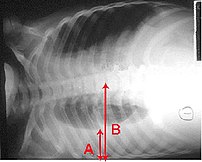
Pleural effusion Chest x-ray of a pleural effu...
Image via Wikipedia

[amazon_link asins=’B01J8WALE0,B00GNOPG8S,1331951720,1893441776,B01MXOPZIB,B01J2IC12U,B01J37411E,B009KT2GWM,B01J2GLUVU’ template=’ProductCarousel’ store=’finmeacur-20′ marketplace=’US’ link_id=’316e2df4-5fab-11e7-aab1-4db380a805de’]


Definition:
Thoracentesis is a procedure used to obtain a sample of fluid from the space around the lungs. Normally, only a thin layer of fluid is present in the area between the lungs and chest wall (show radiograph 1). However, some conditions can cause a large amount of fluid to accumulate. This collection of fluid is called a pleural effusion (show radiograph 2). Thoracentesis is done to collect a sample of the fluid, which can help determine why the pleural effusion developed.
CLICK & SEE
Some infections and diseases cause fluid to accumulate in the space between the lung and the rib cage or between the lung and the diaphragm. This collection of fluid is called a pleural effusion. A pleural effusion might be detected on a chest x-ray. Sampling this fluid is important because it enables doctors to understand what caused the fluid to collect and how to treat the problem. The fluid can be sampled with a needle.

Reasons for Thoracentesis: — A thoracentesis is performed to determine the cause of a pleural effusion. In some cases, a physician may perform thoracentesis to relieve symptoms caused by the pleural effusion, including shortness of breath and low blood oxygen levels. A pleural effusion may be detected during a physical examination or on a chest x-ray.

Pleural effusion can be caused by many different conditions, including infections, heart failure, cancer, or tuberculosis. In some cases, blood or other fluid may be leaking into the pleural space from another part of the body, causing the effusion. By examining the fluid and the types of cells it contains, the cause of the effusion can usually be determined.

In general, there is no reason a thoracentesis cannot be performed. The procedure is more easily performed and complications are fewer when the pleural effusion is large. Special consideration may be necessary in patients who are on respirators.

Patients who have a bleeding disorder or who are on medications that affect blood clotting may need extra care to minimize the risk of bleeding. Patients should tell their healthcare provider if they have a history of bleeding problem or if they are taking medicine that decreases blood clotting. In some cases, a blood test will be taken prior to the procedure to exclude any blood clotting abnormalities caused by disease or medications.

Procedure: A thoracentesis involves the following steps:

*The patient will be placed in a position that allows the doctor to easily access the effusion. Usually, the patient is asked to sit upright during the procedure. It is important to remain still during the procedure so that the fluid does not shift.

*The skin is cleaned with an antibacterial solution in the area where the needle will be inserted.

*A small amount of numbing medicine (a local anesthetic, similar to novocaine) is injected into the area. This medicine helps minimize discomfort during the procedure.

*A slightly larger needle is inserted in the same location. A syringe is attached to this needle and is used to withdraw fluid from around the lung. Patients who have symptoms from the effusion (eg, shortness of breath) may have a large amount of fluid removed, which allows the lung to re-expand.

*The needle is removed and pressure is briefly applied to the insertion site.

How do you prepare for the test?
You will need to sign a consent form giving your doctor permission to perform this test. Some patients have this test done in a doctor’s office, while others are admitted to the hospital for it. Generally your doctor will decide whether you need to be in the hospital based on your medical condition. A chest x-ray or an ultrasound is done before the procedure.

Tell your doctor if you have ever had an allergic reaction to lidocaine or the numbing medicine used at the dentist’s office. If you take aspirin, nonsteroidal anti-inflammatory drugs, or other medicines that affect blood clotting, talk with your doctor. It may be necessary to stop or adjust the dose of these medicines before your test.

What happens when the test is performed?
You wear a hospital gown and sit on a bed or table leaning forward against some pillows. The doctor listens to your lungs with a stethoscope and may tap on your back to find out how much fluid has collected.

Soap is used to disinfect an area of skin on one side of your back. A small needle is used to numb a patch of skin between two of your lower ribs. The numbing medicine usually stings for a second. A needle on an empty syringe is then inserted into the fluid pocket. Usually this pocket is around one inch below the skin surface. You might feel some minor pressure as the needle is inserted. Depending on the quantity of fluid that the doctor plans to remove, either the syringe itself is filled or soft plastic tubing is used to remove fluid into a collection bag or jar. While the doctor is attaching the tubing, he or she might ask you to hum out loud. This humming is for your safety: It prevents you from taking a deep breath, which could expand your lung, causing it to touch the needle.

It sometimes takes 15 minutes or longer to remove the necessary amount of fluid. Most patients feel no discomfort during this time, although a few patients feel some chest pain at the end of the procedure as their lung expands and touches the chest wall. After the fluid is removed, a bandage is placed on your back.

Risk Factors:
This procedure carries a few serious risks, but most patients have no complications. If the needle touches the lung it may create an air leak, which is seen on the x-ray and might require you to stay in the hospital for a few days. Some patients with this complication need to have a plastic tube (called a chest tube) inserted between two ribs. The tube uses vacuum pressure to keep the lung expanded until it has healed.

In most cases, a thoracentesis is performed without complications. Most complications are minor and resolve on their own or are easily treated. Potential complications include the following:

*Pain — Some discomfort may occur when the needle is inserted. Using a local anesthetic helps to reduce the pain. Pain generally resolves once the needle is removed.

*Bleeding — A blood vessel may be nicked as the needle is inserted through the skin and chest wall, causing bleeding. The bleeding is usually minor and stops on its own, although it may cause bruising around the puncture site. In rare cases, bleeding into or around the lung may occur, requiring drainage or surgery.

*Infection
Infection can occur if bacteria are introduced by the needle puncture. Using disinfectant solution to clean the area and using sterile technique during the procedure minimizes this risk.

*Pneumothorax or collapsed lung Occasionally, the needle used to obtain a fluid sample can puncture the lung. The hole created by the puncture usually seals quickly on its own. If it does not, air can build up around the lung, causing the lung to collapse. This is called a pneumothorax. When a pneumothorax occurs, a chest tube may be used to drain the air and allow the lung to re-expand.

A pneumothorax may also occur if the lung fails to expand when fluid is withdrawn. This is considered to be a drainage-related pneumothorax, and is the most common type of pneumothorax to occur when ultrasound is used for needle placement. Drainage-related pneumothorax is most commonly caused by disorders of the surface lining of the lung and not by the puncture needle. Treatment is rarely needed.

Pneumothorax occurs in less than 12 percent of procedures. Those that do occur are usually small and resolve on their own. A chest tube to helps re-expand the lung is necessary only if the pneumothorax is large, continues to expand, or causes symptoms.

*Liver or spleen puncture — In very rare cases, the liver or spleen may be punctured during thoracentesis. Sitting upright and remaining still during the procedure helps to keep the liver and spleen away from the insertion area and minimizes the risk of this complication.

Must you do anything special after the test is over?
You will need to have an x-ray taken after the sampling is completed. Your breathing should feel the same (or better) after the procedure.

How long is it before the result of the test is known?
The fluid may be tested for a variety of things, including infection and cancer. Cells in the fluid will be examined. It may be several days before full results are available.

Where you may get more information:-Your healthcare provider is the best source of information for questions and concerns related to your medical problem. Because no two patients are exactly alike and recommendations can vary from one person to another, it is important to seek guidance from a provider who is familiar with your individual situation.

This discussion will be updated as needed every four months on our web site (www.uptodate.com/patients). Additional topics as well as selected discussions written for healthcare professionals are also available for those who would like more detailed information.

Some of the most pertinent include:
Professional  Level Information:-
Diagnostic thoracentesis 
An overview of medical thoracoscopy
Diagnostic evaluation of a pleural effusion in adults
Imaging of pleural effusions in adults
Management of malignant pleural effusions

A number of web sites have information about medical problems and treatments, although it can be difficult to know which sites are reputable. Information provided by the National Institutes of Health, national medical societies and some other well-established organizations are often reliable sources of information, although the frequency with which they are updated is variable.

*American Thoracic Society
(www.thoracic.org)

*American Lung Association
(lungusa.org)

*National Heart Lung & Blood Institute
(www.nhlbi.nih.gov/index.htm)

*National Library of Medicine
(www.nlm.nih.gov/medlineplus/healthtopics.html)

Resources:
https://www.health.harvard.edu/diagnostic-tests/pleural-fluid-sampling.htm
http://www.uptodate.com/patients/content/topic.do?topicKey=~0aPG4xpnulisDf

Reblog this post [with Zemanta]
Categories
Herbs & Plants (Spices) Human Organ Transplantation

Lung Transplantation

[amazon_link asins=’1482233916,1426967314,3662046792,0521036771,1552125041,145570430X,0323354521,1553696840,1461476356′ template=’ProductCarousel’ store=’finmeacur-20′ marketplace=’US’ link_id=’a1736ec9-7d7d-11e7-8ccf-cb25f941da25′]

Introduction:
Lung transplantation is a surgical procedure in which a patient’s diseased lungs are partially or totally replaced by lungs which come from a donor. While lung transplants carry certain associated risks, they can also extend life expectancy and enhance the quality of life for end-stage pulmonary patients.

CLICK & SEE THE PICTURES

It is a life-preserving therapeutic intervention for a variety of end-stage pulmonary diseases that has been used successfully for the past 20 years. Since the early 1990s, more than 6400 lung transplants have been performed, and lung transplant programs exist in many countries.

The agency for health care policy and research in the United States has concluded that “lung transplantation has evolved as a clinical procedure achieving a favorable risk-benefit ratio and acceptable 1- and 2-year survival rates.”
.Qualifying conditions;-
Lung transplantation is the therapeutic measure of last resort for patients with end-stage lung disease who have exhausted all other available treatments without improvement. A variety of conditions may make such surgery necessary. As of 2005, the most common reasons for lung transplantation in the United States were:

*27% chronic obstructive pulmonary disease or COPD, including emphysema;

*16% idiopathic pulmonary fibrosis;

*14% cystic fibrosis;

*12% idiopathic (formerly known as “primary”) pulmonary hypertension;

* 5% alpha 1-antitrypsin deficiency;

*2% replacing previously transplanted lungs that have since failed;

*24% other causes, including bronchiectasis and sarcoidosis.

Contraindications:-
Despite the severity of a patient’s respiratory condition, certain preexisting conditions may make a person a poor candidate for lung transplantation. These conditions include:

*concurrent chronic illness (e.g. congestive heart failure, kidney disease, liver disease);

*current infections, including HIV and hepatitis;

*current or recent cancer;

*current use of alcohol, tobacco, or illegal drugs;

*age;

*psychiatric conditions;

*history of noncompliance with medical instructions.

History:-
The history of organ transplants began with several attempts that were unsuccessful due to transplant rejection. Animal experimentation by various pioneers, including Vladimir Demikhov and Dominique Metras, during the 1940s and 1950s, first demonstrated that the procedure was technically feasible. James Hardy of the University of Mississippi performed the first human lung transplant in 1963.  Following a left lung transplantation, the patient survived for 18 days. From 1963-1978, multiple attempts at lung transplantation failed because of rejection and problems with anastomotic bronchial healing. It was only after the invention of the heart-lung machine, coupled with the development of immunosuppressive drugs such as cyclosporine, that organs such as the lungs could be transplanted with a reasonable chance of patient recovery.

The first successful transplant surgery involving the lungs was a heart-lung transplant, performed by Dr. Bruce Reitz of Stanford University on a woman who had idiopathic pulmonary hypertension.

*1983: First successful single lung transplant (Tom Hall) by Joel Cooper (Toronto)

*1986: First successful double lung transplant (Ann Harrison) by Joel Cooper (Toronto)

*1988: First successful double lung transplant for cystic fibrosis by Joel Cooper (Toronto)

Transplant requirements:-

Requirements for potential donors:-
There are certain requirements for potential lung donors, due to the needs of the potential recipient. In the case of living donors, this is also in consideration of how the surgery will affect the donor.

*healthy;
*size match; the donated lung or lungs must be large enough to adequately oxygenate the patient, but small enough to fit within the recipient’s chest cavity;
*age;
*blood type.

Requirements for potential recipients:
While each transplant center is free to set its own criteria for transplant candidates, certain requirements are generally agreed upon:

*end-stage lung disease;

*has exhausted other available therapies without success;

*no other chronic medical conditions (e.g. heart, kidney, liver);

*no current infections or recent cancer. There are certain cases where preexisting infection is unavoidable, as with many patients with cystic fibrosis. In such cases, transplant centers, at their own discretion, may accept or reject patients with current infections of B. cepacia or MRSA.

*no HIV or hepatitis;

*no alcohol, smoking, or drug abuse;

*within an acceptable weight range (marked undernourishment or obesity are both associated
*with increased mortality);

*age (single vs. double tx);

*acceptable psychological profile;

*has social support system;

*financially able to pay for expenses;

*able to comply with post-transplant regimen. A lung transplant is a major operation, and following the transplant, the patient must be willing to adhere to a lifetime regimen of medications as well as continuing medical care.

Medical tests for potential transplant candidates:-
Patients who are being considered for placement on the organ transplant list must undergo an extensive series of medical tests in order to evaluate their overall health status and suitability for transplant surgery.

*blood typing; the blood type of the recipient must match that of the donor due to certain antigens that are present on donated lungs. A mismatch of blood type can lead to a strong response by the immune system and subsequent rejection of the transplanted organs;

*tissue typing; ideally, the lung tissue would also match as closely as possible between the donor and the recipient, but the desire to find a highly compatible donor organ must be balanced against the patient’s immediacy of need;

*Chest X-ray – PA & LAT, to verify the size of the lungs and the chest cavity;

*pulmonary function tests;

*CT Scan (High Resolution Thoracic & Abdominal);

*Bone mineral density scan;

*MUGA (Gated cardiac blood pool scan);

*Cardiac stress test (Dobutamine/Thallium scan);

*ventilation/perfusion (V/Q) scan;

*electrocardiogram;

*cardiac catheterization;

*echocardiogram.

Lung allocation score:-
Click to see:-> lung allocation score
Prior to 2005, donor lungs within the United States were allocated by the United Network for Organ Sharing on a first-come, first-serve basis to patients on the transplant list. This was replaced by the current system, in which prospective lung recipients of age of 12 and older are assigned a lung allocation score or LAS, which takes into account various measures of the patient’s health. The new system allocates donated lungs according to the immediacy of need rather than how long a patient has been on the transplant list. Patients who are under the age of 12 are still given priority based on how long they have been on the transplant waitlist. The length of time spent on the list is also the deciding factor when multiple patients have the same lung allocation score.

Patients who are accepted as good potential transplant candidates must carry a pager with them at all times in case a donor organ becomes available. These patients must also be prepared to move to their chosen transplant center at a moment’s notice. Such patients may be encouraged to limit their travel within a certain geographical region in order to facilitate rapid transport to a transplant center.

Types of lung transplant:-

Lobe
A lobe transplant is a surgery in which part of a living donor’s lung is removed and used to replace part of recipient’s diseased lung. This procedure usually involves the donation of lobes from two different people, thus replacing a single lung in the recipient. Donors who have been properly screened should be able to maintain a normal quality of life despite the reduction in lung volume.

Single-lung
Many patients can be helped by the transplantation of a single healthy lung. The donated lung typically comes from a donor who has been pronounced brain-dead.

Double-lung
Certain patients may require both lungs to be replaced. This is especially the case for people with cystic fibrosis, due to the bacterial colonisation commonly found within such patients’ lungs; if only one lung were transplanted, bacteria in the native lung could potentially infect the newly transplanted organ.

Heart-lung
Click to see:->Heart-lung transplant
Some respiratory patients may also have severe cardiac disease which in of itself would necessitate a heart transplant. These patients can be treated by a surgery in which both lungs and the heart are replaced by organs from a donor or donors.

A particularly involved example of this has been termed a “domino transplant” in the media. First performed in 1987, this type of transplant typically involves the transplantation of a heart and lungs into recipient A, whose own healthy heart is removed and transplanted into recipient B.

Procedure:-
While the precise details of surgery will depend on the exact type of transplant, there are many steps which are common to all of these procedures. Prior to operating on the recipient, the transplant surgeon inspects the donor lung(s) for signs of damage or disease. If the lung or lungs are approved, then the recipient is connected to an IV line and various monitoring equipment, including pulse oximetry. The patient will be given general anesthesia, and a machine will breathe for him or her.

It takes about one hour for the pre-operative preparation of the patient. A single lung transplant takes about four to eight hours, while a double lung transplant takes about six to twelve hours to complete. A history of prior chest surgery may complicate the procedure and require additional time.

Lobe:
Single-lung
In single-lung transplants, the lung with the worse pulmonary function is chosen for replacement. If both lungs function equally, then the right lung is usually favored for removal because it avoids having to maneuver around the heart, as would be required for excision of the left lung.

In a single-lung transplant the process starts out after the donor lung has been inspected and the decision to accept the donor lung for the patient has been made. An incision is generally made from under the shoulder blade around the chest, ending near the sternum. An alternate method involves an incision under the breastbone. In the case of a singular lung transplant the lung is collapsed, the blood vessels in the lung tied off, and the lung removed at the bronchial tube. The donor lung is placed, the blood vessels reattached, and the lung reinflated. To make sure the lung is satisfactory and to clear any remaining blood and mucus in the new lung a bronchoscopy will be performed. When the surgeons are satisfied with the performance of the lung the chest incision will be closed.

Double-lung
A double-lung transplant, also known as a bilateral transplant, can be executed either sequentially, en bloc, or simultaneously. Sequential is more common than en bloc.[2] This is effectively like having two separate single-lung transplants done. A less common alternative is the transplantation of both lungs en bloc or simultaneously.

….
Incision scarring from a double lung transplant.

.The transplantation process starts after the donor lungs are inspected and the decision to transplant has been made. An incision is then made from under the patient’s armpit, around to the sternum, and then back towards the other armpit, this is known as a clamshell incision. In the case of a sequential transplant the recipients lung with the poorest lung functions is collapsed, the blood vessels tied off, and cut at the corresponding bronchi. The new lung is then placed and the blood vessels reattached. To make sure the lung is satisfactory before transplanting the other a bronchoscopy is performed. When the surgeons are satisfied with the performance of the new lung, surgery on the second lung will proceed. In 10% to 20% of double-lung transplants the patient is hooked up to a heart-lung machine which pumps blood for the body and supplies fresh oxygen.

Post-operative care:-
Immediately following the surgery, the patient is placed in an intensive care unit for monitoring, normally for a period of a few days. The patient is put on a ventilator to assist breathing. Nutritional needs are generally met via total parenteral nutrition, although in some cases a nasogastric tube is sufficient for feeding. Chest tubes are put in so that excess fluids may be removed. Because the patient is confined to bed, a urinary catheter is used. IV lines are used in the neck and arm for monitoring and giving medications. After a few days, barring any complications, the patient may be transferred to a general inpatient ward for further recovery. The average hospital stay following a lung transplant is generally one to three weeks, though complications may require a longer period of time.

There may be a number of side effects following the surgery. Because certain nerve connections to the lungs are cut during the procedure, transplant recipients cannot feel the urge to cough or feel when their new lungs are becoming congested. They must therefore make conscious efforts to take deep breaths and cough in order to clear secretions from the lungs. Their heart rate responds less quickly to exertion due to the cutting of the vagus nerve that would normally help regulate it. They may also notice a change in their voice due to potential damage to the nerves that coordinate the vocal cords.

Risks Factors:-
As with any surgical procedure, there are risks of bleeding and infection. The newly transplanted lung itself may fail to properly heal and function. Because a large portion of the patient’s body has been exposed to the outside air, sepsis is a possibility, so antibiotics will be given to try to prevent that.

Transplant rejection is a primary concern, both immediately after the surgery and continuing throughout the patient’s life. Because the transplanted lung or lungs come from another person, the recipient’s immune system will “see” it as an invader and attempt to neutralize it. Transplant rejection is a serious condition and must be treated as soon as possible.

Signs of rejection:

*fever;

*flu-like symptoms, including chills, dizziness, nausea, general feeling of illness;

*increased difficulty in breathing;

*worsening pulmonary test results;

*increased chest pain or tenderness.

In order to prevent transplant rejection and subsequent damage to the new lung or lungs, patients must take a regimen of immunosuppressive drugs. Patients will normally have to take a combination of these medicines in order to combat the risk of rejection. This is a lifelong commitment, and must be strictly adhered to. The immunosuppressive regimen is begun just before or after surgery. Usually the regimen includes cyclosporine, azathioprine and corticosteroids, but as episodes of rejection may reoccur throughout a patient’s life, the exact choices and dosages of immunosuppressants may have to be modified over time. Sometimes tacrolimus is given instead of cyclosporine and mycophenolate mofetil instead of azathioprine.

The immunosuppressants that are needed to prevent organ rejection also introduce some risks. By lowering the body’s ability to mount an immune reaction, these medicines also increase the chances of infection. Antibiotics may be prescribed in order to treat or prevent such infections. Certain medications may also have nephrotoxic or other potentially harmful side-effects. Other medications may also be prescribed in order to help alleviate these side effects. There is also the risk that a patient may have an allergic reaction to the medications. Close follow-up care is required in order to balance the benefits of these drugs versus their potential risks.

Chronic rejection, meaning repeated bouts of rejection symptoms beyond the first year after the transplant surgery, occurs in approximately 50% of patients. Such chronic rejection presents itself as bronchiolitis obliterans, or less frequently, atherosclerosis.

Prognosis:-
These statistics are based on data from 2006. The source data made no distinction between living and deceased donor organs, nor was any distinction made between lobar, single, and double lung transplants.

Lung transplant-1 year survival- 84.9%, 5 years survival- 51.6%,10 years survival – 25.6%

Heart-lung transplant-1 year survival 77.8%,5 years survival- 43.6%,10 years survival -27.3%

Transplanted lungs typically last three to five years before showing signs of failure.

Living donor tranplantation:-
Living lobar lung transplantation was developed as a procedure for adult and pediatric patients considered too ill to await cadaveric transplantation. Despite fairly extensive experience, no donor mortality has been reported, and morbidity has been relatively low. Compared to bilateral cadaveric lung transplants, long-term studies have shown that the relatively smaller-sized lobes can provide similar pulmonary function and exercise capacity. Living lobar lung transplantation should be considered in a patient with a clinically deteriorating condition. Although no deaths have been reported in the donor cohort, a risk of death between 0.5% and 1% should be quoted, pending further data. A case series of 128 living lobar lung transplantations performed in 123 patients between 1993 and 2003 was published. The actuarial survival among the living lobar recipients was 70%, 54%, and 45%, at 1, 3, and 5 years, respectively.

Resources:
http://en.wikipedia.org/wiki/Lung_transplantation
http://emedicine.medscape.com/article/429499-followup

Categories
Featured

The Heart of The Matter : Your Genes

[amazon_link asins=’0979625246,1517547539,1500159638,1119095530,0323447333,1416029346,1405122005,1118768256,9401794081′ template=’ProductCarousel’ store=’finmeacur-20′ marketplace=’US’ link_id=’75cb6ac0-113e-11e8-bce2-93305197d98b’]

Scientists have found that a mutated gene involved in making cardiac muscles is responsible for the prevalence of cardiac diseases in India.
…………………….CLICK & SEE
Scientists have exposed a chink in the genetic armour of Indians — an inherited handicap that puts 60 million people in the Indian sub-continent at the risk of heart failure.

The genetic vulnerability to heart diseases, unearthed by a team of Indian researchers, comes in the form of a mutation in one of the seven genes that make the human heart muscles and may, partly, explain the higher incidence of heart diseases among Indians. The mutation is on account of a minor deletion in the gene.

The landmark study, which appeared yesterday in the journal Nature Genetics, found that the defective gene exacerbates by seven times its carrier’s risk of suffering from cardiomyopathy, a frequent cause of death. The defective gene has been circulating only in the people of South Asian origin for the last 33,000 years and puts nearly 4 per cent of the current population at risk.

So far, scientists elsewhere have identified 20 genes associated with cardiac disease. “This is the first ever major gene in the Indian population that has been implicated in heart complications,” says Kumarasamy Thangaraj, lead author of the study and a researcher at the Centre for Cellular and Molecular Biology, Hyderabad.

This genetic mutation is also present in certain ethnic communities of Pakistan, Sri Lanka, Indonesia and Malaysia, all said to be Indian ancestry. Sri Lankans and the Burusho community from northern Pakistan have the highest prevalence of the genetic defect — nine out of every 100 people. This is more than double the average prevalence in India.

Within the country, the mutation is higher than the national average in several states. Bihar tops the list with 8 per cent, followed by Orissa (7 per cent) Kerala and Kashmir (6 per cent each). Significantly, the mutation is absent in the Andaman and Nicobar Islands and the northeastern states.

This isn’t surprising as anthropological evidence to shows that ancestors of these people reached India in another wave of migration precededing the journey of the forefathers of those who people the rest of the country, points out Thangaraj.

Conservative estimates show that nearly 30 million Indians suffer from heart diseases, and it is feared that by 2010 India will carry 60 per cent of the world’s heart disease burden. India is projected to lose nearly 18 million man-years by 2030 (10 times more than the US) due to heart failures.

Perundurai Dhandapany, the first author and a researcher at Madurai Kamaraj University, Tamil Nadu, and Thangaraj commenced the study by analysing the genetic material (DNA) of 800 cardiac patients in cities across the country, including Hyderabad, Madurai, Thiruvananthapuram and Delhi. Subsequently, they screened nearly 6,300 people of 107 ethnic communities across three major religions to see how widespread the mutation is.

The gene in question makes myosin binding protein-C (MYBPC3), critical for maintaining the structure of cardiac muscles. It is essential for regulating cardiac contraction. The genetic defect deadly as it is found to be involved in all three different types of cardiomyopathies with distinctly different symptoms.

The most common one is known as dilative cardiomyopathy and is precipitated by stretched heart muscles that make the heart too weak to pump normally. The second one, called hypertrophic cardiomyopathy, is caused by enlargement of muscle mass in the one of the ventricles. The third, restrictive cardiomyopathy — as the name suggests — is caused by rigid heart muscles.

Cardiomyopathies are dangerous, being the leading cause of sudden cardiac deaths, says Dhandapany. It has been found to be the underlying cause in many sports personalities who have died while playing. It is said that one in every 50 Indians and one in every 250 Indians are susceptible, respectively, to dilative and hypertrophic cardiomyopathies, he says. A prominent personality who died of cardiomypathy complications is former union minister Murasoli Maran.

The researchers say that persons with two copies of the defective gene (received one each from each parent) may be at a higher risk and may even die at a young age. Individuals with the mutation in only one copy can live without symptoms of heart problems up to the age of 45. Beyond that, symptoms may catch up, even leading to death due to a subsequent heart attack.

Since the deletion in the gene leads to the formation of an abnormal protein, such individuals have both abnormal as well as normal proteins. In young people this abnormal protein is degraded efficiently by a cellular machinery called proteasome, and carriers thus remain healthy. But as they get older the machinery becomes inefficient and leads to a build up of abnormal protein, eventually resulting in symptoms of cardiac problems and leading to a sudden heart attack, say the researchers.

Prashant Joshi, professor of medicine at Indira Gandhi Medical College, Nagpur, says the study is commendable as it has been able to locate a genetic mutation so widespread. “Understanding one’s genetic vulnerability to heart diseases always helps as he or she can be told to reduce additional risk factors precipitated by sedentary lifestyle, smoking and alcohol,” says Joshi.

Dhandapany, currently at Mount Sinai Medical Center in New York, says that the origin of the genetic mutation could be traced to Maharashtra as more samples from there were found to be carrying two copies of the defective gene.

Sources: The Telegraph (Kolkata, India)

Reblog this post [with Zemanta]
Categories
Ailmemts & Remedies

Hamartomas

Definition:-
A benign (noncancerous) tumor-like growth consisting of a disorganized mixture of cells and tissues normally found in the area of the body where the growth occurs. It is focal malformation that resembles a neoplasm in the tissue of its origin. This is not a malignant tumor, and it grows at the same rate as the surrounding tissues. It is composed of tissue elements normally found at that site, but which are growing in a disorganized mass. They occur in many different parts of the body and are most often asymptomatic and undetected unless seen on an image taken for another reason.

CLICK & SEE THE PICTURES

Hamartomas result from an abnormal formation of normal tissue, although the underlying reasons for the abnormality are not fully understood. They grow along with, and at the same rate as, the organ from whose tissue they are made, and, unlike cancerous tumors, only rarely invade or compress surrounding structures significantly. The International Statistical Classification of Diseases and Related Health Problems (commonly known by the abbreviation ICD) is a detailed description of known diseases and injuries. … The following codes are used with International Statistical Classification of Diseases and Related Health Problems. … The International Statistical Classification of Diseases and Related Health Problems (commonly known by the abbreviation ICD) is a detailed description of known diseases and injuries. … The following is a list of codes for International Statistical Classification of Diseases and Related Health Problems. … Tumor (American English) or tumour (British English) originally means swelling, and is sometimes still used with that meaning.

A hamartoma, while generally benign, can cause problems due to their location. When located on the skin, especially the face or neck, they can be extremely disfiguring, as in the case of a man with a hamartoma the size of a small orange on his eyelid. They may obstruct practically any organ in the body, such as the eye, the colon, etc. They are particularly likely to cause major health issues when located in the hypothalamus, spleen or kidneys.

.Cowden syndrome
Cowden Syndrome or Cowden Disease is a serious genetic disorder characterized by multiple hamartomas. Usually skin hamartomas exist, and commonly (about 66% of cases) hamartoma of the thyroid gland exists. Additional growths can form in many parts of the body, especially in mucosa, the GI tract, bones, CNS, the eyes, and the genourinary tract. The hamartomas themselves may cause symptoms or even death, but morbidity is more often associated with increased occurrence of malignancies, usually in the breast or thyroid. Cowden syndrome is an inherited disorder characterized by multiple tumor-like growths called hamartomas, and an increased risk of certain cancers. …

Types:-

Lung
The most common hamartomas occur in the lungs. (Click to see different pictures of hamartomas in the lungs) About 5-8% of all solitary lung tumors, about 75% of all benign lung tumors, are hamartomas. They almost always arise from connective tissue and are generally formed of cartilage, fat, and connective tissue cells, although they may include many other types of cells. The great majority of them form in the connective tissue on the outside of the lungs, although about 10% form deep in the linings of the bronchii. They can be worrisome, especially if situated deep in the lung, as it is important and sometimes difficult to distinguish them from malignancies. An X-ray will often not provide definitive diagnosis, and even a CT scan may be insufficient if the hamartoma atypically lacks cartilage and fat cells. Lung hamartomas are more common in men than in women, and may present additional difficulties in smokers.

Some lung hamartomas can compress surrounding lung tissue to a degree, but this is generally not debilitative or even noticed by the patient, especially for the more common peripheral growths. They are treated, if at all, by surgical resection, with an excellent prognosis: generally, the only real danger is the inherent possibility of surgical complications.

Heart.
Cardiac rhabdomyomas are hamartomas comprised of altered cardiac myocytes that contain large vacuoles and glycogen. They are the most common tumor of the heart in children and infants. There is a strong association between cardiac rhabdomyomas and tuberous sclerosis (characterized by hamartomas of the central nervous system, kidneys and skin, as well as pancreatic cysts; 25-50% of patients with cardiac rhabdomyomas will have tuberous sclerosis, and up to 100% of patients with tuberous sclerosis will have cardiac masses by echocardiography. Symptoms depend on the size of the tumor, its location relative to the conduction system, and whether it obstructs blood flow. Symptoms are usually from congestive heart failure; in utero heart failure may occur. If patients survive infancy, their tumors may regress spontaneously; resection in symptomatic patients has good results.

Hypothalamus
One of the most troublesome hamartomas occurs on the hypothalamus. Unlike most such growths, a hypothalamic hamartoma is symptomatic; it most often causes gelastic seizures, and can cause visual problems, other seizures, rage disorders associated with hypothalamic diseases, and early onset of puberty. The symptoms typically begin in early infancy and are progressive, often into general cognitive and/or functional disability. Moreover, resection is usually difficult, as the growths are generally adjacent to, or even intertwined with, the optic nerve; however, the symptoms are resistant to medical control. Luckily, surgical techniques are improving and can result in immense improvement of prognosis.

...Click for Hypothalamic Hamartoma Treatment

Kidneys, spleen, and other vascular organs
One general danger of hamartoma is that they may impinge into blood vessels,(click to see different pictures of Kidneys, spleen, and other vascular organs Hamartoma). resulting in a risk of serious bleeding. Because hamartoma typically lacks elastic tissue, it may lead to the formation of aneurysms and thus possible hemorrhage. Where a hamartoma impinges into a major blood vessel, such as the renal artery, hemorrhage must be considered life-threatening.

Hamartomas of the spleen are uncommon, but can be dangerous. About 50% of such cases manifest abdominal pain and they are often associated with hematologic abnormalities and spontaneous rupture.

Angiomyolipoma of the kidney was previously considered to be a hamartoma or choristoma, but is now known to be neoplastic.

General danger of hamartoma is that they may impinge into blood vessels, resulting in a risk of serious bleeding. Because hamartoma typically lacks elastic tissue, it may lead to the formation of aneurysms and thus possible hemorrhage. Where a hamartoma impinges into a major blood vessel, such as the renal artery, hemorrhage must be considered life-threatening. Image File history File links Spleen. …

Hamartoma of the kidney is also called angiomyolipoma and can be associated with tuberous sclerosis. It is one of the more frequently seen hamartomas. The condition is more prevalent in women than men, and generally occurs in the right kidney. Hamartomas of the spleen are uncommon, but can be dangerous. About 50% of such cases manifest abdominal pain and they are often associated with hematologic abnormalities and spontaneous rupture. Angiomyolipoma is a benign renal lesion. … Tuberous sclerosis, (meaning hard potatoes), is a rare genetic disorder primarily characterized by a triad of seizures, mental retardation, and skin lesions (called adenoma sebaceum). …

Angiomyolipoma is not a hamartoma by definition, because fat and smooth muscles are not normal constituents of renal parenchyma. It is a Choristoma (microscopically normal cells or tissues in abnormal locations).

Cowden syndrome
Cowden syndrome is a serious genetic disorder characterized by multiple hamartomas. Usually skin hamartomas exist, and commonly (about 66% of cases) hamartoma of the thyroid gland exists. Additional growths can form in many parts of the body, especially in mucosa, the GI tract, bones, CNS, the eyes, and the genitourinary tract. The hamartomas themselves may cause symptoms or even death, but morbidity is more often associated with increased occurrence of malignancies, usually in the breast or thyroid.

Causes
Hamartomas result from an abnormal formation of normal tissue, although the underlying reasons for the abnormality are not fully understood. They grow along with, and at the same rate as, the organ from whose tissue they are made, and, unlike cancerous tumors, only rarely invade or compress surrounding structures significantly.

Prognosis
Hamartomas, while generally benign, can cause problems due to their location. When located on the skin, especially the face or neck, they can be extremely disfiguring, as in the case of a man with a hamartoma the size of a small orange on his eyelid. They may obstruct practically any organ in the body, such as the eye, the colon, etc. They are particularly likely to cause major health issues when located in the hypothalamus, spleen or kidneys.

Disclaimer: This information is not meant to be a substitute for professional medical advise or help. It is always best to consult with a Physician about serious health concerns. This information is in no way intended to diagnose or prescribe remedies.This is purely for educational purpose.


Resources:

http://en.wikipedia.org/wiki/Hamartomas
http://www.nationmaster.com/encyclopedia/Hamartoma

Reblog this post [with Zemanta]
Categories
News on Health & Science

Steps on Enlarged Heart ‘Uncovered’

Researchers in the US claim to have got new insight into the mechanisms that underlie an enlarged heart — a finding that could lead to development of new treatment for managing this common cardiac ailment.

………………….....CLICK & SEE
An enlarged heart can lead to heart failure (Image: CNRI, Science Photo Library)

.
According to them, high blood pressure, heart valve disease and heart attacks can lead to a abnormal thickening of the heart muscle, called myocardial hypertrophy, which plays a role in the pathological increase in the heart size.

At the molecular level, signals driving myocardial hypertrophy, like elevated levels of catecholamine hormones, activate the Myocyte Enhancer Factor (MEF) proteins. This alters gene expression in heart muscle cells and induces an adverse developmental paradigm known as “fetal gene response”.

“Previous research has shown that the signalling pathways leading to MEF2 are altered during pathological cardiac hypertrophy. Although we know that enzymes called histone deacetylases (HDACs) control MEF2 activity, it was not clear that HDACs and MEF2 were integrated into a larger signalling unit,” lead author John D Scott said.

To further identify the molecular mechanisms associated with cardiac hypertrophy, Scott and colleagues at the University of Washington studied cardiac A-Kinase Anchoring Proteins (AKAPs), which are known to play a critical role in organising signalling complexes in response to catecholamine hormones and transmitted signals within cells.

The researchers found that AKAP-Lbc functions as a scaffolding protein that selectively directs catecholamine signals to the transcriptional machinery to potentiate the hypertrophic response, the ‘Cell Press‘ journal reported.

“Our study supports a model where AKAP-Lbc facilitates activation of protein kinase D, which in turn phosphorylates the histone deacetylase HDAC5 to promote its export from the nucleus. The reduction in nuclear HDAC5 favoured MEF2 transcription and onset of cardiac hypertrophy,” Scott said.

Sources: The Times Of India

Reblog this post [with Zemanta]
css.php