Categories
Herbs & Plants

Veldt Grape or Devil’s Backbone.

[amazon_link asins=’B01DWP4XCA,B01JZOW4T6,B014LBFPVM,B00NO3RO36,B014JXL124,B01FKPFVY4′ template=’ProductCarousel’ store=’finmeacur-20′ marketplace=’US’ link_id=’accd4e7b-00eb-11e7-8fc3-5929c59c295c’][amazon_link asins=’B01F7H5UIC,B0015R9AOY,B00COPTQVI,B014JXL124,B01L2X3QEA,B00DMEEDNQ’ template=’ProductCarousel’ store=’finmeacur-20′ marketplace=’US’ link_id=’24b6e520-00eb-11e7-93fb-3f0561575136′]


Botanical Name :Cissus quadrangularis
Family: Vitaceae
Genus: Cissus
Species: C. quadrangularis
Kingdom: Plantae
Order: Vitales
Synonyms :
*Cissus succulenta (Galpin) Burtt Davy
*Cissus tetragona Harv.
*Vitis quadrangularis (L.) Wall. ex Wight & Arn.
*Vitis succulenta Galpin

Common Name : Veldt Grape or Devil’s Backbone.

Habitat : It is probably native to India or Sri Lanka, but is also found in Africa, Arabia, and Southeast Asia. It has been imported to Brazil and the southern United States.

Description:

Cissus quadrangularis is a perennial plant of the grape family grows to  a height of 1.5 m and has quadrangular-sectioned branches with internodes 8 to 10 cm long and 1.2 to 1.5 cm wide. Along each angle is a leathery edge. Toothed trilobe leaves 2 to 5 cm wide appear at the nodes. Each has a tendril emerging from the opposite side of the node. Racemes of small white, yellowish, or greenish flowers; globular berries are red when ripe.

You may click to see the pictures……...(001.).…..(01).....(1)……...(2)….  (3)…    (4)..

Medicinal Uses:
Has been used as a medicinal plant since antiquity. The Ayurveda mentions it as a tonic and analgesic, and prescribes its use to help heal broken bones, thus its name asthisamharaka (that which prevents the destruction of bones). Has also been used to treat osteoporosis, asthma, cough, hemorrhoids, and gonorrhea.

It contains a rich source of carotenoids, triterpenoids and ascorbic acid. Compounds that act as receptor antagonists of glucocorticoids have reduced the healing time of broken bones 30 to 50 percent in clinical trials. It has also been used to treat obesity and associated oxidative stress. Its bactericidal effects on Helicobacter pylori hold promise as an effective treatment of gastric ulcers and preventative of stomach cancer in conjunction with NSAID therapy.

You may click to see : Cissus Quadrangularis health benefit  :->(1)
(2)
Resources:
http://en.wikipedia.org/wiki/Cissus_quadrangularis

http://plantsarethestrangestpeople.blogspot.com/2008/08/infomercial-pitchman-cissus.html

Enhanced by Zemanta
Categories
News on Health & Science

Curcumin Relieves Pain and Inflammation for Osteoarthritis Patients

[amazon_link asins=’B01DBTFO98,B0013OVSUS,B01N27M5DO,B0013OXD3I,B00Y4VIHKG,B00WT4D57K,B000BD0RQS,B01HFLCH02,B000X9P5GM’ template=’ProductCarousel’ store=’finmeacur-20′ marketplace=’US’ link_id=’2f634fed-32e0-11e7-a8d9-9f82c1389f03′]

A study shows that a formulation of curcumin can relieve pain and increase mobility in patients with osteoarthritis.  It can also reduce a series of inflammatory markers.
..CLICK & SEE
100 patients with osteoarthritis were divided in two groups — the first group was given the “best available treatment” and the second group was given the same treatment plus 200 mg of the curcumin formulation each day.

According to IFT:
“The results showed that the [curcumin]-treated group had a statistically significant reduction in all primary clinical end-points … These results were complemented by the evaluation of a series of inflammatory markers, soluble vascular cell adhesion molecule (sVCAM)-1, and erythrocyte sedimentation rate [ESR]) … while no significant variation was observed in the ‘best available treatment’ group.”

This could eventually lead to a phase out of NSAID use, at least as a treatment for mild-to-moderate osteoarthritis.

Resources:
*  IFT January 11, 2011
* Alternative

Posted By Dr. Mercola | January 31 2011

Enhanced by Zemanta
Categories
Ailmemts & Remedies

Achilles Tendon Inflammation

Definition :
The Achilles is the tendonous extension of two muscles in the lower leg: gastrocnemius and soleus . In humans, the tendon passes behind the ankle. It is the thickest and strongest tendon in the body. It is about 15 centimetres (6 in) long, and begins near the middle of the calf, but receives fleshy fibers on its anterior surface, almost to its lower end. Gradually becoming contracted below, it is inserted into the middle part of the posterior surface of the calcaneus, a bursa being interposed between the tendon and the upper part of this surface. The tendon spreads out somewhat at its lower end, so that its narrowest part is about 4 centimetres (1.6 in) above its insertion. It is covered by the fascia and the integument, and stands out prominently behind the bone; the gap is filled up with areolar and adipose tissue. Along its lateral side, but superficial to it, is the small saphenous vein. The Achilles’ muscle reflex tests the integrity of the S1 spinal root. The tendon can receive a load stress 3.9 times body weight during walking and 7.7 times body weight when running.
CLICK TO SEE..
Although it’s the largest tendon in the body and can withstand immense force, the Achilles is surprisingly vulnerable. And the most common Achilles tendon injuries are Achilles tendinosis and Achilles tendon rupture. Achilles tendinosis is the soreness or stiffness of the tendon, generally due to overuse. Achilles tendinitis (inflammation of the tendon) was thought to be the cause of most tendon pain, until the late 90s when scientists discovered no evidence of inflammation. Partial and full Achilles tendon ruptures are most likely to occur in sports requiring sudden eccentric stretching, such as sprinting. Maffulli et al. suggested that the clinical label of tendinopathy should be given to the combination of tendon pain, swelling and impaired performance. Achilles tendon rupture is a partial or complete break in the tendon; it requires immobilization or surgery. Xanthoma can develop in the Achilles tendon in patients with familial hypercholesterolemia.
click & see

Achilles tendon, which feels like a very painful sudden kick in the back of the ankle and needs urgent repair. Inflammation of the tendon, or Achilles tendonitis, is more common.

Symptoms:
•Mild pain after exercise or running that gradually gets worse
•Localised pain along the tendon during or a few hours after running, which may be quite severe
•Localised tenderness of the tendon about 3cm above the point where it joins the heel bone, especially first thing in the morning
•Stiffness of the lower leg, again particularly first thing in the morning
•Swelling or thickening around the tendon
There are several conditions that can cause similar symptoms, such as inflammation of a heel bursa (or fluid sac) or a partial tear of the tendon. You should see your doctor to confirm what’s causing your symptoms

Causes and risk factors:
To help prevent another attack, it’s important to know what triggers Achilles tendonitis in the first place.

Triggers may include:
•Overuse of the tendon – the result of a natural lack of flexibility in the calf muscles. Ask your coach about exercises specifically to improve calf muscle flexibility, and ensure your running shoes cushion the heel fully
•Starting up too quickly, especially after a long period of rest from sport – always warm up thoroughly
•Rapidly increasing running speeds or mileage – build your activity slowly, by no more than ten per cent a week
•Adding stair climbing or hill running to a training programme too quickly

•Sudden extra exertion, such as a final sprint

•Calf pain

Diagnosis & Tests:
The doctor will perform a physical exam and look for tenderness along the tendon and for pain in the area of the tendon when you stand on your toes.

Imaging studies can also be helpful. X-rays can help diagnose arthritis, and an MRI will show inflammation in the tendon.

Treatment :

Treatment of Achilles tendonitis depends on the severity of the injury and whether you’re a professional sportsperson. Treatment includes:

•Rest, to allow the inflammation to settle. Any sport that aggravates the tendon should be sped for at least a week, although exercise that doesn’t stress the tendon, such as swimming, may be possible
•Regular pain relief with non-steroidal anti-inflammatory drugs (NSAIDs), such as ibuprofen
•Steroid injections
•Bandaging and orthotic devices, such as shoe inserts and heel lifts, to take the stress off the tendon
•Physiotherapy to strengthen the weak muscle group in the front of the leg and the upward foot flexors
•Surgery (rarely needed) to remove fibrous tissue and repair tears

According to reports by Hakan Alfredson, M.D., and associates of clinical trials in Sweden, the pain in Achilles tendinopathy arises from the nerves associated with neovascularization and can be effectively treated with 1–4 small injections of a sclerosant. In a cross-over trial, 19 of 20 of his patients were successfully treated with this sclerotherapy.


Prognosis :

Conservative therapy usually helps improve symptoms. However, symptoms may return if activities that cause the pain are not limited, or if the strength and flexibility of the tendon is not maintained.
Depending on the severity of the injury, recovery from an Achilles injury can take up to 12–16 months.

Prevention:
Prevention is very important in this disease. Maintaining strength and flexibility in the muscles of the calf will help reduce the risk of tendinitis. Overusing a weak or tight Achilles tendon makes you more likely to develop tendinitis.

Disclaimer: This information is not meant to be a substitute for professional medical advise or help. It is always best to consult with a Physician about serious health concerns. This information is in no way intended to diagnose or prescribe remedies.This is purely for educational purpose.

Resources:
http://www.bbc.co.uk/health/physical_health/conditions/achilles.shtml
http://en.wikipedia.org/wiki/Achilles_tendon
http://www.umm.edu/ency/article/001072all.htm

Categories
Ailmemts & Remedies

Cytopenia

DEFINITION:

Cytopenia is a reduction in the number of blood cells. It takes a number of forms:
*Low red blood cell count: anemia.
*Low white blood cell count: leukopenia or neutropenia (because neutrophils make up at least half of all white cells, they are almost always low in leukopenia).
*Low platelet count: thrombocytopenia.
*Low granulocyte count: granulocytopenia
*Low red blood cell, white blood cell, and platelet counts: pancytopenia..

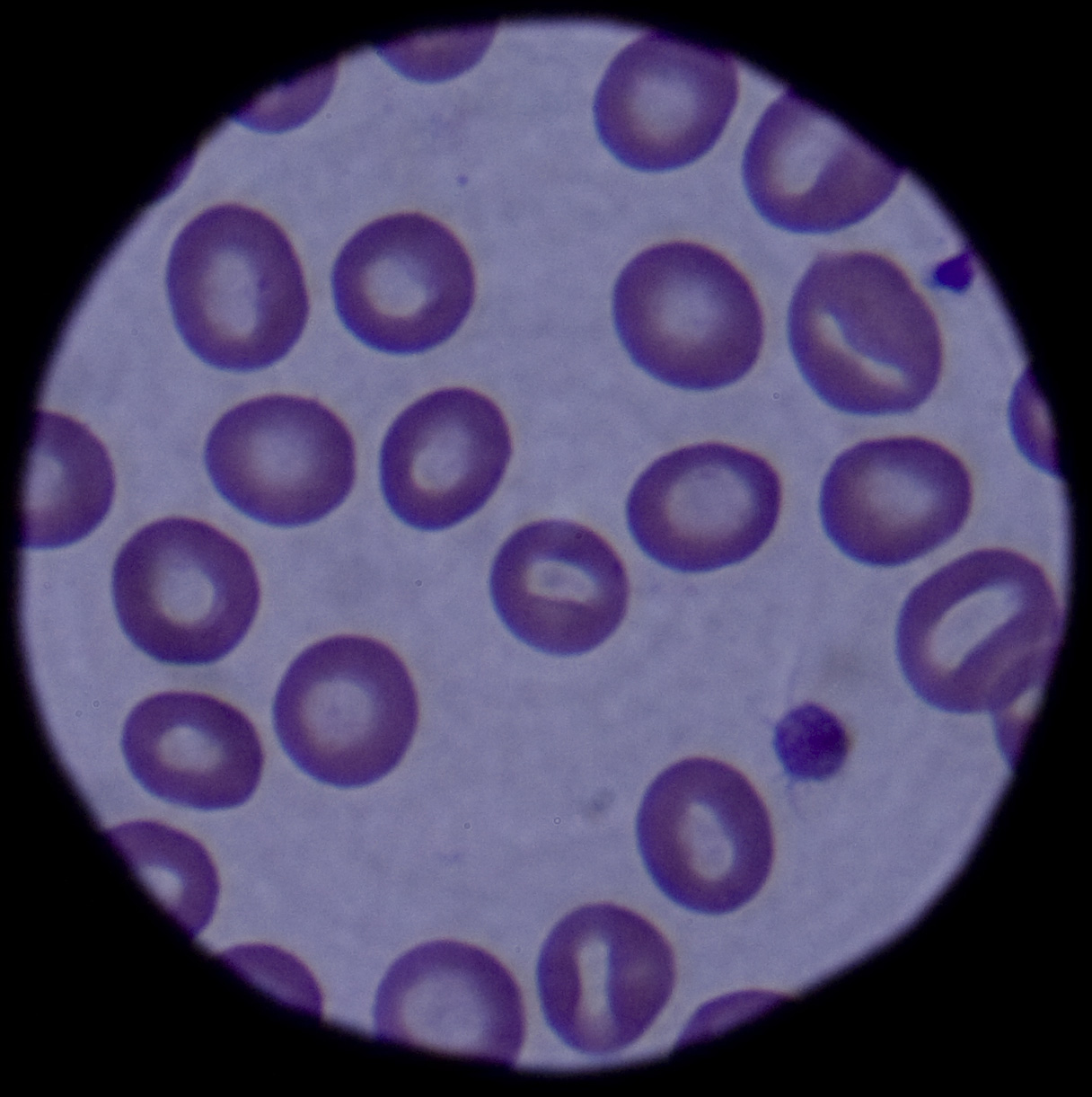
Click to see the picture

Blood cell development. A blood stem cell goes through several steps to become a red blood cell, platelet, or white blood cell.

CLICK & SEE THE PICTURES

Cancer patients may frequently develop cytopenia, a disorder in which the production of one or more blood cell types ceases or is greatly reduced. Cancer and chemotherapy used to treat cancer, and sometimes radiation therapy, may sometimes cause cytopenia.

TYPES:
A deficiency of red blood cells which  is called anemia; a deficiency of white blood cells, or leukocytes, leukopenia or neutropenia (neutrophils make up over half of all white blood cells); and deficiency of platelets is called  thrombocytopenia.

Pancytopenia is the deficiency of all three blood cell types and is characteristic of aplastic anemia, a potentially life-threatening disorder that requires a stem cell transplant.

Blood Cells
The blood consists of three different  types of cells: red blood cells (erythrocytes), white blood cells (leukocytes), and platelets. Erythrocytes contain hemoglobin, the protein that carries oxygen from the lungs to all cells in the body. Proper cell function depends on an adequate oxygen supply. When cells are oxygen deprived, organ function can be seriously impaired.

Leukocytes (white blood cells) protect the body against viral, bacterial, and parasitic infection and detect and remove damaged, dying, or dead tissues. Someone with a deficiency of white blood cells is extremely vulnerable to infection.

The term “leukocyte” refers to all six types of white blood cells; each plays a unique role in the immune system:

1. Basophils circulate in the blood and initiate the inflammatory response.
2.Eosinophils kill infecting parasites and produce allergic reactions.
3.Lymphocytes produce antibodies and regulate immune responses.
4. Mast cells are fixed in tissues and initiate the inflammatory response.
5. Monocytes capture infecting organisms for identification, ingest infecting organisms, and remove damaged or dying cells and cell debris. When monocytes become fixed in tissue, they are called macrophages.
6.Neutrophils identify and kill infecting organisms, and remove dead tissue.

Platelets are essential factors for blood clotting. Sudden blood loss triggers platelet activity at the site of the wound. Exposure to oxygen in the air causes platelets to break apart and combine with a substance called fibrinogen to form fibrin. Fibrin has a thread-like structure and forms a scab, or external clot, as it dries. Platelet deficiency causes one to bruise and bleed easily. Blood does not clot at an open wound, and there is greater risk for internal bleeding.

All blood cells have a lifespan: erythrocytes have a lifespan of about 120 days; leukocytes, 1 to 3 days; and platelets, approximately 10 days. The body continually replenishes the blood supply through a process called hematopoiesis.

Blood Cell Formation—Hematopoiesis, the formation and development of blood cells, occurs in bone marrow. Bone marrow is a nutrient-rich spongy tissue located mainly in the central portions of long flat bones (e.g., sternum, pelvic bones) in adults and all bones in infants.

All blood cells derive from blood-forming stem cells that reside in bone marrow. Stem cells replicate indefinitely and develop into mature, specialized cells. A hormone produced in the kidneys, erythropoietin, stimulates blood stem cells to produce all three types of blood cells.

CAUSES & RISK FACTORS:-

Chemotherapy and radiation therapy both reduce the number of blood-forming stem cells in cancer patients, but chemotherapeutic agents have a greater adverse effect because they suppress bone marrow function in several ways.The degree of damage is related to the particular drug(s) and the dose.

Chemotherapeutic agents can produce deficiencies in all blood cell types by

* damaging blood-forming stem cells,
* suppressing the kidneys? production of erythropoietin (hormone that stimulates blood cell production), and
* triggering red cell destruction (hemolysis) by inducing an immune response that causes the body to mistakenly identify erythrocytes as foreign bodies and destroy them.

Malignant tumors can cause anemia and other cytopenias when they directly invade bone marrow and suppress marrow function. Malignant cells also can migrate from tumors in other parts of the body to bone marrow. Tumors also can replace normal blood-forming stem cells with abnormal clones.

SIGN & SYMPTOMS:-

Anemia
A deficiency in erythrocytes reduces the amount of oxygen reaching all cells in the body, thus impairing all tissue and organ function. Severe fatigue is the most common symptom of anemia and is experienced by approximately 75% of chemotherapy patients. Patients find it more disabling than other treatment side effects, including nausea and depression.

Anemia also produces these symptoms:

* Confusion
* Dizziness
* Headache
* Lightheadedness
* Loss of concentration
* Pallor (pale skin, nail beds, gums, linings of eyelids)
* Rapid heart rate (tachycardia)
* Shortness of breath (dyspnea)

Neutropenia
Patients with a white blood cell deficiency experience frequent and/or severe bacterial, viral, and/or fungal infections; fever; and mouth and throat ulcers.

Complications—Bacteremia, the form of sepsis characterized by the presence of bacteria in the blood, can develop in immunocompromised patients who have neutropenia. Fever, rapid heart rate, and quick shallow breathing are signs of early sepsis, usually a reversible condition.

Untreated bacteremia can lead to severe sepsis, in which one or more organs become dysfunctional. Septic shock is severe sepsis with low blood pressure. The risk for death increases with the development of septic shock. Even aggressive treatment can fail to reverse the condition.

Thrombocytopenia
Platelet deficiency causes patients to bruise and bleed easily. Bleeding occurs most often in the mucous membranes lining the mouth, nose, colon, and vagina. Tiny reddish-purple skin lesions (petechiae), evidence of pinpoint hemorrhages, may appear on the skin or in the mouth.

Pancytopenia
Patients who are deficient in all blood cell types experience signs and symptoms associated with each, but bleeding from the nose and gums, and easy bruising usually appear first. Symptoms of anemia (e.g., fatigue, shortness of breath) are also common. Patients may look and feel well, otherwise, despite the seriousness of their condition.

Anemia
People with anemia (reduced red cell production) are advised to rest and eat foods high in iron (meat, fish, poultry, lentils, legumes, iron-enriched grains and flours).

If immediate remedy is necessary, treatment may include medication that helps restore the red blood supply and a transfusion of packed red blood cells.

Epoetin alpha (Epogen®, Procrit®)is a synthetic erythropoietin (normally produced by the kidneys) that stimulates stem cells to produce red blood cells. Restoration of the red blood cell supply with medication is gradual.

Darbepoetin alfa (Aranesp®) also stimulates red blood cell production but requires fewer doses and less disruption of daily living.

In March 2007, the Food and Drug Administration (FDA) issued a warning about these medications in response to studies indicating that they may increase the risk for blood clots, strokes, and heart attacks in some patients (e.g., patients who have kidney disease).

Thrombocytopenia
People with an abnormally low platelet count should avoid bruising or breaking the skin, and should carefully brush their teeth. A persistently decreased platelet count may be treated with a transfusion of platelets.

Neutropenia
The patient with a low white blood cell count is advised to  do the following:

*Avoid contact with people who are ill,
*Monitor closely for signs of infection (e.g., fever), and
*Take antibiotics when appropriate.

Medication, a colony-stimulating factor (CSF), may be prescribed to speed the development of white blood cells and shorten the period of susceptibility to infection.

Growth Factors
Growth factors are synthetic versions of substances involved in stimulating red and white blood cell production. Physicians exercise caution when prescribing these medications for people with tumors that involve the bone marrow, because growth factors might stimulate malignant cell growth.

These medications include the following:

Epoetin alpha (Procrit®, Epogen®; stimulates red blood cell production)
G-CSF (granulocyte colony-stimulating factor; e.g., filgrastim [Neupogen®]; stimulates neutrophil production)
GM-CSF (granulocyte-macrophage colony-stimulating factor; stimulates production of several white blood cells, including macrophages)

Leukocytes and other cells that contain granules are also called granulocytes.

Side effects
Fever, fatigue, dizziness, diarrhea, nausea, vomiting, weakness, and paresthesia (prickling sensation) are side effects associated with epoetin alpha.

Bone pain, malaise, headache, flu-like symptoms, muscle ache, redness at the injection site, and skin rash may occur with GM-CSF.

G-CSF commonly produces bone pain.

MEDICATIONS:-

Medications used to treat bacterial infection and other illnesses also can contribute to immune system suppression.

Some of these are :

* Antacids: cimetidine (Tagamet®)
* Antibiotics: chloramphenical (Chloromycetin®), sulfonamide (Thiosulfil®, Gantanol®); cephalosporin (Cephalaxin®), vancomycin (Vancocin®)
* Anticonvulsants: phenytoin/hydantoin (Dilantin®), felbamate (Felbatol®), carbamazepine (Tegretol®)
* Antimalarials: chloroquine (Aralin®)
* Antivirals: ganciclovir (Vitrasert®), zidovudine (AZT®)
* Cardiac drugs: diltiazem (Cardizem®), nifedipine (Procardia®), verapamil (Calan®)
* Diabetes drugs: glipizide (Glucotrol®), glyburide (Micronase®)
* Hyperthyroid drug: propylthiouracil
* NSAIDs (nonsteroidal anti-inflammatory drugs): phenylbutazone (Butazolidine®), indomethacin (Indocin®, Indochron E-R®)—Due to potentially severe gastrointestinal and cardiovascular side effects, NSAIDs should only be used as instructed.
* Rheumatoid arthritis drugs: auranofin (Ridaura®), aurothioglucose (Solganal®), gold sodium thiomalate (Myochrisine®)

Bone Marrow and Stem Cell Transplantation:-
The treatment of choice for the pancytopenic patient with a matched bone marrow donor is stem cell transplantation. The goal of transplantation is to restore blood-forming stem cells to the marrow.

Disclaimer: This information is not meant to be a substitute for professional medical advise or help. It is always best to consult with a Physician about serious health concerns. This information is in no way intended to diagnose or prescribe remedies.This is purely for educational purpose.

Resources:
http://www.oncologychannel.com/cytopenia/index.shtml
http://en.wikipedia.org/wiki/Cytopenia
http://www.cancer.umn.edu/cancerinfo/NCI/CDR378089.html

Enhanced by Zemanta
Categories
Diagnonistic Test

Percutaneous Transhepatic Cholangiography (PTCA)

[amazon_link asins=’B0006EUSVG’ template=’ProductCarousel’ store=’finmeacur-20′ marketplace=’US’ link_id=’d3ed266b-9cfe-11e7-a024-43c133717570′]


Definition:

Percutaneous transhepatic cholangiography (PTHC or PTC) is a radiologic technique used to visualize the anatomy of the biliary tract. A contrast medium is injected into a bile duct in the liver, after which X-rays are taken. It allows access to the biliary tree in cases where endoscopic retrograde cholangiopancreatography (ERCP) has been unsuccessful. Initially reported in 1937, the procedure became popular after a 1952 report in the English-language literature.

CLICK & SEE
It is an x-ray test that can help show whether there is a blockage in the liver or the bile ducts that drain it. Since the liver and its drainage system do not normally show up on x-rays, the doctor doing the x-ray needs to inject a special dye directly into the drainage system of the liver. This dye, which is visible on x-rays, should then spread out to fill the whole drainage system. If it does not, that means there is a blockage. This type of blockage might result from a gallstone or a cancer in the liver.

It is predominatly now performed as a therapeutic technique. There are less invasive means of imaging the biliary tree including transabdominal ultrasound, MRCP, computed tomography and endoscopic ultrasound. If the biliary system is obstructed, PTC may be used to drain bile until a more permanent solution for the obstruction is performed (e.g. surgery). Additionally, self expanding metal stents can be placed across malignant biliary strictures to allow palliative drainage. Percutaneous placement of metal stents can be utilised when therapeutic ERCP has been unsuccessful, anatomy is altered precluding endoscopic access to the duodenum, or where there has been separation of the segmental biliary drainage of the liver, allowing more selective placement of metal stents. It is generally accepted that percutanous biliary procedures have higher complication rates than therapeutic ERCP. Complications encountered include infection, bleeding and bile leaks.
Why the test is performed?
Bile is a by-product of protein metabolism. It is created in the liver and excreted into the intestines via the bile ducts. If bile cannot be removed from the body, it collects in the blood and is seen as a yellow discoloration of the skin and eyes (jaundice).

Also, the pancreas creates digestive fluids which drain via a common bile duct into the intestine, and thus obstruction can prevent the drainage of the fluids and may cause pancreatitis (inflammation of the pancreas).

A PTCA test can help identify whether a blockage is causing the jaundice and pancreatitis.

How do you prepare for the test?
Tell your doctor if you have ever had an allergic reaction to lidocaine or the numbing medicine used at the dentist’s office. Also tell your doctor if you could be pregnant. If you have diabetes and take insulin, discuss this with your doctor before the test.

Most people need to have a blood test done some time before the procedure, to make sure they are not at high risk for bleeding complications. If you take aspirin, nonsteroidal anti-inflammatory drugs, or other medicines that affect blood clotting, talk with your doctor. It may be necessary to stop or adjust the dose of these medicines before your test.

You will be told not to eat anything on the morning of the test so that your stomach is empty. This is a safety measure in the unlikely case you have a complication, such as bleeding, that might require repair surgery.

What happens when the test is performed?
You lie on a table wearing a hospital gown. An IV (intravenous) line is inserted into a vein in case you need medicines or fluid during the procedure. An area over your right ribcage is cleaned with an antibacterial soap. Then the radiologist may take a picture of your abdomen with an overhead camera. Medicine is injected through a small needle to numb the skin and the tissue underneath the skin in the area where the dye is to be injected. You may feel some brief stinging from the numbing medicine.

A separate needle is then inserted between two of your ribs on your right side. A small amount of xray dye is injected, and some pictures are taken that are visible on a video screen. Your doctor adjusts the placement of the needle until it is clear that the dye is flowing easily through the ducts (drainage tubes) inside your liver.

Because taking the x-ray pictures sometimes requires a significant amount of time, the doctor replaces the needle with a softer plastic tube. First, the syringe holding the dye is detached from the top of the needle, leaving the needle in place. The doctor then gently pushes a thin wire through the needle and into the duct where the needle has been sitting. Next the needle is pulled out, sliding over the outside end of the wire. The wire is left with one end inside the liver to hold the position where the needle had been. A thin plastic tube similar to an IV line is slid along the wire, like a long bead on a string, until it is in the same place where the needle was. The wire is then pulled out, and the dye syringe is attached to the tube.

More dye is injected through the plastic tube, and pictures are taken with the video camera as the dye spreads inside the liver. If there is no blockage, the dye drains out of the liver through the bile ducts and begins to show up on the x-ray in the area of your small intestine. Once all of the needed pictures have been taken, the plastic tube is pulled out, and a small bandage is placed over your side. The whole test usually takes less than an hour.

Risk Factors:
It is possible to have serious bleeding from this test. In some cases, blood leaks to the outside surface of the liver and causes a buildup of blood there. In other cases, blood can leak directly into the liver’s drainage system, in which case it might start showing up in your intestine, causing a bloody bowel movement. It is less likely that you could develop an infection after the test. The only soreness you are likely to have is at the skin surface where the needle went in. This should last for only a day or two.

In rare cases, the dye used in the test can damage your kidneys. This kidney effect is almost always temporary, but some people have permanent damage.

As with all x-rays, there is a small exposure to radiation. In large amounts, exposure to radiation can cause cancers or (in pregnant women) birth defects. The amount of radiation from the video x-ray in this test is very small-too small to be likely to cause any harm. (The people performing the test on you will wear lead shields, since they would otherwise be exposed to this radiation over and over, which could be more of a danger.)

Must you do anything special after the test is over?
Call your doctor right away if you have pain in your right abdomen or shoulder, fever, dizziness, or a change in your stool color to black or red.

How long is it before the result of the test is known?
You may be told a few early results of your test as soon as the test is done. It takes a day or two for the radiologist to review the x-rays more thoroughly and to give your doctor a full report.

RESULTS:-

Normal Result:-The bile ducts are normal in size and appearance for the age of the patient.

Abnormal Results:-The results may show that the ducts are enlarged, which may indicate the ducts are blocked. The blockage may be caused by infection, scarring, or stones. It may also indicate cancer in the bile ducts, liver, pancreas, or region of the gallbladder.

You may click & See:
*Blocked bile ducts
*Cholangitis (infection in common bile duct)
Special considerations:-
A PTCA may be done if an endoscopic retrograde cholangiopancreatography ( ERCP) cannot be performed or has failed in the past.

An MRCP (magnetic resonance cholangiopancreatography) is a newer, non-invasive imaging method, based on MRI, which provides similar views of the bile ducts.

Resources:
https://www.health.harvard.edu/fhg/diagnostics/percutaneous-transhepatic-cholangiography.shtml
http://en.wikipedia.org/wiki/Percutaneous_transhepatic_cholangiography
http://www.healthline.com/adamcontent/percutaneous-transhepatic-cholangiogram

Reblog this post [with Zemanta]
css.php