Categories
Diagnonistic Test

Bone Marrow Biopsy

[amazon_link asins=’0443081107,B006NO3EZK,0340740892,B017MDO9MC,B004GXATC6,3805538634,3642622968,0497001667,B00ET6DRV0′ template=’ProductCarousel’ store=’finmeacur-20′ marketplace=’US’ link_id=’b7bb6803-454d-11e7-b04d-afa9c178e4cb’]

Introduction: Bone marrow is the spongy material found in the center of most large bones in the body. The different cells that make up blood are made in the bone marrow. Bone marrow produces red blood cells, white blood cells, and platelets. Along with a biopsy (the sampling of mostly solid tissue or bone), an aspiration (the sampling of mostly liquid) is often done at the same time.

…………….....CLICK & SEE

Doctors can diagnose many problems that cause anemia, some infections, and some kinds of leukemia or lymphoma cancers by examining a sample of your bone marrow (the tissue where blood cells are made). A bone marrow biopsy is the procedure to collect such a sample. It is done using a large needle inserted through the outside surface of a bone and into the middle of the bone, where the marrow is.

Why the procedure is performed: A bone marrow aspiration and biopsy procedure is done for many reasons.

*The test allows the doctor to evaluate your bone marrow function. It may aid in the diagnosis of low numbers of red blood cells (anemia), low numbers of white blood cells (leukopenia), or low numbers of platelets (thrombocytopenia), or a high number of these types of blood cells.

*The doctor can also determine the cause of some infections, diagnose tumors, determine how far a disease, such as lymphoma, has progressed, and evaluate the effectiveness of chemotherapy or other bone marrow active drugs.

*Where the procedure is performed: Bone marrow aspirations and biopsies can be performed in doctor’s offices, outpatient clinics, and hospitals. The procedure itself takes 10-20 minutes.

Preperation for the test:
You will need to sign a consent form giving your doctor permission to perform this test. Because you will probably receive some pain medicines or anti-anxiety medications that can make you drowsy, you will need to arrange a ride home.

Tell your doctor if you have ever had an allergic reaction to lidocaine or the numbing medicine used at the dentist’s office. Also talk with your doctor before the test if you are taking insulin, or if you take aspirin, nonsteroidal anti-inflammatory drugs, or other medicines that affect blood clotting. It may be necessary to stop or adjust the dose of these medicines before your test. Most people need to have a blood test done some time before the procedure to make sure they are not at high risk for bleeding complications.

*You may receive instructions about not eating food or drinking liquids before the procedure.

*Be sure to tell your doctor about any prescription medications, over-the-counter medications, as well as herbal supplements you are taking.

*Notify your doctor about all allergies, previous reactions to medications, if you have had any bleeding problems in the past, or if you are pregnant.

*Before the procedure, you will be asked to change into a patient gown.

*Your vital signs-blood pressure, heart rate, respiratory rate, and temperature-will be measured.

*Depending on your doctor, you may have an IV placed or your blood drawn.

*You may be given some medicine to help you relax.

*You may be asked to position yourself on your stomach or your side depending on the site the doctor chooses to use.

Risk Factors:
You will be asked to sign a consent form before the procedure. You will be notified of the alternatives as well as the potential risks and complications of this procedure.

Risks are minimal.

Possible risks include these:

*Persistent bleeding and infection

*Pain after the procedure

*A reaction to the local anesthetic or sedative

Having a sample taken is not harmful for your bone or bone marrow. Injury of nearby tissue from the biopsy is very uncommon. You might have some buttock soreness for a few days, and you may have some bruising at the biopsy site. A few individuals have an allergy or a side effect from the pain medicine or anti-anxiety medicine.

What happens when the test is performed?
Most patients have this test done by a hematologist in a clinic procedure area. You wear a hospital gown during the procedure. A sedative may be injected at this time. (If you are prescribed a sedative in pill form, you will be instructed to take it ahead of time.)

*Most patients have bone marrow sampled from the pelvis. You lie on your stomach and the doctor feels the bones at the top of your buttock. An area on your buttock is cleaned with soap. A local anesthetic is injected to numb the skin and the tissue underneath the skin in the sampling area. This causes some very brief stinging.

*The doctor will choose a place to withdraw bone marrow. Often this is the hip (pelvic bone), but it also can be done from the breastbone (sternum), lower leg bone (tibia), or backbone (vertebra).

*The chosen site will be cleaned with a special soap (iodine solution) or alcohol. After the skin is clean, sterile towels will be placed around the area. It is important that you do not touch this area once it has become sterile.

*Local anesthetic, usually lidocaine, will be injected with a tiny needle at the site. Initially, there may be a little sting followed by a burning sensation. After a few minutes, the site will become numb. A needle is then placed through the skin and into the bone. You may feel a pressure sensation.

*For the bone marrow aspiration, a small amount of bone marrow is then pulled into a syringe.

*A bone marrow biopsy is then usually performed. A somewhat larger needle is then put in the same place and a small sample of bone and marrow is taken up into the needle.

*After taking the liquid sample, the doctor carefully moves the needle a little bit further into the bone marrow to collect a second sample of marrow called a core biopsy. This core biopsy is a small solid piece of bone marrow, with not just the liquid and cells but also the fat and bone fibers that hold them together. After the needle is pulled out, this solid sample can be pushed out of the needle with a wire so that it can be examined under a microscope. Pressure is applied to your buttock at the biopsy location for a few minutes, until you are not at risk of bleeding. A bandage is placed on your buttock.
Must you do anything special after the test is over?
You will feel sleepy from the medicines used to reduce pain and anxiety.
After the local anesthetic wears off over the next few hours, you may have some discomfort at the biopsy site. Your doctor will advise you about pain medication.Once these medicines have worn off (a few hours after the test), you can return to normal activities, but you should not drive or drink alcohol for the rest of the day.

You should keep the bandage on for 48 hours, and then it should be removed.

After the test:
The samples taken from your bone marrow will be sent to a laboratory and the pathologist for analysis. Several tests are done including looking at the bone marrow under a microscope. The results of these tests will usually be available in a few days. Your doctor will give you instructions for follow-up.

When to Seek Medical Care:
Call your doctor if you notice signs of spreading redness, continued bleeding, fever, worsening pain, or if you have other concerns after this procedure.

Go to a hospital’s emergency department if these conditions develop:

*If your bleeding will not stop with direct pressure
*If you see thick discharge from the wound
*If you have a persistent fever
*If you feel lightheaded

How long is it before the result of the test is known?
Some parts of your bone marrow biopsy report may be available within a day, but some tests require special stains or tests that can take longer, in some cases up to one week.

Resources:
https://www.health.harvard.edu/fhg/diagnostics/bone-marrow-biopsy.shtml
http://www.emedicinehealth.com/bone_marrow_biopsy/article_em.htm

Categories
Diagnonistic Test Health Problems & Solutions

Cardiac Catheterization

[amazon_link asins=’0323340393,1451127405,0323476716,1605478857,081512614X,1572225378,032308057X,1416039996,0323079024′ template=’ProductCarousel’ store=’finmeacur-20′ marketplace=’US’ link_id=’62c28cfe-b0f6-11e7-aa33-4b0807246ed5′]

Alternative Name: Catheterization – cardiac; Heart catheterization

Definition:
Cardiac catheterization with coronary angiogram takes pictures of the blood vessels in your heart, to evaluate the health of your heart and detect any narrowing of the blood vessels or other problems. The catheterization is performed by a cardiologist (or sometimes a radiologist) who is a specialist in doing this type of test.
CLICK & SEE
This process involves passing a catheter (a thin flexible tube) into the right or left side of the heart. In general, this procedure is performed to obtain diagnostic information about the heart or its blood vessels or to provide treatment in certain types of heart conditions.

Cardiac catheterization can be used to determine pressure and blood flow in the heart’s chambers, collect blood samples from the heart, and examine the arteries of the heart with an x-ray technique called fluoroscopy. Fluoroscopy provides immediate (“real-time”) visualization of the x-ray images on a screen and provides a permanent record of the procedure.
Why the Test is Performed ?
Cardiac catheterization is usually performed to evaluate heart valves, heart function and blood supply, or heart abnormalities in newborns. It may also be used to determine the need for heart surgery.

Therapeutic catheterization may be used to repair certain types of heart defects, open a stenotic heart valve, and open blocked arteries or grafts in the heart.
How the Test is Performed:
You lie on your back as a medical technician connects you to a heart monitor. An intravenous (IV) line is inserted into one of the blood vessels in your arm, neck, or groin after the site has been cleansed and numbed with a local anesthetic.. You may be given a sedative through the IV so that you are relaxed during the test.

First, the doctor injects a local anesthetic into the skin. This might sting momentarily. After the skin is numb, the cardiologist inserts a catheter (a thin, hollow plastic tube) into a large artery-usually in your groin but possibly in your arm or wrist.

Using live x-rays displayed on a video monitor as a guide, your doctor moves the catheter along the artery until it reaches your aorta (the large blood vessel that carries blood from your heart to the rest of your body). The tip of the catheter is pushed up the aorta until it reaches the heart and then gently pushed into the coronary arteries that supply blood directly to your heart muscle.When the tip of the catheter reaches one of the coronary arteries, the doctor injects contrast dye through the catheter. The dye illuminates the artery, allowing the doctor to see if it is blocked or narrowed. The procedure is repeated to visualize the other coronary arteries.

X-ray pictures are taken while the dye travels down the arteries. The arteries look like thick lines on the x-ray; a narrowing or blockage in an artery appears as a thinner line (see Figure 1). Your doctor might also inject some contrast medium into the left ventricle of your heart to show how forcefully your heart is pumping. The entire procedure usually takes from one to several hours.

How you Prepare for the Test:
Food and fluid are restricted 6 to 8 hours before the test. The procedure takes place in the hospital and you will be asked to wear a hospital gown. Sometimes, admission the night before the test is required. Otherwise, you will be admitted as an outpatient or an inpatient the morning of the procedure.

Your health care provider should explain the procedure and its risks. A witnessed, signed consent for the procedure is required.

Tell your doctor if you are allergic to seafood, if you have had a bad reaction to contrast material in the past, if you are taking Viagra, or if you might be pregnant.

During this procedure, local anesthetics (numbing agents) are used to minimize pain. Tell the cardiologist if you have ever had an allergic reaction to a local anesthetic or to contrast dyes. Also let your doctor know if you could be pregnant, since the x-rays used during this procedure can damage a fetus.

. Tell the cardiologist if you’re taking a nonsteroidal anti-inflammatory drug (NSAID) or other medicines that affect blood clotting and could increase the chance of bleeding from the procedure. You should also tell your doctor if you take insulin shots or blood sugar-lowering pills so that you can take steps to avoid dangerously low blood sugar, or hypoglycemia.

How the Test Will Feel?
The study is carried out in a laboratory by a trained cardiologist or radiologist and technicians or nurses.

You will be awake and able to follow instructions during the catheterization. A mild sedative is usually given 30 minutes before the procedure to help you relax. The procedure may last from 1 to several hours.

You may feel some discomfort at the site where the IV is placed. Local anesthesia will be used to numb the site, so the only sensation should be one of pressure at the site. You may experience some discomfort from having to remain still for a long time.

After the test, the catheter is removed. You might feel a firm pressure at the insertion site, used to prevent bleeding. If the IV is placed in your groin, you will usually be asked to lie flat on your back for a few hours after the test to avoid bleeding. This may cause some mild back discomfort.

Risk Factors:
There are several potential risks. First, the catheter can irritate the heart, in rare cases causing a disturbance in the heart rhythm. Should this happen, the doctor can immediately use devices and medicines to restore a normal heart rhythm. The catheter occasionally can cause the coronary artery to go into spasm, temporarily reducing the blood flow and causing chest pain. For this reason, alert the doctors and nurses if you develop any chest discomfort, trouble breathing, or any other problem during the test.

In addition, the contrast medium can sometimes impair kidney function. This effect is almost always temporary, but some people have permanent damage. Another possible complication is bleeding at the place where the catheter was inserted. If blood collects under the skin, it can form a large painful bruise called a hematoma. This usually resolves on its own, without requiring additional treatment. Occasionally, people are allergic to the contrast dye and develop a rash, hives, or difficulty breathing after the dye is injected. If this should occur, the medical staff in the catheterization laboratory have medicines available to treat the allergic reaction.

The amount of radiation from this test is too small to be likely to cause harm.
Cardiac catheterization carries a slightly increased risk when compared with other heart tests. However, the test is very safe when performed by an experienced team.

Generally, the risk of serious complications ranges from 1 in 1,000 to 1 in 500. The risks include the following:
*Cardiac arrhythmias
*Cardiac tamponade
*Trauma to the artery caused by hematoma
*Low blood pressure
*Reaction to contrast medium
*Hemorrhage
*Stroke
*Heart attack
Must you do anything special after the test is over?
You should lie flat for a few hours after this procedure. Often, a small plug or stitch is used to prevent bleeding from the artery that was entered to perform the catheterization. If you received a sedative, you might feel sleepy and shouldn’t drive or drink alcohol for one day after the catheterization.

What Abnormal Results Mean

The procedure can identify heart defects or disease, such as coronary artery disease, valve problems, ventricular aneurysms, or heart enlargement.

The procedure also may be performed for the following:
*Primary pulmonary hypertension
*Pulmonary valve stenosis
*Pulmonary embolism
*Tetralogy of Fallot
*Transposition of the great vessels
*Tricuspid regurgitation
*Ventricular septal defect

How long is it before the result of the test is known?
Your doctor will have your results as soon as the test is completed. In particular, the doctor can tell you if you have any blockages in the coronary arteries, how many and how severe they are, and the best way to treat them. In some cases, your doctor is even able to remove any blockages immediately by performing an intervention known as a coronary angioplasty, a procedure that uses a tiny inflatable balloon to reopen the artery.

Resources:
https://www.health.harvard.edu/fhg/diagnostics/cardiac-catheterization.shtml
http://www.nlm.nih.gov/medlineplus/ency/article/003419.htm

Reblog this post [with Zemanta]
Categories
Diagnonistic Test

Pleural Fluid Sampling (or Thoracentesis)

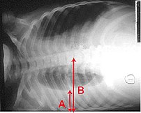
Pleural effusion Chest x-ray of a pleural effu...
Image via Wikipedia

[amazon_link asins=’B01J8WALE0,B00GNOPG8S,1331951720,1893441776,B01MXOPZIB,B01J2IC12U,B01J37411E,B009KT2GWM,B01J2GLUVU’ template=’ProductCarousel’ store=’finmeacur-20′ marketplace=’US’ link_id=’316e2df4-5fab-11e7-aab1-4db380a805de’]


Definition:
Thoracentesis is a procedure used to obtain a sample of fluid from the space around the lungs. Normally, only a thin layer of fluid is present in the area between the lungs and chest wall (show radiograph 1). However, some conditions can cause a large amount of fluid to accumulate. This collection of fluid is called a pleural effusion (show radiograph 2). Thoracentesis is done to collect a sample of the fluid, which can help determine why the pleural effusion developed.
CLICK & SEE
Some infections and diseases cause fluid to accumulate in the space between the lung and the rib cage or between the lung and the diaphragm. This collection of fluid is called a pleural effusion. A pleural effusion might be detected on a chest x-ray. Sampling this fluid is important because it enables doctors to understand what caused the fluid to collect and how to treat the problem. The fluid can be sampled with a needle.

Reasons for Thoracentesis: — A thoracentesis is performed to determine the cause of a pleural effusion. In some cases, a physician may perform thoracentesis to relieve symptoms caused by the pleural effusion, including shortness of breath and low blood oxygen levels. A pleural effusion may be detected during a physical examination or on a chest x-ray.

Pleural effusion can be caused by many different conditions, including infections, heart failure, cancer, or tuberculosis. In some cases, blood or other fluid may be leaking into the pleural space from another part of the body, causing the effusion. By examining the fluid and the types of cells it contains, the cause of the effusion can usually be determined.

In general, there is no reason a thoracentesis cannot be performed. The procedure is more easily performed and complications are fewer when the pleural effusion is large. Special consideration may be necessary in patients who are on respirators.

Patients who have a bleeding disorder or who are on medications that affect blood clotting may need extra care to minimize the risk of bleeding. Patients should tell their healthcare provider if they have a history of bleeding problem or if they are taking medicine that decreases blood clotting. In some cases, a blood test will be taken prior to the procedure to exclude any blood clotting abnormalities caused by disease or medications.

Procedure: A thoracentesis involves the following steps:

*The patient will be placed in a position that allows the doctor to easily access the effusion. Usually, the patient is asked to sit upright during the procedure. It is important to remain still during the procedure so that the fluid does not shift.

*The skin is cleaned with an antibacterial solution in the area where the needle will be inserted.

*A small amount of numbing medicine (a local anesthetic, similar to novocaine) is injected into the area. This medicine helps minimize discomfort during the procedure.

*A slightly larger needle is inserted in the same location. A syringe is attached to this needle and is used to withdraw fluid from around the lung. Patients who have symptoms from the effusion (eg, shortness of breath) may have a large amount of fluid removed, which allows the lung to re-expand.

*The needle is removed and pressure is briefly applied to the insertion site.

How do you prepare for the test?
You will need to sign a consent form giving your doctor permission to perform this test. Some patients have this test done in a doctor’s office, while others are admitted to the hospital for it. Generally your doctor will decide whether you need to be in the hospital based on your medical condition. A chest x-ray or an ultrasound is done before the procedure.

Tell your doctor if you have ever had an allergic reaction to lidocaine or the numbing medicine used at the dentist’s office. If you take aspirin, nonsteroidal anti-inflammatory drugs, or other medicines that affect blood clotting, talk with your doctor. It may be necessary to stop or adjust the dose of these medicines before your test.

What happens when the test is performed?
You wear a hospital gown and sit on a bed or table leaning forward against some pillows. The doctor listens to your lungs with a stethoscope and may tap on your back to find out how much fluid has collected.

Soap is used to disinfect an area of skin on one side of your back. A small needle is used to numb a patch of skin between two of your lower ribs. The numbing medicine usually stings for a second. A needle on an empty syringe is then inserted into the fluid pocket. Usually this pocket is around one inch below the skin surface. You might feel some minor pressure as the needle is inserted. Depending on the quantity of fluid that the doctor plans to remove, either the syringe itself is filled or soft plastic tubing is used to remove fluid into a collection bag or jar. While the doctor is attaching the tubing, he or she might ask you to hum out loud. This humming is for your safety: It prevents you from taking a deep breath, which could expand your lung, causing it to touch the needle.

It sometimes takes 15 minutes or longer to remove the necessary amount of fluid. Most patients feel no discomfort during this time, although a few patients feel some chest pain at the end of the procedure as their lung expands and touches the chest wall. After the fluid is removed, a bandage is placed on your back.

Risk Factors:
This procedure carries a few serious risks, but most patients have no complications. If the needle touches the lung it may create an air leak, which is seen on the x-ray and might require you to stay in the hospital for a few days. Some patients with this complication need to have a plastic tube (called a chest tube) inserted between two ribs. The tube uses vacuum pressure to keep the lung expanded until it has healed.

In most cases, a thoracentesis is performed without complications. Most complications are minor and resolve on their own or are easily treated. Potential complications include the following:

*Pain — Some discomfort may occur when the needle is inserted. Using a local anesthetic helps to reduce the pain. Pain generally resolves once the needle is removed.

*Bleeding — A blood vessel may be nicked as the needle is inserted through the skin and chest wall, causing bleeding. The bleeding is usually minor and stops on its own, although it may cause bruising around the puncture site. In rare cases, bleeding into or around the lung may occur, requiring drainage or surgery.

*Infection
Infection can occur if bacteria are introduced by the needle puncture. Using disinfectant solution to clean the area and using sterile technique during the procedure minimizes this risk.

*Pneumothorax or collapsed lung Occasionally, the needle used to obtain a fluid sample can puncture the lung. The hole created by the puncture usually seals quickly on its own. If it does not, air can build up around the lung, causing the lung to collapse. This is called a pneumothorax. When a pneumothorax occurs, a chest tube may be used to drain the air and allow the lung to re-expand.

A pneumothorax may also occur if the lung fails to expand when fluid is withdrawn. This is considered to be a drainage-related pneumothorax, and is the most common type of pneumothorax to occur when ultrasound is used for needle placement. Drainage-related pneumothorax is most commonly caused by disorders of the surface lining of the lung and not by the puncture needle. Treatment is rarely needed.

Pneumothorax occurs in less than 12 percent of procedures. Those that do occur are usually small and resolve on their own. A chest tube to helps re-expand the lung is necessary only if the pneumothorax is large, continues to expand, or causes symptoms.

*Liver or spleen puncture — In very rare cases, the liver or spleen may be punctured during thoracentesis. Sitting upright and remaining still during the procedure helps to keep the liver and spleen away from the insertion area and minimizes the risk of this complication.

Must you do anything special after the test is over?
You will need to have an x-ray taken after the sampling is completed. Your breathing should feel the same (or better) after the procedure.

How long is it before the result of the test is known?
The fluid may be tested for a variety of things, including infection and cancer. Cells in the fluid will be examined. It may be several days before full results are available.

Where you may get more information:-Your healthcare provider is the best source of information for questions and concerns related to your medical problem. Because no two patients are exactly alike and recommendations can vary from one person to another, it is important to seek guidance from a provider who is familiar with your individual situation.

This discussion will be updated as needed every four months on our web site (www.uptodate.com/patients). Additional topics as well as selected discussions written for healthcare professionals are also available for those who would like more detailed information.

Some of the most pertinent include:
Professional  Level Information:-
Diagnostic thoracentesis 
An overview of medical thoracoscopy
Diagnostic evaluation of a pleural effusion in adults
Imaging of pleural effusions in adults
Management of malignant pleural effusions

A number of web sites have information about medical problems and treatments, although it can be difficult to know which sites are reputable. Information provided by the National Institutes of Health, national medical societies and some other well-established organizations are often reliable sources of information, although the frequency with which they are updated is variable.

*American Thoracic Society
(www.thoracic.org)

*American Lung Association
(lungusa.org)

*National Heart Lung & Blood Institute
(www.nhlbi.nih.gov/index.htm)

*National Library of Medicine
(www.nlm.nih.gov/medlineplus/healthtopics.html)

Resources:
https://www.health.harvard.edu/diagnostic-tests/pleural-fluid-sampling.htm
http://www.uptodate.com/patients/content/topic.do?topicKey=~0aPG4xpnulisDf

Reblog this post [with Zemanta]
Categories
Ailmemts & Remedies Featured

Ankle Sprain? Fret not

Ankle joints, undependable as they are, can easily slip. As joints they are fragile, and, unlike the hip, are not surrounded by protective strong muscles. Instead the bones are held in place by ligaments — the rope-like ends of muscles — that are not as strong. They are also more prone to displacement, stretching and tears. This means around 25,000 people sprain their ankles every day just in the US.
CLICK & SEE THE PICTURES
The ankle is a hinge joint, meant to move upwards and downwards. If torsion or twisting movement occurs accidentally, the ligaments fail to hold. The ankle can then be forced out of its normal position. As the body is unprepared, the joint can “give way” resulting in a sprain.

The entire weight of the body rests on this weak pair of ankle joints. Acute injury usually means that the person either falls or hobbles away painfully.

Age is no bar, and both the young and old can “sprain” their ankles. An ankle sprain is more likely to occur if:

• The footwear is unsuitable, with hard unyielding soles, or spiky or rocker-bottom heels.

The foot is placed awkwardly while landing after a jump.

• A fall causes the ankle to twist.

• One walks or exercises on an uneven surface resulting in loss of balance.

• A person is overweight.

• The ankle has been injured before and is unstable, causing a repetition of the injury.

If the sprain is mild, after a few painful minutes it is possible to put the foot back on the ground and walk. With a more severe injury, the foot may be swollen. There may be a bluish red discolouration over the joint. Weight bearing may cause such excruciating agony that it is impossible to walk. If there is a popping sound or weight bearing is impossible, the ligament may have been torn. In the process, a small piece of bone may have been dislocated or chipped off.

In many mild sprains “home remedies” work wonders. This consists of PRICE — Protection, Rest, Ice Compression and Elevation:

• Immediately apply ice. It reduces the swelling and inflammation, reduces muscle spasm and numbs pain.

• Protect the joint from further dislocation with elastocrepe bandages and an ankle support. These are available in medical stores.

• Avoid bearing weight by using a crutch or leaning on a cane.

• Rest as much as possible.

• Elevate the leg so that the ankle is above the level of the heart. Keep it like that as long as possible. This will reduce the swelling.

• Take mild painkillers like Aspirin, or NSAIDs (non-steroidal anti-inflammatory agents) like Ibuprofen.

If the sprain is mild, with these measures the pain will disappear in three days. If discomfort persists, it is better to consult a physician, as the ligaments may be torn or the bone chipped. An X-ray or a scan may be needed to evaluate the joint.

The first stage of recovery takes around a week. The ankle should be rested as much as possible. For the next two weeks, passive exercises should be done to increase the strength and restore flexibility. This is very important because if the ligaments are not moved they can become stiff and fixed.

Plaster of Paris (POP) can be applied as a cast if the sprain is severe. It keeps the joint fixed and helps rapid recovery. Casts can also be made with new lightweight materials and fitted to facilitate walking.

Surgery is rarely required, but can be performed if pain and instability persist after months of using elastocrepe bandages and exercises. Arthroscopy can be done with a scope to visualise the joint and remove any loose fragments of bone or cartilage. If the ligaments are badly damaged, reconstruction of the joint can be undertaken.

Athletes sometimes tape their ankles or wear an elastic guard prior to exercise to prevent a sprain from occurring. This is not effective unless the joint is already known to be unstable.

If the sprain is unrecognised or neglected, the joint can become chronically unstable and troublesome. A sprain is diagnosed as chronic if it lasts for more than four weeks. This is because by then the muscles have become weak and the ligaments lax. This predisposes the joint to instability and repeated injury.

The best way to prevent ankle sprains is to maintain good strength, muscle balance and flexibility.

• Don’t start exercising with “cold” stiff muscles. Warm ups are essential to prevent dislocations.

• Run in well-lit areas. That way, uneven surfaces, stones and obstructions can be seen well.

• Pay attention to footwear. Wear sports shoes for games. Formal stilettos, spiky heels and elevated shoes can cause loss of balance and are better avoided.

• If the ankle suddenly pains, stop the activity and check to see what is wrong.

To learn more You may click to-> ……………………….(1)....(2).….(3)……(4)

Sources: The Telegraph (Kolkata, India)

Categories
Herbs & Plants

Feverfew

[amazon_link asins=’B00020HR1I,B0083HAPL6,B0153IQXR2,B00028PJ2E,B0002IHF6M,B0016B9Z44,B003TXNVZQ,B0009EXLE4,B0013OVXWQ’ template=’ProductCarousel’ store=’finmeacur-20′ marketplace=’US’ link_id=’50302d4c-47a5-11e7-aa6b-b5c09c5b1b74′]

Botanical Name:Tanacetum parthenium
Family: Asteraceae
Kingdom: Plantae
Division: Magnoliophyta
Class: Magnoliopsida
Order: Asterales
Genus: Tanacetum
Species: T. parthenium
Other Names: Altamisa, Amargosa, Bachelor’s Button, Feverfew, Flirtwort, Manzanilla, Featherfew, Featherfoil, Wild Chamomile

synonyms: Chrysanthemum parthenium (L.) Bernh. and Pyrethrum parthenium (L.) Sm.

Parts Used: Leaves and flowers in extract, infusion, and dried in capsules.
Habitat: Native to southwest Europe and was brought to America originally as an ornamental. It is commercially cultivated in Japan, Africa and Europe. Greek and European herbalists traditionally used it to reduce fevers.

Description:    Feverfew is a hearty perennial that will produce an abundant supply of blossoms. It prefers full sun or partial shade and well-drained average soil.Feverfew is a traditional medicinal herb which is found in many old gardens, and is also occasionally grown for ornament. The plant grows into a small bush up to around 18 inches high, with citrus-scented leaves and is covered by flowers reminiscent of daisies. It spreads rapidly, and they will cover a wide area after a few years.

You may click to see  pictures of Feverfew

The leaves have a refreshing aromatic aroma. Growing to 2 1/2 feet the stem is upright, erect, hairy, finely furrowed and branching. Strongly aromatic leaves are alternate, hairless, toothed, light green about 4 inches long, and divided into broad, lobed segments. The lower leaves are bipinnate with oval shaped leaflets. Many daisy-like flower heads (composite) bloom June-August, with white ray flowers surrounding nearly flat yellow centers, growing to about 1 inch across. Gather entire plant in bloom, dry for later use.

Cultivation: A very easily grown plant, it succeeds in an ordinary garden soil, plants can even be grown in walls.

Medicinal Uses:   Feverfew is edible and medicinal. has a good reputation as alternative medicine and extensive research has proved it to be of special benefit in the treatment of certain types of migraine headaches and rheumatism or arthritis. The plant is rich in sesquiterpene lactones, the principal one being parthenolide. Parthenolide helps prevent excessive clumping of platelets and inhibits the release of certain chemicals, including serotonin and some inflammatory mediators. Constituents of Feverfew are Volatile oils, containing pinene and several pinene derivatives, bornylacetate and angelate, costic acid, b-farnesine and spiroketalenol ethers. Other constituents include essential oils, flavonoid glycosides, pinene derivatives and costic acid. Feverfew should be taken regularly to receive maximum benefit and protection from migraines. The leaves and flowering heads are antiinflammatory, antispasmodic, aperient, bitter, carminative, emmenagogue, sedative, stimulant, stomachic, vasodilator and vermifuge. An infusion made from the whole plant is used in the treatment of arthritis, colds, fevers, as a sedative and to regulate menses. Also used as a foot bath for swollen feet. Applied externally as a tincture, the plant is used in the treatment of bruises. Chewing several leaves a day has proven to be effective in preventing some migraine headaches. Feverfew’s sedative properties make it useful in hysterical complaints, nervousness, low spirits, and is a general tonic. Also said to be good as a syrup for coughs, wheezing and breathing difficulties. The dried flower buds are said to have the same properties as pyrethrum, and used as an insecticide. An essential oil from the plant is used in perfumery.

Parthenion is the Greek word for girl. Feverfew is Elizabethan English and comes from febrifuge, an old medical term for a medicine that reduces fever. Feverfew is an effective remedy for migraine. Parthenolide appears to inhibit the release of the hormone serotonin that triggers migraine. It has also been shown to reduce fever, hence the name Feverfew.

Feverfew has been used for reducing fever, for treating headaches, arthritis and digestive problems. It is hypothesized that by inhibiting the release of serotonin and prostaglandins, both of which are believed to aid the onset of migraines, feverfew limits the inflammation of blood vessels in the head. This would, in theory, stop the blood vessel spasm which is believed to contribute to headaches. The active ingredients in feverfew include parthenolide and tanetin. Capsules or tablets of feverfew generally contain at least 205 mcg. parthenolide; however, it might take four to six weeks before they become effective, and feverfew is not a remedy for acute migraine attacks. Parthenolide has also been found recently in 2005 to induce cell death in leukemia cancer stem cells.

Recently, feverfew has been used by Aveeno skincare brand to calm red and irritated skin.

The herb has a long history of use in traditional and folk medicine as a treatment for disorders often controlled by aspirin, such as fever, headaches and some of the accompanying symptoms such as nausea and depression.

Recently feverfew has been gaining fame as a effective treatment for migraine headaches. It may also help ease diseases caused by chronic inflammation such as arthritis. It is an aromatic plant with a strong and lasting odor, it has been used externally as an insect repellent and for treating insect bites.

It is the combination of ingredients in the feverfew plant that brings such effective relief. It works to inhibit the release of two inflammatory substances, serotonin and prostaglandins, both believed to contribute to the onset of migraines. By inhibiting these amines as well as the production of the chemical histamine, the herb controls inflammation that constricts the blood vessels in the head, and prevents blood vessel spasms which may contribute to headaches.

The plant is rich in sesquiterpene lactones, the principal one being parthenolide. Other constituents include essential oils, flavonoid glycosides, pinene derivatives and costic acid. Feverfew should be taken regularly to receive maximum benefit and protection from migraines.

The tea, drunk cold, may also relieve skin perspiration associated with migraines, and has been used to stimulate appetite, and improve digestion and kidney function.

Clinical tests have shown the use of feverfew may reduce of frequency and severity of headaches. It may be more effective than other nonsteroidal antiinflammatories (NSAIDS), like aspirin. Additional benefits include lower blood pressure, less stomach irritation and a renewed sense of well-being.

It may also relieve dizziness, tinnitus, and painful or sluggish menstruation. Its extracts have been claimed to relieve asthma, coughs, dermatitis and worms.

Common Use: The herb has historically been used as remedy for headache, inflammation and as a general substitute for ailments treated with aspirin. Its most popular use is for the prevention of migraine headaches and associated symptoms. Pregnant women should not use the herb, and some people have developed mouth ulcers or experienced loss of taste from eating the fresh leaves.

The herb has been used since Roman times to induce menstruation. It is given in difficult births to aid expulsion of the placenta. It has not been shown to cause uterine contractions, but because of its history in promoting menstruation pregnant women should probably not use it.

In South America where feverfew is naturalized, it has been effective for colic, stomachahe, morning sickness and kidney pains. In Costa Rica, it has also been employed as a digestive aid and emmenagogue. Mexicans have used it as a sitz bath to regulate menstruation as well as an antispasmodic and tonic.

Feverfew is useful for cats as an alternative to aspirin, which is toxic to felines. Use a glycerin-based tincture or a cooled tea with a dose of 12-20 drops of the tincture or ? tsp of a strong tea for each 20 pounds of the animal?s weight, twice daily. Pets can be bathed in a cooled tea as a flea rinse.

CAUTION:Adverse effects include: gastrointestinal distress, mouth ulcers, and antiplatelet actions. Feverfew should not be used during pregnancy because of the stimulant action on the womb. The fresh leaves may cause mouth ulcers in sensitive people.

Recipe
Infusion: TO 1 oz. of dry herb add a pint of boiling water, allowed to cool, take in half cup doses 3 times a day.
The dried flowers and plant are used as a flavoring in cooking to give food a deliciously aromatic bitter taste.

Disclaimer:The information presented herein is intended for educational purposes only. Individual results may vary, and before using any supplements, it is always advisable to consult with your own health care provider.
Resources:
http://www.kcweb.com/herb/feverfew.htm
http://en.wikipedia.org/wiki/Feverfew
http://www.herbalextractsplus.com/feverfew.cfm

http://www.piam.com/mms_garden/plants.html

http://www.pfaf.org/user/Plant.aspx?LatinName=Tanacetum+parthenium

css.php