Categories
Diagnonistic Test

Amniocentesis

[amazon_link asins=’B010WHYAI8,B000OI0GZC,B00G6AB71Y,B016HW4KMS,B072F1XDPG,B01BQA2DLQ’ template=’ProductCarousel’ store=’finmeacur-20′ marketplace=’US’ link_id=’7256bb3f-c696-11e7-a513-3108b805401d’]

Definition:
Amniocentesis (also referred to as amniotic fluid test or AFT), is a medical procedure used in prenatal diagnosis of chromosomal abnormalities and fetal infections, in which a small amount of amniotic fluid, which contains fetal tissues, is extracted from the amnion or amniotic sac surrounding a developing fetus, and the fetal DNA is examined for genetic abnormalities
Tests of fetal cells found in this fluid can reveal the presence of Down syndrome or other chromosome problems in the baby. Amniocentesis can also show whether the lungs of the baby are mature enough to allow it to survive if it were elivered right away.

CLICK & SEE

Amniocentesis is often recommended for pregnant women over age 35, women who have an abnormal “triple screen” blood test during pregnancy, or women who have (or whose husbands have) a family history of certain diseases or birth defects.

How do you prepare for the test?

You should have a serious discussion with your obstetrician regarding whether to have amniocentesis. Amniocentesis may be done anytime between the 14th and 20th weeks of pregnancy to test for fetal abnormalities. To check on fetal lung development, the test may be done late in the third trimester.

Tell your doctor ahead of time if you have ever had an allergic reaction to lidocaine or the numbing medicine used at the dentist’s office.

Just before the test, you should empty your bladder.

How the test is performed ?
Before the actual procedure, a local anesthetic is sometimes given to relieve the pain when inserting the needle used to withdraw the fluid. A needle is usually inserted through the mother’s abdominal wall through the wall of the uterus into the amniotic sac. With the aid of ultrasound-guidance, a physician aims towards an area of the sac that is away from the fetus and extracts approximately 20ml of amniotic fluid for testing. The puncture heals, and the amniotic sac replenishes the liquid over a day or so. After the amniotic fluid is extracted, the fetal cells are separated from it. The cells are grown in a culture medium, then fixed and stained. Under a microscope the chromosomes are examined for abnormalities. The most common abnormalities detected are Down syndrome, Edward syndrome [Trisomy 18] and Turner syndrome [Monosomy X]. Amniocentesis is most safely performed after the 14th-16th week of pregnancy, does not need to be done before then due to risk it can to to the babys limbs. Usually genetic counseling is offered prior to amniocentesis.

What happens when the test is performed?
You wear a hospital gown and lie on your back on a table. An ultrasound is done to show the location of the fetus and placenta. Your lower abdomen is cleaned with an antibacterial soap. In some cases, the doctor uses a small needle to inject a numbing medicine just under the skin, so you do not feel the amniocentesis sampling needle later. (Because the sampling needle does not cause much more stinging than the numbing medicine itself, not every doctor includes this step.)

The hollow sampling needle is several inches long and is inserted through the skin and abdominal muscle and then through the wall of the uterus. A syringe attached to the needle is used to collect a sample of fluid.

The baby’s heart tones and the mother’s blood pressure and heart rate are checked at the beginning and end of the procedure. The whole procedure takes close to 30 minutes.

Risk Factors:
Although the procedure is routine, possible complications include infection of the amniotic sac from the needle, and failure of the puncture to heal properly, which can result in leakage or infection. Serious complications can result in miscarriage. Other possible complications include preterm labor and delivery, respiratory distress, postural deformities, fetal trauma and alloimmunisation (rhesus disease). Studies from the 1970s originally estimated the risk of amniocentesis-related miscarriage at around 1 in 200 (0.5%). A more recent study (2006) has indicated this may actually be much lower, perhaps as low as 1 in 1,600 (0.06%). In contrast, the risk of miscarriage from chorionic villus sampling (CVS) is believed to be approximately 1 in 100, although CVS may be done up to four weeks earlier, and may be preferable if the possibility of genetic defects is thought to be higher

Most women experience a few hours of mild pelvic cramping, and a few will have slight vaginal bleeding. About 1 in 100 women will have a temporary leak of amniotic fluid through the vagina; this usually causes no problem.

There is a small risk of miscarriage associated with amniocentesis; this occurs in about 1 in every 200 to 400 cases, depending in part on the timing of the test and the experience level of the physician performing it. Other risks (such as infection or injury to the fetus that does not cause miscarriage) are extremely rare.

What must you do after the test is over?
If the test confirms that you are Rh incompatible with the fetus, you will need to receive an injection of a medicine called Rh immune globulin (Rhogam) to protect the baby from complications.

Let your doctor know immediately if you are having any vaginal bleeding, fluid leakage, or strong abdominal pain.

Time to know the result:
Chromosome analysis of the fluid sample takes two weeks or more. The results of some tests may be available sooner.

Amniocentesis and stem cells:
Recent studies discovered that in amniotic fluid there are a lot of multipotent stem cell, mesenchymal, hematopoietic, neural,epithelial and endothelial stem cell[1][2][3]. Amniotic stem cells don’t have ethical problem. In fact, in harvesting embryonic stem cells, a human embryo is destroyed, and so it’s considered it immoral. Another potential benefit of using amniotic stem cells over those obtained from embryos is that they side-step ethical concerns among pro-life activists by obtaining pluripotent lines of undifferentiated cells without harm to a fetus or destruction of an embryo.

Artificial heart valves, working tracheas, as well as muscle, fat, bone, heart, neural and liver cells have all been engineered through use of amniotic stem cells [4]. Tissues obtained from amniotic cell lines show enormous promise for patients suffering from congenital diseases/malformations of the heart, liver, lungs, kidneys, and cerebral tissue

You may click to see:->HOW TO – Isolate amniotic stem cells from a placenta, at home

Resources:
https://www.health.harvard.edu/fhg/diagnostics/amniosentesis.shtml
http://en.wikipedia.org/wiki/Amniocentesis

http://healthlibrary.epnet.com/GetContent.aspx?token=7e9094f4-c284-4b3a-8f7c-867fd12b36ee&chunkiid=14762

Reblog this post [with Zemanta]
Categories
News on Health & Science

A Possible Cure for Diabetes Ignored by Big Pharma

Twelve years ago, Professor Irving Weissman discovered a treatment that could have saved the lives of thousands of women with advanced breast cancer. Pharmaceutical companies weren’t interested in developing the therapy at the time.

Though interest in his methods are finally being ignited, Weissman regrets the wasted time. In a set of lectures, Weissman repeatedly expressed frustration that while many of his discoveries in the field of stem cell research seemed to hold remarkable potential for life-saving treatments, commercial or regulatory hurdles have prevented his scientific findings from benefiting patients.

One example is Weissman’s research on type I diabetes, in which he demonstrated the ability to fully cure type I diabetes in mice using stem cells. But even though his experiments avoided political controversy by using adult stem cells, which do not come from embryos, Weissman ran into a road block when pharmaceutical companies refused to sponsor clinical trials.

Weissman believes that the pharmaceutical companies put profit over principle, preferring to keep diabetes sufferers dependent on costly insulin than to cure them once and for all.

Sources: Columbia Spectator January 23, 2009

Reblog this post [with Zemanta]
Categories
News on Health & Science

New Way to Repair Heart Attack Damage

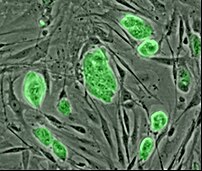
Mouse embryonic stem cells with fluorescent ma...
Image via Wikipedia

[amazon_link asins=’B004HEYPLU,B0081S5OOA,B01K2PKHLE,B004X73FAI’ template=’ProductCarousel’ store=’finmeacur-20′ marketplace=’US’ link_id=’3ef22f7f-48bc-11e7-8a93-db3c53a38b14′]

British scientists have developed a stem cell treatment that could dramatically boost the body’s ability to repair itself. The treatment,  which makes the bone marrow release a flood of stem cells into the bloodstream, could heal serious tissue damage caused by heart attacks and even repair broken bones.

Scientists already use stem cell therapy to treat leukemia patients, getting the marrow to release a type of stem cell that can only make fresh blood cells. British researchers said, “They have found a way to get the bone marrow to release two other types of stem cell that can repair bone, blood vessels and cartilage.”

“The bone marrow of treated mice released 100 times as many stem cells, which help to regenerate tissue”, said Sara Rankin who led the research team at Imperial College. “We hope that by releasing extra stem cells, as we were able to do in mice in our study, we could potentially call up extra numbers of whichever stem cells the body needs, in order to boost its ability to mend itself and accelerate the repair process,” she said.

The group hopes to begin trials later this year to investigate how effective it is at repairing tissue damage in rodents. “All the evidence suggests these cells will make a significant difference to the natural repair process,” Rankin said.

Peter Weissberg, medical director at the British Heart Foundation, which co-funded the research along with the Welcome Trust, said: “It now seems increasingly likely that the bone marrow also contains cells that have the capacity to repair damaged internal organs, such as the heart and blood vessels, but that too few of them are released to be effective.”

He further added, “This research has identified some important molecular pathways involved in mobilizing these cells. It may be possible to develop a drug that interacts with these pathways to encourage the right number and type of stem cells to enter the circulation and repair damage to the heart.”

Sources:
The Times Of India

Categories
News on Health & Science

Stem Cell Therapy May End Transplants

myocardial infarction - Myokardinfarkt - scheme
Image via Wikipedia

British scientists have developed a new technique that can rebuild a severely damaged heart, and one day, might replace the need for transplantation.

Researchers at Imperial College London revealed that stem cell heart surgery can help repair damaged hearts using progenitor cells derived from patients’ own cardiac muscle.

They have discovered a way to extract, grow in the laboratory and then graft on a patient’s own muscle-building cells which then can be used to patch up the heart and increase its pumping power. Moreover, it can increase the quality of life for people who suffer a heart attack.

“This could transform the care for patients who have had heart attacks or have heart disease,” the Telegraph quoted Nicholas Boon, president of the British Cardiovascular Society as saying. “Because the cell therapy uses a patient’s own cells, it negates the risks or complications associated with other treatment options such as rejection linked to transplantation,” he said

Sources: The Times Of India

Reblog this post [with Zemanta]
Categories
Ailmemts & Remedies

Umbilical Hernia

Definition :
An umbilical hernia is an outward bulging (protrusion) of the abdominal lining or part of the abdominal organ(s) through the area around the belly button

CLICK & SEE THE PICTURES
An umbilical hernia is a protrusion of the peritoneum and fluid, omentum, or a portion of abdominal organ(s) through the umbilical ring. The umbilical ring is the fibrous and muscle tissue around the navel (belly-button). Small hernias usually close spontaneously without treatment by age 1 or 2. Umbilical hernias are usually painless and are common in infants.

UMBILICAL Hernias, and nearby hernias called “Paraumbilical Hernias” develop in and around the area of the umbilicus (belly button or navel). A Congenital (present since birth) weakness in the naval area exists. This was the area at which the vessels of the fetal and infant umbilical cord exited through the muscle of the abdominal wall. After birth, although the umbilical cord disappears (leaving just the dimpled belly-button scar), the weakness underneath may persist. Hernias can occur in this area of weakness at any time from birth through late adulthood. The signs and symptoms include pain at or near the navel area as well as the development of an associated bulge or navel deformity. This bulge pushes out upon the skin beneath or around the navel, distorting the normal contour and architecture in or around the navel (creating an ‘OUTIE’ instead of a normal ‘INNIE‘).
Although often appearing at or just after birth, these hernias can also occur at any time during later life. In INFANTS, these hernias may gradually close by age 3 or 4 and surgery can often be delayed until then, unless the hernias are causing problems or enlarging. This decision should be made after examination by a Pediatrician or skilled Surgeon. In ADULTS however, umbilical hernias cannot “heal”, and do gradually increase in size and often become problematic. Incarceration or Strangulation may occur….CLICK & SEE

Umbilical hernia is a congenital malformation, especially common in infants of African descent, and more frequent in boys. An Acquired umbilical hernia directly results from increased intra-abdominal pressure and are most commonly seen in obese individuals.

Causes:

Children:
Umbilical hernias are fairly common. Such a hernia is obvious at birth, as it pushes the belly button outward. This is more obvious when the infant cries, becauses increased pressure results in more noticable bulging.

In infants, the defect is not usually treated surgically. In most cases, by age 3 the umbilical hernia shrinks and closes without treatment.

Umbilical hernia repair may be necessary for children for the following reasons:

*The herniated tissue is stuck in the protruding position, or if blood supply is affected
*The defect has not closed by age 3 or 4
*The defect is very large or unacceptable to parents for cosmetic reasons
*An umbilical hernia in an infant occurs when the muscle through which blood vessels pass to feed the developing fetus doesn’t close completely.

Adults:
Umbilical or para-umbilical hernias are relatively common in adults. They are more common in overweight people and in women, especially after pregnancy. Most surgeons recommend they be surgically repaired, as they tend to get bigger ov

Without surgery, there is a risk that some abdominal contents, typically a bit of fat or intestine, will get stuck (incarcerated) in the hernia defect and become impossible to push back in, which is typically painful. If the blood supply is compromised (strangulation), urgent surgery is needed.

Incarcerated abdominal tissue may cause nausea, vomiting, and abdominal distension.

Any patient with a hernia that cannot be reduced, or pushed back in, while lying down and relaxed should seek urgent medical attention.

Symptoms
A hernia can vary in width from less than 1 centimeter to more than 5 centimeters.

There is a soft swelling over the belly button that often bulges when the baby sits up, cries, or strains. The bulge may be flat when the infant lies on the back and is quiet.

Risks Factors:
Risks for any anesthesia include the following:
*Strangulation of bowel tissue is rare but serious, and needs immediate surgery.
*Reactions to medications
*Breathing problems, pneumonia
*Heart problems

Risks for any surgery include the following:
*Bleeding
*Infection
*Risks specific to umbilical hernia surgery include injury to bowel, which is rare.

Diagnosis:
The doctor can find the hernia during a physical exam.

Treatment
Usually, no treatment is needed unless the hernia continues past age 3 or 4. In very rare cases, bowel or other tissue can bulge out and lose its blood supply (become strangulated). This is an emergency needing surgery.

Most umbilical hernia repairs are done on an outpatient basis, but some may require a short hospital stay if the hernia is very large. After surgery, the patient’s vital signs are monitored and he or she will remain in the recovery area until stable. Medication is supplied for pain as necessary. Patients, or parents if the patient is a child, are taught to care for the incision at home. Full activity can be resumed in 2-4 weeks.

Prognosis:

Most umbilical hernias get better without treatment by the time the child is 3 – 4 years old. Those that do not close may need surgery. Umbilical hernias are usually painless.

Expect successful repair of the hernia. The long-term prognosis is excellent. Very rarely the hernia will recur. Recurrence is more common if a larger hernia (more than 3 cm) is repaired without a mesh.

Recovery
Most umbilical hernia repairs are done on an outpatient basis, but some may require a short hospital stay if the hernia is very large.

After surgery, the health care team will monitor the patient’s vital signs. The patient will stay in the recovery area until stable. Pain medication is prescribed as needed.

Patients, or parents if the patient is a child, are taught to care for the surgical cut at home. Full activity can be resumed in 2-4 weeks.

Disclaimer: This information is not meant to be a substitute for professional medical advise or help. It is always best to consult with a Physician about serious health concerns. This information is in no way intended to diagnose or prescribe remedies.This is purely for educational purpose.

Resources:
http://hernia.tripod.com/types.html
http://www.nlm.nih.gov/medlineplus/ency/article/000987.htm
http://www.nlm.nih.gov/medlineplus/ency/article/002935.htm
http://en.wikipedia.org/wiki/Umbilical_hernia

css.php