Categories
News on Health & Science

Grow Your Own Teeth

[amazon_link asins=’B008FPN92I,B01N484BXZ,B001FWXTA0,B01N1JTO4S,B077DXMWJ8,B002W8SAGY,B00B4IIA8G,B00B4IJZNA’ template=’ProductCarousel’ store=’finmeacur-20′ marketplace=’US’ link_id=’a3051115-09bc-11e8-a6d8-d7afee85ebc4′]

Scientists have made teeth from stem cells in a world first that could make dentures a thing of the past.


They looked like normal teeth, were sensitive to pain and chewed food easily.
While the experiments were on mice, they pave the way for people to ‘grow their own teeth’ as required.

The new tooth at full size, seen at the back of the mouse’s mouth.
The technique could also be adapted to other organs, allowing hearts, lungs and kidneys to be grown inside the body to replace parts worn by age or damaged by disease.
The Japanese study focused on stem cells – ‘master cells’ with the ability to turn into other cell types.

The researchers from the Tokyo University of Science identified two types of stem cell, which together contain all the instructions for a fully grown tooth.
The cells were grown in the laboratory for five days until they formed a tiny tooth ‘bud’.
This was then transplanted deep into the jawbone of a mouse that had had a tooth removed.
Five weeks later, the tip of the tooth broke through the gum. And after seven weeks, it was fully-grown, the journal Proceedings of the National Academy of Sciences reports.
The researchers, who repeated the experiment many times, also showed that the new, bioengineered teeth were fully-functional.

Dr Kazuhisa Nakao said: ‘Every bio- engineered tooth erupted through the gum and had every tooth component such as dentine, enamel, pulp, blood vessels, nerve fibres, crown and root.’
Importantly, the rodent recipients had no trouble eating.
The cells used were take from mouse embryos, but the researchers believe it should be possible to make teeth from other types of cell as well.
They are now looking for suitable cells in people. Possibilities include skin cells and cells from the pulp inside teeth.

They also have to work out how to control the size of the bio-engineered teeth, as those grown in the experiments were slightly smaller than usual.
The process would also have to be speeded up if it was to be used on people as human teeth take years to form.

CLICK @ SEE

However, the pioneering technology could one day allow those with teeth missing to fill the gaps in their smile without having to resort to false teeth, bridges or synthetic implants.
Experts believe that using ‘living’ teeth rather than artificial ones would be better for oral health and may also provide a more natural ‘bite’. Bio- engineered teeth are likely to cost around £2,000 each – a similar price to the implants used at the moment.
But Britain’s 11 million denture wearers should not throw away their fixative creams and gels quite yet.

The technology is still at a very early stage and the Japanese researchers believe it will not be widely used by dentists for at least 15 years. Despite this, British experts said it was an important landmark.

Professor Robin Lovell-Badge, a stem cell researcher at the National Institute for Medical Research in London, said the work was ‘excellent’ and highlighted the promise of using bio-engineering to make complex structures.

But he cautioned that the researchers had yet to find cells suitable for use in people.
Professor Damien Walmsley, of the British Dental Association, said: ‘If you lose a tooth at the moment, one of the options is a metal implant. If you could have a natural replacement, that would be good.’

Natural-looking replacements-also have massive psychologicalbenefits for self-conscious patients.
The technique of creating cell ‘buds’ could be applied more widely to grow other organs, such as hearts, kidneys and livers, inside the body.
Lead researcher Professor Takashi Tsuji said: ‘The ultimate goal of regenerative therapy is to develop fully-functioning bioengineered organs that can replace lost or damaged organs following disease, injury or ageing.
‘Our study makes a substantial contribution to the development of bio- engineering technology for future organ replacement therapy.’

Source:Mailonline. Dated: Aug.4.2009.

Reblog this post [with Zemanta]
Categories
News on Health & Science

Scientists Find New Way to Fix a Broken Heart

[amazon_link asins=’1501120123,1520519958,B0733VP18J,157921228X,B00EI0QN2W,160550131X,B079RM5CWJ,B00LFW4G8O,B00HIMN5O6′ template=’ProductCarousel’ store=’finmeacur-20′ marketplace=’US’ link_id=’29ca3987-1143-11e8-9f27-070680d876b0′]

A new way to mend damage to the heart has been found by scientists.

CLICK & SEE THE PICTURES

The boffins have devised a method to coax heart muscle cells into re-entering the cell cycle, allowing the differentiated adult cells to divide and regenerate healthy heart tissue after a heart attack, according to studies in mice and rats by Children’s Hospital Boston reported in the July 24th issue of the journal Cell.

If the same mechanisms identified by the researchers can be shown to work in the human heart, it opens up real possibilities for new and more efficient ways to treat people with heart disease, reports The BBC.

Theoretically, it could be used to treat heart attack patients, those with heart failure and children with congenital heart defects.

The key ingredient is a growth factor known as neuregulin1 (NRG1).

Previously, it was believed that the heart was incapable of repairing itself. During prenatal development, heart muscle cells (cardiomyocytes) proliferate but were thought to lose that ability shortly after birth. But, recent research has indicated that the adult cells do have some ability to replace themselves at a low level.

And, the new study provides evidence that this is true – and that NRG1 can ramp up the process significantly.

The Boston team tested the ability of various molecules to spur cell division in cultured cardiomyocytes, including several factors known to drive proliferation of the cells during prenatal development. NRG1 produced the most significant effect, and it was repeated when the factor was injected into adult mice.

When administered to animals who had suffered a heart attack, it promoted regeneration of heart muscle, and improved the overall function of the organ.

Writing in the journal, they said: “We have identified the major elements of a new approach to promote myocardial regeneration.

“Many efforts and important advances have been made toward the goal of developing stem-cell based strategies to regenerate damaged tissues in the heart as well as in other organs.

“The work presented here suggests that stimulating differentiated cardiomyocytes to proliferate may be a viable alternative.”

Professor Jeremy Pearson, associate medical director of the British Heart Foundation, said: “This fascinating study shows, remarkably, that a significant fraction of adult heart cells in mice can be made to replicate and help to repair damaged hearts.

“If the same mechanisms identified by the researchers can be shown to work in the human heart, it opens up real possibilities for new and more efficient ways to treat people with heart disease.”

Click to see:->

Mending Broken Heart

Can Seaweed Mend a Broken Heart?

Source: The Times Of India

Reblog this post [with Zemanta]
Categories
Ailmemts & Remedies

Macular Degeneration (AMD OR ARMD)

Definition:
Macular degeneration is a medical condition usually of older adults which results in a loss of vision in the center of the visual field (the macula) because of damage to the retina. It occurs in “dry” and “wet” forms. It is a major cause of blindness in the elderly (>50 years). Macular degeneration can make it difficult or impossible to read or recognize faces, although enough peripheral vision remains to allow other activities of daily life.

CLICK TO SEE THE PICTURES.>...(01)...…….(1).…………..(2).……..(3)………..(4)

Human eye cross section view

Macular degeneration doesn’t cause total blindness, but it worsens your quality of life by blurring or causing a blind spot in your central vision. Clear central vision is necessary for reading, driving, recognizing faces and doing detail work.

The deterioration occurs in the macula (MAK-u-luh), which is in the center of the retina — the layer of tissue on the inside back wall of your eyeball.

The inner layer of the eye is the retina, which contains nerves that communicate sight, and behind the retina is the choroid, which contains the blood supply to the retina. In the dry (nonexudative) form, cellular debris called drusen accumulate between the retina and the choroid, and the retina can become detached. In the wet (exudative) form, which is more severe, blood vessels grow up from the choroid behind the retina, and the retina can also become detached. It can be treated with laser coagulation, and with medication that stops and sometimes reverses the growth of blood vessels.

Although some macular dystrophies affecting younger individuals are sometimes referred to as macular degeneration, the term generally refers to age-related macular degeneration (AMD or ARMD).

Signs:
Drusen
Pigmentary alterations
Exudative changes: hemorrhages in the eye, hard exudates, subretinal/sub-RPE/intraretinal fluid
Atrophy: incipient and geographic
Visual acuity drastically decreasing (two levels or more) ex: 20/20 to 20/80.

Symptoms:
Dry macular degeneration usually develops gradually and painlessly. You may notice these vision changes:

* The need for increasingly bright light when reading or doing close work
* Increasing difficulty adapting to low light levels, such as when entering a dimly lit restaurant
* Increasing blurriness of printed words
* A decrease in the intensity or brightness of colors
* Difficulty recognizing faces
* Gradual increase in the haziness of your overall vision
* Blurred or blind spot in the center of your visual field combined with a profound drop in the sharpness (acuity) of your central vision
CLICK FOR PICTURE

Normal Vision.……………………………...Vision with AMD

Your vision may falter in one eye while the other eye remains fine for years. You may not notice any or much change because your good eye compensates for the weak one. Your vision and lifestyle begin to be dramatically affected when this condition develops in both eyes.

Hallucinations

Additionally, some people with macular degeneration may experience visual hallucinations as their vision loss becomes more severe. These hallucinations may include unusual patterns, geometric figures, animals or even faces. You might be afraid to discuss these symptoms with your doctors or friends and family for fear you’ll be considered crazy. However, such hallucinations aren’t a sign of mental illness. In fact, they’re so common that there’s a name for this phenomenon — Charles Bonnet syndrome.

The Amsler Grid Test is one of the simplest and most effective methods for patients to monitor the health of the macula. The Amsler Grid is essentially a pattern of intersecting lines (identical to graph paper) with a black dot in the middle. The central black dot is used for fixation (a place for the eye to stare at). With normal vision, all lines surrounding the black dot will look straight and evenly spaced with no missing or odd looking areas when fixating on the grid’s central black dot. When there is disease affecting the macula, as in macular degeneration, the lines can look bent, distorted and/or missing. See a video on how to use an Amsler grid here:  and watch an animation showing the Amsler grid with macular degeneration here: .

Macular degeneration by itself will not lead to total blindness. For that matter, only a very small number of people with visual impairment are totally blind. In almost all cases, some vision remains. Other complicating conditions may possibly lead to such an acute condition (severe stroke or trauma, untreated glaucoma, etc.), but few macular degeneration patients experience total visual loss. The area of the macula comprises about 5% of the retina and is responsible for about 35% of the visual field. The remaining 65% (the peripheral field) remains unaffected by the disease

The loss of central vision profoundly affects visual functioning. It is not possible, for example, to read without central vision. Pictures which attempt to depict the central visual loss of macular degeneration with a black spot do not really do justice to the devastating nature of the visual loss. This can be demonstrated by printing letters 6 inches high on a piece of paper and attempting to identify them while looking straight ahead and holding the paper slightly to the side. Most people find this surprisingly difficult to do.

There is a loss off contrast sensitivity, so that contours, shadows and color vision are less vivid. The loss in contrast sensitivity can be quickly and easily measured by a contrast sensitivity test performed either at home or by an eye specialist.

Similar symptoms with a very different etiology and different treatment can be caused by Epiretinal membrane or macular pucker or leaking blood vessels in the eye..

When to see a doctor
See your eye doctor — particularly after age 50 — if:

* You notice changes in your central vision
* Your ability to see colors and fine detail becomes impaired

One way to monitor your eyes to determine if you may need to visit your eye doctor is to check your vision regularly using an Amsler grid. This simple test may help you detect changes in your sight that you otherwise may not notice.

Here’s how to perform the test:

* Hold the grid 14 inches (about 36 centimeters) in front of you in good light. Use your corrective glasses or reading glasses if you normally wear them.
* Cover one eye.
* Look directly at the center dot with your uncovered eye.
* While looking at this dot, determine whether all of the lines of the grid appear straight, uninterrupted and have the same contrast.
* Repeat the above steps with your other eye.
* If any part of the grid is missing or looks wavy, blurred or dark, contact your eye doctor immediately.

Causes:
The exact cause of dry macular degeneration is unknown, but the condition develops as the eye ages. The initial site of change is not in the light-sensitive cells of the macula, but in the retinal pigment epithelium (RPE), a single layer of cells located just behind the retina close to the back wall of your eye.

Your macula is an area about two-tenths of an inch (5 millimeters) in diameter at the center of your retina. This small part of your eye is responsible for clear vision, particularly in your direct line of sight.

The macula consists of millions of densely packed light-sensitive cells called cones and rods. Cones and rods have two segments: An inner segment controls cell functions and produces proteins responsive to light, and an outer segment stores and makes use of these proteins.

As they absorb light, outer segment proteins become degraded and eventually are shed as waste. Meanwhile, the inner segments continuously provide replacements for the outer segments. One function of the cells of the RPE is to remove the outer segments that are shed.

As the eye ages, cells in the RPE begin to deteriorate (atrophy) and lose their pigment. As a consequence, the RPE becomes less efficient in removing outer segment waste. When that happens, the normally uniform reddish color of the macula (as seen with an ophthalmoscope) takes on a mottled appearance. Drusen — yellow, fat-like deposits — begin to appear under the cones and rods. As the drusen and mottled pigmentation continue to develop, your vision gradually deteriorates.

Based on this progression, dry macular degeneration is categorized in three stages:

* Early stage. Several small drusen or a few medium-sized drusen are detected on the macula in one or both eyes. Generally, there’s no vision loss in the earliest stage.
* Intermediate stage. Many medium-sized drusen or one or more large drusen are detected in one or both eyes. At this stage, your central vision may start to blur and you may need extra light for reading or doing detail work.
* Advanced stage. Several large drusen, as well as extensive breakdown of light-sensitive cells in the macula, are detected. These features cause a well-defined spot of blurring in your central vision. The blurred area may become larger and more opaque over time.

Macular degeneration almost always starts out as the dry form. Dry macular degeneration may initially affect only one eye but, in most cases, both eyes eventually become involved.

Risk factors:
Contributing factors for development of macular degeneration include:

* Age. In the United States, macular degeneration is the leading cause of severe vision loss in people age 60 and older.
* Family history of macular degeneration. If someone in your family had macular degeneration, your odds of developing macular degeneration are higher. In recent years, researchers have identified some of the genes associated with macular degeneration. In the future, genetic screening tests may be helpful for assessing early risk of the disease.
* Race. Macular degeneration is more common in whites than it is in other groups, especially after age 75.
* Sex. Women are more likely than men to develop macular degeneration, and because they tend to live longer, women are more likely to experience the effects of severe vision loss from the disease.
* Cigarette smoking. Exposure to cigarette smoke doubles your risk of macular degeneration. Cigarette smoking is the single most preventable cause of macular degeneration.
*Stargardt’s disease (STGD, also known as Juvenile Macular Degeneration) is an autosomal recessive retinal disorder characterized by a juvenile-onset macular dystrophy, alterations of the peripheral retina, and subretinal deposition of lipofuscin-like material. A gene encoding an ATP-binding cassette (ABC) transporter was mapped to the 2-cM (centiMorgan) interval at 1p13-p21 previously shown by linkage analysis to harbor the STGD gene. This gene, ABCR, is expressed exclusively and at high levels in the retina, in rod but not cone photoreceptors, as detected by in situ hybridization. Mutational analysis of ABCR in STGD families revealed a total of 19 different mutations including homozygous mutations in two families with consanguineous parentage. These data indicate that ABCR is the causal gene of STGD/FFM.
*Drusen CMSD studies indicate that drusen are similar in molecular composition to plaques and deposits in other age-related diseases such as Alzheimer’s disease and atherosclerosis.
While there is a tendency for drusen to be blamed for the progressive loss of vision, drusen deposits can, however, be present in the retina without vision loss. Some patients with large deposits of drusen have normal visual acuity. If normal retinal reception and image transmission are sometimes possible in a retina when high concentrations of drusen are present, then even if drusen can be implicated in the loss of visual function, there must be at least one other factor that accounts for the loss of vision. Retinitis Pigmentosa (RP) is a genetically linked dysfunction of the retina and is related to mutation of the ATP Synthase Gene 63.
* Obesity. Being severely overweight increases the chance that early or intermediate macular degeneration will progress to the more severe form of the disease.
* Light-colored eyes. People with light-colored eyes appear to be at greater risk than do those with darker eyes.
* Exposure to sunlight. Although the retina is more sensitive to shorter wavelengths of light, including ultraviolet (UV) light, only a small percentage of ultraviolet light actually reaches the retina. Most ultraviolet light is filtered by the transparent outer surface of your eye (cornea) and the natural crystalline lens in your eye. Some experts believe that long-term exposure to ultraviolet light may increase your risk of developing macular degeneration, but this risk has not been proved and remains controversial.
* Low levels of nutrients. This includes low blood levels of minerals, such as zinc, and of antioxidant vitamins, such as A, C and E. Antioxidants may protect your cells from oxygen damage (oxidation), which may partially be responsible for the effects of aging and for the development of certain diseases such as macular degeneration.
* Cardiovascular diseases. These include high blood pressure, stroke, heart attack and coronary artery disease with chest pain (angina).
*High fat intake is associated with an increased risk of macular degeneration in both women and men. Fat provides about 42% of the food energy in the average American diet. A diet that derives closer to 20-25% of total food energy from fat is probably healthier. Reducing fat intake to this level means cutting down greatly on consumption of red meats and high-fat dairy products such as whole milk, cheese, and butter. Eating more cold-water fish (at least twice weekly), rather than red meats, and eating any type of nuts may help macular degeneration patients.
*Oxidative stress: It has been proposed that age related accumulation of low molecular weight, phototoxic, pro-oxidant melanin oligomers within lysosomes in the retinal pigment epithelium may be partly responsible for decreasing the digestive rate of photoreceptor outer rod segments (POS) by the RPE. A decrease in the digestive rate of POS has been shown to be associated with lipofuscin formation – a classic sign associated with macular degeneration.
*Fibulin-5 mutation Rare forms of the disease are caused by geneic defects in fibulin-5, in an autosomal dominant manner. In 2004 Stone et al. performed a screen on 402 AMD patients and revealed a statistically significant correlation between mutations in Fibulin-5 and incidence of the disease. Furthermore the point mutants were found in the Calcium binding sites of the cbEGF domains of the protein. there is no structural basis for the effects of the mutations.

Diagnosis:
Diagnostic tests for macular degeneration may include:

*An eye examination. One of the things your eye doctor looks for while examining the inside of your eye is the presence of drusen and mottled pigmentation in the macula. The eye examination includes a simple test of your central vision and may include testing with an Amsler grid. If you have macular degeneration, when you look at the grid some of the straight lines may seem faded, broken or distorted. By noting where the break or distortion occurs — usually on or near the center of the grid — your eye doctor can better determine the location and extent of your macular damage.

Regular screening examinations can detect early signs of macular degeneration before the disease leads to vision loss.
*Angiography. To evaluate the extent of the damage from macular degeneration, your eye doctor may use fluorescein angiography. In this procedure, fluorescein dye is injected into a vein in your arm and photographs are taken of the back of the eye as the dye passes through blood vessels in your retina and choroid. Your doctor then uses these photographs to detect changes in macular pigmentation or to identify small macular blood vessels.

Your doctor may also suggest a similar procedure called indocyanine green angiography. Instead of fluorescein, a dye called indocyanine green is used. This test provides information that complements the findings obtained through fluorescein angiography.
* Optical coherence tomography. This noninvasive imaging test helps identify and display areas of retinal thickening or thinning. Such changes are associated with macular degeneration. This test can also reveal the presence of abnormal fluid in and under the retina or the RPE. It’s often used to help monitor the response of the retina to macular degeneration treatments.

Treatment:
There’s no treatment available to reverse dry macular degeneration. But this doesn’t mean you’ll eventually lose all of your sight. Dry macular degeneration usually progresses slowly, and many people with the condition are able to live relatively normal, productive lives, especially if only one eye is affected. Dry macular degeneration can, however, develop into the more rapidly progressive wet type of macular degeneration at any time.

Taking a high-dose formulation of antioxidants and zinc may reduce progression of dry macular degeneration to advanced macular degeneration. The National Eye Institute-sponsored Age-Related Eye Disease Study (AREDS) showed that a daily supplement of 500 milligrams (mg) of vitamin C, 400 international units (IU) of vitamin E, 15 mg of beta carotene (often as vitamin A — up to 25,000 IU), 80 mg of zinc (as zinc oxide) and 2 mg of copper (as cupric oxide) reduced the risk of progressing to moderate or severe vision loss by up to 25 percent.

Life Style & Home Remedies:
Macular degeneration doesn’t affect your side (peripheral) vision and usually doesn’t cause total blindness. But it can rob you of your central vision — which is important for driving, reading and recognizing people’s faces. A low-vision center may be able to assess your visual capabilities and suggest certain optical and household devices that can be helpful for some near-vision tasks. Ask your eye doctor if there are any low-vision centers in your area.

There are ways to cope with impaired vision. Below are a few suggestions:

* Use caution when driving. First, check with your doctor to see if driving is still safe based on your current visual acuity. When you do drive, there are certain situations to avoid. For example, don’t drive at night, in heavy traffic or in bad weather.
* Seek help traveling. Use public transportation or ask family members to help, especially with night driving.
* Travel with others. Contact your local area agency on aging for a list of vans and shuttles, volunteer driving networks or ride shares.
* Get good glasses. Optimize the vision you have with the right glasses, and keep an extra pair in the car.
* Use magnifiers. Large-print books and magazines can help you read more easily.
* View with large type on the Internet. Look for Web sites that use large-sized type fonts, or change the font size on your display.
* Obtain specialized appliances. Some clocks, radios, telephones and other appliances have extra-large numbers.
* Have proper light in your home. This will help with reading and other activities.
* Remove home hazards. Eliminate throw rugs and other possible tripping hazards in your home.
* Ask friends and family members for help. Tell them about your vision problems so that they can help you perform certain tasks and help you recognize people.
* Don’t become socially isolated. A common frustration of people with macular degeneration is the inability to recognize other people and greet them by name. If this happens to you, try asking people you know to say hi and tell you their names when you meet them on the street or in other situations so that you can greet them back.
* Take advantage of online networks. The Internet is a good source for support groups and resources for people with macular degeneration.

Alternative Medicine:
Some people have turned to complementary or alternative therapies, such as bilberry, ginkgo and shark cartilage, in the belief that they can help prevent the progression of macular degeneration.

However, there’s no conclusive evidence that any of these products are effective for macular degeneration, and some may interact with other medications you’re taking. Check with your doctor before taking any dietary or herbal supplement.

Prevention
The Age-Related Eye Disease Study showed that a combination of high-dose beta-carotene, vitamin C, vitamin E, and zinc can reduce the risk of progressing from early to advanced AMD by about 25 percent.  Studies are underway with the goal of reducing lipofuscin accumulation.

Studies have found that Lutein and zeaxanthin (Carotenoid nutrients found in green vegetables such as Kale, Spinach, Collards, spices such as Saffron, and egg yolk) protect against and possibly reverse macular degeneration and Retinitis pigmentosa.  Studies found that antioxidants disrupt the link of two processes that cause macular degeneration and extend the lifetime of irreplaceable photoreceptors and other retinal cells (Lutein is known to have antioxidant properties).

Eating spinach or collard greens five times a week decreases the risk of AMD by 43%

Studies reported in the British Journal of Ophthalmology suggest that while beneficial for those in advanced stages, antioxidant supplements can be counterproductive for people with early stages of AMD as antioxidants can potentially negate the beneficial effects of Omega-3 fats. It has been found that Omega-3 fatty acids can prevent or even halt the progress of degeneration. However, moderation of oily fishes in patients’ diets is suggested as they can lead to a build up of pollutants such fishes may contain.

The following measures may help you avoid macular degeneration:
*Eat foods containing antioxidants.
*Take antioxidant and zinc supplements.
* Eat fish.
*Stop smoking.
*Manage your other diseases.
*Get regular eye exams.
*Screen your vision regularly.

If you have some vision loss because of macular degeneration, your eye doctor can prescribe optical devices called low-vision aids that will help you see better for close-up work. Or your doctor may refer you to a low-vision specialist. In addition, a wide variety of support services and rehabilitation programs are available that may help you adjust your lifestyle.
Impact:
Macular degeneration can advance to legal blindness and inability to drive. It can also result in difficulty or inability to read or see faces.

Adaptive devices can help people read. These include magnifying glasses, special eyeglass lenses, desktop and portable electronic devices, and computer screen readers such as JAWS for Windows.

Composer Josef Tal checks a manuscript (2006)Accessible publishing also aims to provide a variety of fonts and formats for published books to make reading easier. This includes much larger fonts for printed books, patterns to make tracking easier, audiobooks and DAISY books with both text and audio.

Because the peripheral vision is not affected, people with macular degeneration can learn to use their remaining vision to continue most activities. Assistance and resources are available in every country and every state in the U.S. Classes for “independent living” are given and some technology can be obtained from a state department of rehabilitation. You can also search for macular degeneration on the internet and contact one of the non-profit organizations for assistance.

Disclaimer: This information is not meant to be a substitute for professional medical advise or help. It is always best to consult with a Physician about serious health concerns. This information is in no way intended to diagnose or prescribe remedies.This is purely for educational purpose.
Resources:
http://www.mayoclinic.com/health/macular-degeneration/DS00284
http://en.wikipedia.org/wiki/Macular_degeneration

Enhanced by Zemanta
Categories
Ailmemts & Remedies

Cytopenia

DEFINITION:

Cytopenia is a reduction in the number of blood cells. It takes a number of forms:
*Low red blood cell count: anemia.
*Low white blood cell count: leukopenia or neutropenia (because neutrophils make up at least half of all white cells, they are almost always low in leukopenia).
*Low platelet count: thrombocytopenia.
*Low granulocyte count: granulocytopenia
*Low red blood cell, white blood cell, and platelet counts: pancytopenia..

Click to see the picture

Blood cell development. A blood stem cell goes through several steps to become a red blood cell, platelet, or white blood cell.

CLICK & SEE THE PICTURES

Cancer patients may frequently develop cytopenia, a disorder in which the production of one or more blood cell types ceases or is greatly reduced. Cancer and chemotherapy used to treat cancer, and sometimes radiation therapy, may sometimes cause cytopenia.

TYPES:
A deficiency of red blood cells which  is called anemia; a deficiency of white blood cells, or leukocytes, leukopenia or neutropenia (neutrophils make up over half of all white blood cells); and deficiency of platelets is called  thrombocytopenia.

Pancytopenia is the deficiency of all three blood cell types and is characteristic of aplastic anemia, a potentially life-threatening disorder that requires a stem cell transplant.

Blood Cells
The blood consists of three different  types of cells: red blood cells (erythrocytes), white blood cells (leukocytes), and platelets. Erythrocytes contain hemoglobin, the protein that carries oxygen from the lungs to all cells in the body. Proper cell function depends on an adequate oxygen supply. When cells are oxygen deprived, organ function can be seriously impaired.

Leukocytes (white blood cells) protect the body against viral, bacterial, and parasitic infection and detect and remove damaged, dying, or dead tissues. Someone with a deficiency of white blood cells is extremely vulnerable to infection.

The term “leukocyte” refers to all six types of white blood cells; each plays a unique role in the immune system:

1. Basophils circulate in the blood and initiate the inflammatory response.
2.Eosinophils kill infecting parasites and produce allergic reactions.
3.Lymphocytes produce antibodies and regulate immune responses.
4. Mast cells are fixed in tissues and initiate the inflammatory response.
5. Monocytes capture infecting organisms for identification, ingest infecting organisms, and remove damaged or dying cells and cell debris. When monocytes become fixed in tissue, they are called macrophages.
6.Neutrophils identify and kill infecting organisms, and remove dead tissue.

Platelets are essential factors for blood clotting. Sudden blood loss triggers platelet activity at the site of the wound. Exposure to oxygen in the air causes platelets to break apart and combine with a substance called fibrinogen to form fibrin. Fibrin has a thread-like structure and forms a scab, or external clot, as it dries. Platelet deficiency causes one to bruise and bleed easily. Blood does not clot at an open wound, and there is greater risk for internal bleeding.

All blood cells have a lifespan: erythrocytes have a lifespan of about 120 days; leukocytes, 1 to 3 days; and platelets, approximately 10 days. The body continually replenishes the blood supply through a process called hematopoiesis.

Blood Cell Formation—Hematopoiesis, the formation and development of blood cells, occurs in bone marrow. Bone marrow is a nutrient-rich spongy tissue located mainly in the central portions of long flat bones (e.g., sternum, pelvic bones) in adults and all bones in infants.

All blood cells derive from blood-forming stem cells that reside in bone marrow. Stem cells replicate indefinitely and develop into mature, specialized cells. A hormone produced in the kidneys, erythropoietin, stimulates blood stem cells to produce all three types of blood cells.

CAUSES & RISK FACTORS:-

Chemotherapy and radiation therapy both reduce the number of blood-forming stem cells in cancer patients, but chemotherapeutic agents have a greater adverse effect because they suppress bone marrow function in several ways.The degree of damage is related to the particular drug(s) and the dose.

Chemotherapeutic agents can produce deficiencies in all blood cell types by

* damaging blood-forming stem cells,
* suppressing the kidneys? production of erythropoietin (hormone that stimulates blood cell production), and
* triggering red cell destruction (hemolysis) by inducing an immune response that causes the body to mistakenly identify erythrocytes as foreign bodies and destroy them.

Malignant tumors can cause anemia and other cytopenias when they directly invade bone marrow and suppress marrow function. Malignant cells also can migrate from tumors in other parts of the body to bone marrow. Tumors also can replace normal blood-forming stem cells with abnormal clones.

SIGN & SYMPTOMS:-

Anemia
A deficiency in erythrocytes reduces the amount of oxygen reaching all cells in the body, thus impairing all tissue and organ function. Severe fatigue is the most common symptom of anemia and is experienced by approximately 75% of chemotherapy patients. Patients find it more disabling than other treatment side effects, including nausea and depression.

Anemia also produces these symptoms:

* Confusion
* Dizziness
* Headache
* Lightheadedness
* Loss of concentration
* Pallor (pale skin, nail beds, gums, linings of eyelids)
* Rapid heart rate (tachycardia)
* Shortness of breath (dyspnea)

Neutropenia
Patients with a white blood cell deficiency experience frequent and/or severe bacterial, viral, and/or fungal infections; fever; and mouth and throat ulcers.

Complications—Bacteremia, the form of sepsis characterized by the presence of bacteria in the blood, can develop in immunocompromised patients who have neutropenia. Fever, rapid heart rate, and quick shallow breathing are signs of early sepsis, usually a reversible condition.

Untreated bacteremia can lead to severe sepsis, in which one or more organs become dysfunctional. Septic shock is severe sepsis with low blood pressure. The risk for death increases with the development of septic shock. Even aggressive treatment can fail to reverse the condition.

Thrombocytopenia
Platelet deficiency causes patients to bruise and bleed easily. Bleeding occurs most often in the mucous membranes lining the mouth, nose, colon, and vagina. Tiny reddish-purple skin lesions (petechiae), evidence of pinpoint hemorrhages, may appear on the skin or in the mouth.

Pancytopenia
Patients who are deficient in all blood cell types experience signs and symptoms associated with each, but bleeding from the nose and gums, and easy bruising usually appear first. Symptoms of anemia (e.g., fatigue, shortness of breath) are also common. Patients may look and feel well, otherwise, despite the seriousness of their condition.

Anemia
People with anemia (reduced red cell production) are advised to rest and eat foods high in iron (meat, fish, poultry, lentils, legumes, iron-enriched grains and flours).

If immediate remedy is necessary, treatment may include medication that helps restore the red blood supply and a transfusion of packed red blood cells.

Epoetin alpha (Epogen®, Procrit®)is a synthetic erythropoietin (normally produced by the kidneys) that stimulates stem cells to produce red blood cells. Restoration of the red blood cell supply with medication is gradual.

Darbepoetin alfa (Aranesp®) also stimulates red blood cell production but requires fewer doses and less disruption of daily living.

In March 2007, the Food and Drug Administration (FDA) issued a warning about these medications in response to studies indicating that they may increase the risk for blood clots, strokes, and heart attacks in some patients (e.g., patients who have kidney disease).

Thrombocytopenia
People with an abnormally low platelet count should avoid bruising or breaking the skin, and should carefully brush their teeth. A persistently decreased platelet count may be treated with a transfusion of platelets.

Neutropenia
The patient with a low white blood cell count is advised to  do the following:

*Avoid contact with people who are ill,
*Monitor closely for signs of infection (e.g., fever), and
*Take antibiotics when appropriate.

Medication, a colony-stimulating factor (CSF), may be prescribed to speed the development of white blood cells and shorten the period of susceptibility to infection.

Growth Factors
Growth factors are synthetic versions of substances involved in stimulating red and white blood cell production. Physicians exercise caution when prescribing these medications for people with tumors that involve the bone marrow, because growth factors might stimulate malignant cell growth.

These medications include the following:

Epoetin alpha (Procrit®, Epogen®; stimulates red blood cell production)
G-CSF (granulocyte colony-stimulating factor; e.g., filgrastim [Neupogen®]; stimulates neutrophil production)
GM-CSF (granulocyte-macrophage colony-stimulating factor; stimulates production of several white blood cells, including macrophages)

Leukocytes and other cells that contain granules are also called granulocytes.

Side effects
Fever, fatigue, dizziness, diarrhea, nausea, vomiting, weakness, and paresthesia (prickling sensation) are side effects associated with epoetin alpha.

Bone pain, malaise, headache, flu-like symptoms, muscle ache, redness at the injection site, and skin rash may occur with GM-CSF.

G-CSF commonly produces bone pain.

MEDICATIONS:-

Medications used to treat bacterial infection and other illnesses also can contribute to immune system suppression.

Some of these are :

* Antacids: cimetidine (Tagamet®)
* Antibiotics: chloramphenical (Chloromycetin®), sulfonamide (Thiosulfil®, Gantanol®); cephalosporin (Cephalaxin®), vancomycin (Vancocin®)
* Anticonvulsants: phenytoin/hydantoin (Dilantin®), felbamate (Felbatol®), carbamazepine (Tegretol®)
* Antimalarials: chloroquine (Aralin®)
* Antivirals: ganciclovir (Vitrasert®), zidovudine (AZT®)
* Cardiac drugs: diltiazem (Cardizem®), nifedipine (Procardia®), verapamil (Calan®)
* Diabetes drugs: glipizide (Glucotrol®), glyburide (Micronase®)
* Hyperthyroid drug: propylthiouracil
* NSAIDs (nonsteroidal anti-inflammatory drugs): phenylbutazone (Butazolidine®), indomethacin (Indocin®, Indochron E-R®)—Due to potentially severe gastrointestinal and cardiovascular side effects, NSAIDs should only be used as instructed.
* Rheumatoid arthritis drugs: auranofin (Ridaura®), aurothioglucose (Solganal®), gold sodium thiomalate (Myochrisine®)

Bone Marrow and Stem Cell Transplantation:-
The treatment of choice for the pancytopenic patient with a matched bone marrow donor is stem cell transplantation. The goal of transplantation is to restore blood-forming stem cells to the marrow.

Disclaimer: This information is not meant to be a substitute for professional medical advise or help. It is always best to consult with a Physician about serious health concerns. This information is in no way intended to diagnose or prescribe remedies.This is purely for educational purpose.

Resources:
http://www.oncologychannel.com/cytopenia/index.shtml
http://en.wikipedia.org/wiki/Cytopenia
http://www.cancer.umn.edu/cancerinfo/NCI/CDR378089.html

Enhanced by Zemanta
Categories
Ailmemts & Remedies

Polycythemia

Definition:
Polycythemia is the increase of the RBC count, hemoglobin, and total RBC volume, accompanied by an increase in total blood volume. This must be distinguished from relative erythrocytosis secondary to fluid loss or decreased intake; this distinction can be made easily on a clinical basis. Polycythemia accompanies increased total blood volume, whereas relative erythrocytosis does not. Two basic categories of polycythemia are recognized:
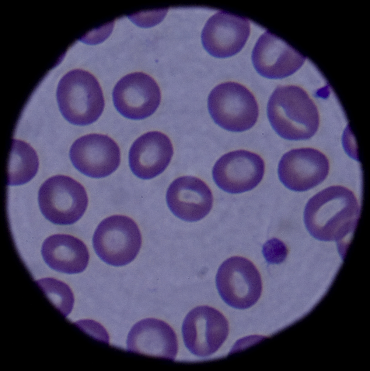
...click to see…the picture..
* Primary polycythemias are due to factors intrinsic to red cell precursors and include the diagnoses of primary familial and congenital polycythemia (PFCP) and polycythemia vera (PV).
* Secondary polycythemias are caused by factors extrinsic to red cell precursors.

In normal hematopoiesis, myeloid stem cells give rise to erythrocytes, platelets, granulocytes, eosinophils, basophils, and monocytes. The production of each lineage is a function of cell proliferation, differentiation, and apoptosis. These various stages of differentiation rely on multiple interrelated processes. Protein growth factors, known as cytokines, stimulate proliferation of the multilineage cells (eg, interleukin [IL]-3, granulocyte-macrophage colony-stimulating activity [GM-CSF]). Other factors primarily stimulate the growth of committed progenitors (eg, GM-CSF, macrophage colony-stimulating factor [M-CSF], erythropoietin [Epo]).

Erythropoiesis is a carefully ordered sequence of events. Initially occurring in fetal hepatocytes, the process is taken over by the bone marrow in the child and adult. Although multiple cytokines and growth factors are dedicated to the proliferation of the RBC, the primary regulator is Epo. Red cell development is initially regulated by stem cell factor (SCF), which commits hematopoietic stem cells to develop into erythroid progenitors. Subsequently, Epo continues to stimulate the development and terminal differentiation of these progenitors. In the fetus, Epo is produced by monocytes and macrophages found in the liver. After birth, Epo is produced in the kidneys; however, Epo messenger RNA (mRNA) and Epo protein are also found in the brain and in RBCs, suggesting that some paracrine and autocrine function is present as well.  CLICK & SEE THE PICTURES

Erythropoiesis escalates as increased expression of the EPO gene produces higher levels of circulating Epo. EPO gene expression is known to be affected by multiple factors, including hypoxemia, transition metals (Co2+, Ni2+, Mn2+), and iron chelators. However, the major influence is hypoxia, including factors of decreased oxygen tension, RBC loss, and increased oxygen affinity of hemoglobin. In fact, Epo production has been observed to increase as much as 1000-fold in severe hypoxia.

Absolute polycythemia
The overproduction of red blood cells may be due to a primary process in the bone marrow (a so-called myeloproliferative syndrome), or it may be a reaction to chronically low oxygen levels or, rarely, a malignancy.

Primary polycythemia (Polycythemia vera)
Primary polycythemia, often called polycythemia vera (PCV), polycythemia rubra vera (PRV), or erythremia, occurs when excess red blood cells are produced as a result of an abnormality of the bone marrow.  Often, excess white blood cells and platelets are also produced. Polycythemia vera is classified as a myeloproliferative disease. Symptoms include headaches, vertigo, and an abnormally enlarged spleen and/or liver. In some cases, affected individuals may have associated conditions including high blood pressure or the formation of blood clots. Transformation to acute leukemia is rare. Phlebotomy is the mainstay of treatment. A hallmark of polycythemia is an elevated hematocrit, with Hct > 55% seen in 83% of cases. Mutations in JAK2 are found in 95% of cases, though also present in other myeloproliferative disorders.

Secondary polycythemia
Secondary polycythemia is caused by either natural or artificial increases in the production of erythropoietin, hence an increased production of erythrocytes. In secondary polycythemia, there may be 6 to 8 million and occasionally 9 million erythrocytes per cubic millimeter (microliter) of blood. Secondary polycythemia resolves when the underlying cause is treated.

Secondary polycythemia in which the production of erythropoietin increases appropriately is called physiologic polycythemia. This physiologic (meaning normal) polycythemia is a normal adaptation to living at high altitudes (see altitude sickness). Many athletes train at high altitude to take advantage of this effect — a legal form of blood doping. Similarly, athletes with primary polycythemia may have a competitive advantage due to greater stamina.

Other causes of secondary polycythemia include smoking, renal or liver tumors, hemangioblastomas in the central nervous system, heart or lung diseases that result in hypoxia, and endocrine abnormalities including pheochromocytoma and adrenal adenoma with Cushing’s syndrome. People whose testosterone levels are high because of the use anabolic steroids, including athletes who abuse steroids and people whose doctors put them on doses that are too high, as well as people who take erythropoietin may develop secondary polycythemia.

Secondary polycythemia can be induced directly by phlebotomy to withdraw some blood, concentrate the erythrocytes, and return them to the body.

Chuvash polycythemia
Chuvash polycythemia refers to a familial form of erythrocytosis different from classical polycythemia vera. This involved patients from Chuvashia and is associated with a C598T mutation in the von Hippel-Lindau gene (VHL).[6] A cluster of patients with Chuvash polycythemia have been found in other populations, such as on the Italian island of Ischia, located in the Bay of Naples.

Relative polycythemia
Relative polycythemia is an apparent rise of the erythrocyte level in the blood; however, the underlying cause is reduced blood plasma. Relative polycythemia is often caused by loss of body fluids, such as through burns, dehydration and stress.

Polycythemia vera
Earlier diagnostic criteria for polycythemia vera included the following (based on the Polycythemia Vera Study Group Diagnostic Criteria):1

* Red cell mass greater than 36 mL/kg for men and greater than 32 mL/kg for women
* Arterial oxygen saturation greater than 92%
* Splenomegaly or 2 of the following:
o Thrombocytosis greater than 400 X 109/L
o Leukocytosis greater than 12 X 109/L
o Leukocyte alkaline phosphatase activity greater than 100 U/L in adults (reference range, 30-120 U/L) without fever or infection
o Serum vitamin B-12 greater than 900 pg/mL (reference range, 130-785 pg/mL)
o Unsaturated vitamin B-12 binding capacity greater than 2200 pg/mL

The reference range for the clinician’s laboratory should be cross-correlated. The diagnostic criteria have undergone scrutiny and several revisions in recent years. In 2001, the World Health Organization (WHO) proposed a classification system for chronic myeloid neoplasms.2 The diagnosis of polycythemia vera fell under the broader category of chronic myeloproliferative diseases. This set of criteria quickly lost favor because of lack of validation3 and the discovery of JAK2 mutations in adult patients.4,5,6,7,8,9

Currently, the diagnosis of polycythemia vera is based on the 2008 WHO criteria, which has integrated molecular diagnostics into the evaluation and screening for polycythemia vera.10 A diagnosis of polycythemia vera is made when both major and one minor criterion are present or when the first major criterion is present with any two minor criteria.

The current criteria include the following:

* Major criteria
1. Hemoglobin level of more than 18.5 g/dL in men (>16.5 g/dL in women) or other evidence of increased red cell volume

or

Hemoglobin or hematocrit level higher than 99th percentile of method-specific reference range for age, sex, altitude, of residence

or

Hemoglobin level of more than 17 g/dL in men (>15 g/dL in women) if associated with a documented and sustained increase of at least 2 g/dL from an individual’s baseline value that can not be attributed to correction of iron deficiency

or

Elevated red cell mass greater than 25% above mean normal predicted value
2. Presence of JAK2V617F or similar mutation (eg, JAK2 exon 12 mutation)
* Minor criteria
1. Bone marrow trilineage myeloproliferation
2. Subnormal serum erythropoietin levels
3. Endogenous erythroid colony growth

Pathophysiology

Primary polycythemia

The disease is considered to be a form of the myeloproliferative syndromes that include polycythemia vera, essential thrombocythemia, agnogenic myeloid metaplasia, and myelofibrosis. The clonality of polycythemia vera is well established and was first demonstrated by Adamson et al in 1976.11 Subsequent studies suggest hypersensitivity of the myeloid progenitor cells to growth factors, including Epo, IL-3, SCF, GM-CSF, and insulinlike growth factor (IGF)–1, whereas other studies show defects in programmed cell death.

Until recently, the pathophysiology of polycythemia vera was unclear. In 2005, significant progress in the understanding of polycythemia vera was made with the discovery of an activating mutation in the tyrosine kinase JAK2 (JAK2V617F ), which now appears to cause most primary cases in adults.4,5,6,7,8 Several other mutations of JAK2 have since been described (eg, exon 12, JAK2H538-K539delinsI ).9,12 The JAK2 mutations are thought to possibly cause hypersensitivity to Epo via the Epo receptor, although the effects of this mutation remain to be fully characterized.

Familial clustering suggests a genetic predisposition. Whether these mutations are responsible for the development of polycythemia vera in pediatric patients is unclear. Some groups have reported lower rates of JAK2 mutations in children compared with adults,13,14,15 whereas other groups have seen similar rates with complete or near complete presence of JAK2V617F and other JAK2 mutations.12

PFCP is caused by a hypersensitivity of erythroid precursors to Epo. Several mutations (approximately 14) have been identified in the Epo receptor (EPOR) gene; however, EPOR mutations have not been identified in all PFCP kindreds. Most identified EPOR mutations (11) cause truncation of the c-terminal cytoplasmic receptor domain of the receptor. These truncated receptors have heightened sensitivity to circulating Epo due to a lack of negative feedback regulation.16

Secondary polycythemia

Secondary polycythemia may result from functional hypoxia induced by lung disease, heart disease, increased altitude (hemoglobin increase of 4% for each 1000-m increase in altitude), congenital methemoglobinemia, and other high–oxygen affinity hemoglobinopathies stimulating increased Epo production. Secondary polycythemia may also result from increased Epo production secondary to benign and malignant Epo-secreting lesions. Secondary polycythemia may also be a benign familial polycythemia.

Chuvash polycythemia, a congenital polycythemia first recognized in an endemic Russian population, has mutations in the von Hippel-Lindau (VHL) gene, which is associated with a perturbed oxygen dependent regulation of Epo synthesis.

Secondary polycythemia of the newborn is fairly common and is a result of either chronic or acute fetal hypoxia or delayed cord clamping and stripping of the umbilical cord.17
Frequency
United States

Primary polycythemia is rare; the overall prevalence of polycythemia vera is 2 cases per 100,000 people. The median age is 60 years. Only 0.1% of cases of polycythemia vera are observed in individuals younger than 20 years. Fewer than 50 cases of pediatric polycythemia vera have been reported in the literature. Secondary polycythemia is seen in 1-5% of all newborns in the United States.
International

Polycythemia vera has a similar incidence in Western Europe as in the United States, and occurrence rates are very low in Africa and Asia (as low as 2 cases per million per year in Japan).

Mortality/Morbidity
Death rates for children are unavailable. The complications found in polycythemia vera are related to 2 primary factors. The first includes complications related to hyperviscosity. The second involves bone marrow–related complications. Untreated, the median survival time for these patients is 18 months. However, if patients are treated, survival is greatly extended, as many as 10-15 years with phlebotomy alone. The causes of death in adults are as follows:

* Thrombosis/thromboembolism (30-40%)
* Acute myelogenous leukemia (19%)
* Other malignancies (15%)
* Hemorrhage (2-10%)
* Myelofibrosis/myeloid metaplasia (4%)
* Other (25%)

In the neonatal period, polycythemia-induced hyperviscosity can lead to altered blood flow and subsequently affect organ function. Infants with polycythemia are at increased risk for necrotizing enterocolitis, renal dysfunction, hypoglycemia, and increased pulmonary vascular resistance with resultant hypoxia and cyanosis. Although initially thought to cause neurologic dysfunction, the decrease in cerebral blood flow seen in newborns with polycythemia is a physiologic response and does not appear to cause cerebral ischemia.17
Race

In the United States, higher rates of polycythemia vera are observed in the Ashkenazi Jewish population, and lower rates are seen in blacks.
Sex

The male-to-female ratio is 1.2-2.2:1 in adults and 1:1 in children.
Age

The median age for polycythemia vera is 60 years. Only 0.1% of polycythemia cases occur in people younger than 20 years.
Clinical
History

The clinical features associated with polycythemia are a direct result of the increase in red cell mass, which causes an expansion of blood volume. Signs of hyperviscosity and increased metabolism accompany polycythemia. A thorough history must be obtained for a history of cardiac, pulmonary (including sleep apnea), hepatic or renal disease in the patient and a complete family history for evidence of familial polycythemia.

Symptoms include the following:

* Headache
* Weight loss
* Weakness or malaise
* Dizziness
* Pruritus
* Bruising
* Ruddy or red appearance of the skin
* Diaphoresis/dyspnea
* Visual disturbance
* Paresthesias
* Arthropathies
* GI – Fullness, thirst, abdominal discomfort, constipation

Physical

A thorough physical must be completed and include specific evaluation for signs and symptoms of underlying disease that may cause secondary polycythemia; it must include pulse oximetry, careful cardiac and pulmonary evaluation, and evaluation for signs of renal or hepatic disease.

Signs of polycythemia include the following:

* Rubor, especially facial rubor
* Skin plethora
* Hypertension, both systolic and diastolic
* Hepatomegaly
* Splenomegaly
* Conjunctival plethora (engorged vessels in the bulbar conjunctiva)
* Ecchymosis
* Cardiac hypertrophy (rarely observed)

Causes

* Primary polycythemia

o In the past, the pathophysiology was unclear, and primary polycythemias were thought to be due to both inherited and acquired mutations in erythroid progenitors, leading to abnormal red cell proliferation. However, in 2005, an activating mutation found in the tyrosine kinase JAK2 was implicated as the causative factor in polycythemia vera (PV). Five separate groups identified this mutation in approximately 80% (56-97% reported) of patients with polycythemia vera.
o This acquired V617F mutation in JAK2 leads to constitutively activated JAK2. Activated JAK2 induces erythropoietin (Epo) hypersensitivity; although not yet completely delineated, it is thought to act through an activating EPOR.
o Additional JAK2 mutations have been identified in exon 12,9 JAK2H538-K539delinsI ,18 and others.19
o Primary familial and congenital polycythemia (PFCP), which is commonly found to have mutations in the Epo receptor (EPOR) gene. Approximately 14 mutations have been identified.
* Secondary polycythemia
o Congenital causes include high affinity hemoglobin and 2,3-Bisphosphoglycerate (2,3-BPG) deficiency.
o Chuvash polycythemia, a congenital polycythemia first recognized in an endemic Russian population, has mutations in the von Hippel-Lindau (VHL) gene, which is associated with a perturbed oxygen-dependent regulation of Epo synthesis.
o Acquired causes included hypoxemia and Epo-secreting tumors.
o Polycythemia of the newborn usually results from a poor intrauterine environment or hypoxic insult during labor or delivery.

Treatment
Medical Care

* Primary polycythemia: The goals of therapy are to maximize survival while minimizing the complications of therapy as well as of the disease itself.
o Phlebotomy and myelosuppressive chemotherapy are the cornerstones of therapy and have produced a median survival time of 9-14 years after the beginning of treatment. The goal of phlebotomy is to maintain normal red cell mass and blood volume, with a target hematocrit of 45%. The mean survival time of adult patients treated solely with phlebotomy is 13.9 years; however, a high risk of thromboembolic complications is observed.
o In the past, patients have been treated with chlorambucil and other alkylating agents such as pipobroman and busulfan. However, these patients exhibited the highest rates of secondary malignancy including acute leukemia, lymphocytic lymphomas, and skin and GI carcinomas. The rates of malignancy appear lower with busulfan than with the other alkylating agents. Currently, these agents are rarely used.
o Patients treated with phosphorus-32 (32 P) tolerate treatment well and have prolonged periods of remission. However, these patients also exhibit increased rates of acute leukemias (10-15%). The mean survival time with32 P treatment is 10.9 years.
o Studies suggest that the use of interferon alfa decreases the need for phlebotomy and decreases the risk of thrombotic events. Its use is limited by side effects, cost, and route of administration.
o Recent studies using hydroxyurea as a myelosuppressive agent also show promise, reducing the need for phlebotomy. However, similarly to those treated with chlorambucil, these patients also experience higher rates of malignancy. Early clinical studies using imatinib are currently underway and are thus far inconclusive.
o Current recommendations for treatment of young patients rely primarily on phlebotomy because the thrombosis is far less likely to occur in children and the long-term risks of leukemia over a longer life span are increased.
o In the past, the use of anticoagulants, including antiplatelet drugs such as aspirin and dipyridamole (Persantine) had demonstrated increased risk of bleeding without an associated decrease in thrombotic events; therefore, anticoagulants have not previously been recommended. However, a large European study, results of which were published in the New England Journal of Medicine by Landolfi et al (2004),20 showed a decrease in thrombotic events in those patients receiving low-dose aspirin therapy and recommended aspirin therapy for those patients for whom no contraindications existed. This issue continues to remain under debate in the field of polycythemia treatment.
* Secondary polycythemia: Phlebotomy is used for symptomatic hyperviscosity. The goal is to treat the underlying cause of polycythemia.

Surgical Care
Surgery is not typically indicated. Occasionally, splenectomy is performed late in the course of the disease if massive splenomegaly causes adverse effects such as early satiety, anemia, or thrombocytopenia from sequestration.

Please note that these patients have a high risk of complications during surgical procedures.
Consultations

Consult a neurologist and neurosurgeon if evidence of a stroke is present.
Diet

Diet is unrestricted.
Activity

Contact sports and other activities should be limited for individuals in hypercoagulable and hypocoagulable states.
Medication

Current recommendations for treatment of young patients rely primarily on phlebotomy.
Antineoplastic agents

The following medications are not approved for pediatric polycythemia but are extrapolated from other pediatric treatment regimens, including leukemia and myelodysplastic syndrome.

Interferon alfa 2a and 2b (Roferon-A [alfa-2a], Intron A [alfa-2b])

A recombinant purified protein used IV for CML, hairy cell leukemia, and Kaposi sarcoma. Inhibits cellular growth and alters cell differentiation.

Dosing
Adult
CML: 9 million U/d IM/SC; initiate with 3 million U/d, increase by 3 million U every third day; not to exceed 9 million U/d
Pediatric: 2.5-5 million U/d IM/SC

Interactions:Theophylline may increase toxicity; cimetidine may increase antitumor effects; zidovudine and vinblastine may increase toxicity.

Contraindictions : Documented hypersensitivity.

Precautions:
Pregnancy
C – Fetal risk revealed in studies in animals but not established or not studied in humans; may use if benefits outweigh risk to fetus.

Precautions
Caution in brain metastases, severe hepatic or renal insufficiencies, seizure disorders, multiple sclerosis, or compromised CNS; use has been associated with depression, suicidal ideation and suicide attempts, and GI hemorrhage

Chlorambucil (Leukeran)
Antineoplastic alkylating agent of nitrogen mustard type used for CLL, giant follicular lymphoma, Hodgkin lymphoma, and lymphosarcoma.

Dosing:
Adult
0.1-0.2 mg/kg/d PO; adjust dose according to blood count

Pediatric
Not established; limited data available

Intractions:Live virus vaccines (eg, MMR) may result in severe or fatal infection when used in immunosuppressed patients

Contraindictions :Documented hypersensitivity; previous resistance to medication

Precautions:
Pregnancy
D – Fetal risk shown in humans; use only if benefits outweigh risk to fetus

Precautions
Caution in history of seizure disorders or current bone marrow suppression

Busulfan (Myleran)
Potent cytotoxic drug that, at recommended dosage, causes profound myelosuppression. As alkylating agent, mechanism of action of active metabolites may involve cross-linking of DNA, which may interfere with growth of normal and neoplastic cells.

Dosing:
Adult
4-8 mg/d PO; may administer up to 12 mg/d; maintenance dosing range is 1-4 mg/d to 2 mg/wk; discontinue regimen when WBC reaches 10,000-20,000 cells/mL; resume therapy when WBC reaches 50,000/mL

Pediatric
0.06-0.12 mg/kg/d or 1.8-4.6 mg/m2/d PO; titrate dose to maintain WBC >40,000/mL; reduce dose by 50% if WBC is 30,000-40,000/mL; discontinue if WBC <20,000/mL

Pipobroman (Vercyte, Vercite)
The mechanism of action is not fully understood; however, the drug is considered to be an alkylating agent. Pipobroman has been used with some success for treatment of polycythemia vera and chronic granulocytic leukemia. The product was discontinued by the manufacturer in the United States in 1996 but is available in Europe.

Dosing
Adult

1 mg/kg/d PO initially for at least 30 d; if refractory, may increase to 1.5-3 mg/kg/d
Maintenance: 0.1-0.2 mg/kg/d PO; typically initiated when hematocrit has decrease by 50-55%
Pediatric

<15 years: Not established
>15 years: Administer as in adults

Follow-up
Inpatient & Outpatient Medications

* Allopurinol for hyperuricemia or gout
* Iron supplementation to manage the increased red cell production that may produce a functional iron deficiency that can cause red cell rigidity and increase the risk of stroke
* Folate
* Cimetidine for pruritus and upper GI distress

Complications

* Vascular occlusive events – Splenic infarcts, thrombosis (cerebral, portal vein, pulmonary embolus)
* Hemorrhage
* Marrow fibrosis resulting in pancytopenia
* Malignancy – Acute myelogenous leukemia (AML), chronic lymphocytic leukemia (CLL), chronic myelogenous leukemia (CML), lymphoma
* Hyperuricemia – Renal stones, nephropathy, gout
* Budd-Chiari syndrome

Prognosis

* The median survival time for patients with polycythemia vera (PV) is 18 months for untreated patients and 9-14 years for treated patients.

Patient Education

* Inform patients that they are prone to surgical complications and are at high risk in trauma situations secondary to coagulopathies.

Miscellaneous
Medicolegal Pitfalls<%2

css.php