Categories
Ailmemts & Remedies Pediatric

Spina bifida

Definition:
Spina bifida is a type of birth defect called a neural tube defect. It occurs when the bones of the spine (vertebrae) don’t form properly around part of the baby’s spinal cord. Spina bifida can be mild or severe….CLICK & SEE

Spina bifida malformations fall into three categories: spina bifida occulta, spina bifida cystica with meningocele, and spina bifida cystica with myelomeningocele. The most common location of the malformations is the lumbar and sacral areas. Myelomeningocele is the most significant and common form, and this leads to disability in most affected individuals. The terms spina bifida and myelomeningocele are usually used interchangeably.

Spina bifida meningocele and myelomeningocele are among the most common birth defects, with a worldwide incidence of about 1 in every 1000 births. The occulta form is much more common, but only rarely causes neurological symptoms.

Clasification:....CLICK & SEE
Spina bifida occulta:
Occulta is Latin for “hidden”. This is the mildest form of spina bifida. In occulta, the outer part of some of the vertebrae is not completely closed. The splits in the vertebrae are so small that the spinal cord does not protrude. The skin at the site of the lesion may be normal, or it may have some hair growing from it; there may be a dimple in the skin, or a birthmark.

Many people with this type of spina bifida do not even know they have it, as the condition is asymptomatic in most cases. The incidence of spina bifida occulta is approximately 10-20% of the population, and most people are diagnosed incidentally from spinal X-rays. A systematic review of radiographic research studies found no relationship between spina bifida occulta and back pain. More recent studies not included in the review support the negative findings.

However, other studies suggest spina bifida occulta is not always harmless. One study found that among patients with back pain, severity is worse if spina bifida occulta is present.

Incomplete posterior fusion is not a true spina bifida, and is very rarely of neurological significance.

Meningocele:
A posterior meningocele  or meningeal cyst  is the least common form of spina bifida. In this form, the vertebrae develop normally, but the meninges are forced into the gaps between the vertebrae. As the nervous system remains undamaged, individuals with meningocele are unlikely to suffer long-term health problems, although cases of tethered cord have been reported. Causes of meningocele include teratoma and other tumors of the sacrococcyx and of the presacral space, and Currarino syndrome.

A meningocele may also form through dehiscences in the base of the skull. These may be classified by their localisation to occipital, frontoethmoidal, or nasal. Endonasal meningoceles lie at the roof of the nasal cavity and may be mistaken for a nasal polyp. They are treated surgically. Encephalomeningoceles are classified in the same way and also contain brain tissue.

Myelomeningocele:
This type of spina bifida often results in the most severe complications. In individuals with myelomeningocele, the unfused portion of the spinal column allows the spinal cord to protrude through an opening. The meningeal membranes that cover the spinal cord form a sac enclosing the spinal elements. The term Meningomyelocele is also used interchangeably.

Myeloschisis:
Spina bifida with myeloschisis is the most severe form of myelomeningocele. In this type, the involved area is represented by a flattened, plate-like mass of nervous tissue with no overlying membrane. The exposure of these nerves and tissues make the baby more prone to life-threatening infections such as meningitis.

The protruding portion of the spinal cord and the nerves that originate at that level of the cord are damaged or not properly developed. As a result, there is usually some degree of paralysis and loss of sensation below the level of the spinal cord defect. Thus, the more cranial the level of the defect, the more severe the associated nerve dysfunction and resultant paralysis may be. People may have ambulatory problems, loss of sensation, deformities of the hips, knees or feet, and loss of muscle tone.

Signs and symptoms:
Physical complications:

*Leg weakness and paralysis
*Orthopedic abnormalities (i.e., club foot, hip dislocation, scoliosis)
*Bladder and bowel control problems, including incontinence, urinary tract infections, and poor renal function
*Pressure sores and skin irritations
*Abnormal eye movement

68% of children with spina bifida have an allergy to latex, ranging from mild to life-threatening. The common use of latex in medical facilities makes this a particularly serious concern. The most common approach to avoid developing an allergy is to avoid contact with latex-containing products such as examination gloves and condoms and catheters that do not specify they are latex free, and many other products, such as some commonly used by dentists.

The spinal cord lesion or the scarring due to surgery may result in a tethered spinal cord. In some individuals, this causes significant traction and stress on the spinal cord and can lead to a worsening of associated paralysis, scoliosis, back pain, and worsening bowel and/or bladder function

Neurological complications:
Many individuals with spina bifida have an associated abnormality of the cerebellum, called the Arnold Chiari II malformation. In affected individuals, the back portion of the brain is displaced from the back of the skull down into the upper neck. In about 90% of the people with myelomeningocele, hydrocephalus also occurs because the displaced cerebellum interferes with the normal flow of cerebrospinal fluid, causing an excess of the fluid to accumulate.  In fact, the cerebellum also tends to be smaller in individuals with spina bifida, especially for those with higher lesion levels.

The corpus callosum is abnormally developed in 70-90% of individuals with spina bifida myelomeningocele; this impacts the communication processes between the left and right brain hemispheres. Further, white matter tracts connecting posterior brain regions with anterior regions appear less organized. White matter tracts between frontal regions have also been found to be impaired.

Cortex abnormalities may also be present. For example, frontal regions of the brain tend to be thicker than expected, while posterior and parietal regions are thinner. Thinner sections of the brain are also associated with increased cortical folding. Neurons within the cortex may also be displaced.

Executive function:
Several studies have demonstrated difficulties with executive functions in youth with spina bifida, with greater deficits observed in youth with shunted hydrocephalus. Unlike typically developing children, youths with spina bifida do not tend to improve in their executive functioning as they grow older. Specific areas of difficulty in some individuals include planning, organizing, initiating, and working memory. Problem-solving, abstraction, and visual planning may also be impaired.  Further, children with spina bifida may have poor cognitive flexibility. Although executive functions are often attributed to the frontal lobes of the brain, individuals with spina bifida have intact frontal lobes; therefore, other areas of the brain may be implicated.

Individuals with spina bifida, especially those with shunted hydrocephalus, often have attention problems. Children with spina bifida and shunted hydrocephalus have higher rates of ADHD than typically developing children (31% vs. 17%). Deficits have been observed for selective attention and focused attention, although poor motor speed may contribute to poor scores on tests of attention.  Attention deficits may be evident at a very early age, as infants with spina bifida lag behind their peers in orienting to faces.

Academic skills:
Individuals with spina bifida may struggle academically, especially in the subjects of mathematics and reading. In one study, 60% of children with spina bifida were diagnosed with a learning disability.  In addition to brain abnormalities directly related to various academic skills, achievement is likely affected by impaired attentional control and executive functioning. Children with spina bifida may perform well in elementary school, but begin to struggle as academic demands increase.

Children with spina bifida are more likely than their typically developing peers to have dyscalculia. Individuals with spina bifida have demonstrated stable difficulties with arithmetic accuracy and speed, mathematical problem-solving, and general use and understanding of numbers in everyday life. Mathematics difficulties may be directly related to the thinning of the parietal lobes (regions implicated in mathematical functioning) and indirectly associated with deformities of the cerebellum and midbrain that affect other functions involved in mathematical skills. Further, higher numbers of shunt revisions are associated with poorer mathematics abilities. Working memory and inhibitory control deficiencies have been implicated for math difficulties, although visual-spatial difficulties are not likely involved. Early intervention to address mathematics difficulties and associated executive functions is crucial.

Individuals with spina bifida tend to have better reading skills than mathematics skills. Children and adults with spina bifida have stronger abilities in reading accuracy than in reading comprehension. Comprehension may be especially impaired for text that requires an abstract synthesis of information rather than a more literal understanding. Individuals with spina bifida may have difficulty with writing due to deficits in fine motor control and working memory.

Causes:
The exact cause of this birth defect isn’t known. Experts think that genes and the environment are part of the cause. For example, women who have had one child with spina bifida are more likely to have another child with the disease. Women who are obese or who have diabetes are also more likely to have a child with spina bifida.

Spina bifida is sometimes caused by the failure of the neural tube to close during the first month of embryonic development (often before the mother knows she is pregnant). Some forms are known to occur with primary conditions that cause raised central nervous system pressure, which raises the possibility of a dual pathogenesis.

In normal circumstances, the closure of the neural tube occurs around the 23rd (rostral closure) and 27th (caudal closure) day after fertilization. However, if something interferes and the tube fails to close properly, a neural tube defect will occur. Medications such as some anticonvulsants, diabetes, having a relative with spina bifida, obesity, and an increased body temperature from fever or external sources such as hot tubs and electric blankets may increase the chances of delivery of a baby with a spina bifida.

Extensive evidence from mouse strains with spina bifida indicates that there is sometimes a genetic basis for the condition. Human spina bifida, like other human diseases, such as cancer, hypertension and atherosclerosis (coronary artery disease), likely results from the interaction of multiple genes and environmental factors.

Research has shown the lack of folic acid (folate) is a contributing factor in the pathogenesis of neural tube defects, including spina bifida. Supplementation of the mother’s diet with folate can reduce the incidence of neural tube defects by about 70%, and can also decrease the severity of these defects when they occur. It is unknown how or why folic acid has this effect.

Spina bifida does not follow direct patterns of heredity like muscular dystrophy or haemophilia. Studies show a woman having had one child with a neural tube defect such as spina bifida has about a 3% risk of having another affected child. This risk can be reduced with folic acid supplementation before pregnancy. For the general population, low-dose folic acid supplements are advised (0.4 mg/day)

Treatment:
There is no known cure for nerve damage caused by spina bifida. To prevent further damage of the nervous tissue and to prevent infection, pediatric neurosurgeons operate to close the opening on the back. The spinal cord and its nerve roots are put back inside the spine and covered with meninges. In addition, a shunt may be surgically installed to provide a continuous drain for the excess cerebrospinal fluid produced in the brain, as happens with hydrocephalus. Shunts most commonly drain into the abdomen or chest wall. However, if spina bifida is detected during pregnancy, then open or minimally-invasive fetal surgery can be performed.

In childhood:
Most individuals with myelomeningocele will need periodic evaluations by a variety of specialists:

*Physiatrists coordinate the rehabilitation efforts of different therapists and prescribe specific therapies, adaptive equipment, or medications to encourage as high of a functional performance within the community as possible.

*Orthopedists monitor growth and development of bones, muscles, and joints.

*Neurosurgeons perform surgeries at birth and manage complications associated with tethered cord and hydrocephalus.

*Neurologists treat and evaluate nervous system issues, such as seizure disorders.

*Urologists to address kidney, bladder, and bowel dysfunction – many will need to manage their urinary systems with a program of catheterization. Bowel management programs aimed at improving elimination are also designed.

*Ophthalmologists evaluate and treat complications of the eyes.

*Orthotists design and customize various types of assistive technology, including braces, crutches, walkers, and wheelchairs to aid in mobility. As a general rule, the higher the level of the spina bifida defect, the more severe the paralysis, but paralysis does not always occur. Thus, those with low levels may need only short leg braces, whereas those with higher levels do best with a wheelchair, and some may be able to walk unaided.

*Physical therapists, occupational therapists, psychologists, and speech/language pathologists aid in rehabilitative therapies and increase independent living skills.

Transition to adulthood:
Although many children’s hospitals feature integrated multidisciplinary teams to coordinate healthcare of youth with spina bifida, the transition to adult healthcare can be difficult because the above healthcare professionals operate independently of each other, requiring separate appointments and communicate among each other much less frequently. Healthcare professionals working with adults may also be less knowledgeable about spina bifida because it is considered a childhood chronic health condition.  Due to the potential difficulties of the transition, adolescents with spina bifida and their families are encouraged to begin to prepare for the transition around ages 14–16, although this may vary depending on the adolescent’s cognitive and physical abilities and available family support. The transition itself should be gradual and flexible. The adolescent’s multidisciplinary treatment team may aid in the process by preparing comprehensive, up-to-date documents detailing the adolescent’s medical care, including information about medications, surgery, therapies, and recommendations. A transition plan and aid in identifying adult healthcare professionals are also helpful to include in the transition process.

Further complicating the transition process is the tendency for youths with spina bifida to be delayed in the development of autonomy, with boys particularly at risk for slower development of independence. An increased dependence on others (in particular family members) may interfere with the adolescent’s self-management of health-related tasks, such as catheterization, bowel management, and taking medications.  As part of the transition process, it is beneficial to begin discussions at an early age about educational and vocational goals, independent living, and community involvement.

Prevention:
There is neither a single cause of spina bifida nor any known way to prevent it entirely. However, dietary supplementation with folic acid has been shown to be helpful in reducing the incidence of spina bifida. Sources of folic acid include whole grains, fortified breakfast cereals, dried beans, leaf vegetables and fruits.

Folate fortification of enriched grain products has been mandatory in the United States since 1998. The U.S. Food and Drug Administration, Public Health Agency of Canada  and UK recommended amount of folic acid for women of childbearing age and women planning to become pregnant is at least 0.4 mg/day of folic acid from at least three months before conception, and continued for the first 12 weeks of pregnancy.  Women who have already had a baby with spina bifida or other type of neural tube defect, or are taking anticonvulsant medication should take a higher dose of 4–5 mg/day.

Certain mutations in the gene VANGL1 are implicated as a risk factor for spina bifida: These mutations have been linked with spina bifida in some families with a history of spina bifida.

Pregnancy screening:
Open spina bifida can usually be detected during pregnancy by fetal ultrasound. Increased levels of maternal serum alpha-fetoprotein (MSAFP) should be followed up by two tests – an ultrasound of the fetal spine and amniocentesis of the mother’s amniotic fluid (to test for alpha-fetoprotein and acetylcholinesterase). AFP tests are now mandated by some state laws (including California). and failure to provide them can have legal ramifications. In one case a man born with spina bifida was awarded a $2 million settlement after court found his mother’s OBGYN negligent for not performing these tests. Spina bifida may be associated with other malformations as in dysmorphic syndromes, often resulting in spontaneous miscarriage. In the majority of cases, though, spina bifida is an isolated malformation.

Genetic counseling and further genetic testing, such as amniocentesis, may be offered during the pregnancy, as some neural tube defects are associated with genetic disorders such as trisomy 18. Ultrasound screening for spina bifida is partly responsible for the decline in new cases, because many pregnancies are terminated out of fear that a newborn might have a poor future quality of life. With modern medical care, the quality of life of patients has greatly improved.

Resources:
http://en.wikipedia.org/wiki/Spina_bifida
http://www.webmd.com/parenting/baby/tc/spina-bifida-topic-overview

Categories
Ailmemts & Remedies Pediatric

Microcephaly

Definition:
Microcephaly (my-kroh-SEF-uh-lee) is a rare  neurodevelopmental disorder in which the circumference of the head is more than two standard deviations smaller than average for the person’s age and sex. Microcephaly may be congenital or it may develop in the first few years of life. The disorder may stem from a wide variety of conditions that cause abnormal growth of the brain, or from syndromes associated with chromosomal abnormalities. Two copies of a loss-of-function mutation in one of the microcephalin genes causes primary microcephaly.

click to see the pictures….>.….…(01)..(1).……..(2)..….…(3)..…….(4)....

Sometimes detected at birth, microcephaly usually is the result of the brain developing abnormally in the womb or not growing as it should after birth.

Microcephaly can be caused by a variety of genetic and environmental factors. Children with microcephaly often have developmental issues. Generally there’s no treatment for microcephaly, but early intervention may help enhance your child’s development and improve quality of life.

Symptoms:
The primary sign of microcephaly is:

*A head size significantly smaller than that of other children of the same age and sex.

Head size is measured as the distance around the top of the child’s head (circumference). Using standardized growth charts, the measurement is compared with other children’s measurements in percentiles. Some children just have small heads, which may measure in the third, second or even first percentiles. In children with microcephaly, head size measures significantly below the first percentile.

These characteristics may accompany severe microcephaly:

*Backward sloping forehead
*Large ears
*Visual impairment


Depending on the severity of the accompanying syndrome, children with microcephaly may have:

*mental retardation,
*delayed motor functions and speech,
*facial distortions,
*dwarfism or short stature,
*hyperactivity,
*seizures,
*difficulties with coordination and balance, and
*other brain or neurological abnormalities.

Some children with microcephaly will have normal intelligence and a head that will grow bigger, but they will track below the normal growth curves for head circumference.


Causes:

It is most often caused by genetic abnormalities that interfere with the growth of the cerebral cortex during the early months of fetal development. It is associated with Down’s syndrome, chromosomal syndromes, and neurometabolic syndromes. :

Babies born with microcephaly will have a smaller than normal head that will fail to grow as they progress through infancy.

Microcephaly usually is the result of abnormal brain development, which can occur in the womb (congenital) or in infancy. Microcephaly may be genetic. Other causes may include:

*Craniosynostosis.
The premature fusing of the joints (sutures) between the bony plates that form an infant’s skull keeps the brain from growing. Treating craniosynostosis usually means your infant needs surgery to separate the fused bones. If there’s no underlying brain abnormality, the surgery allows the brain adequate space to grow and develop.

*Chromosomal abnormalities.
Down syndrome and other conditions may result in microcephaly.

*Decreased oxygen to the fetal brain (cerebral anoxia).
Certain complications of pregnancy or delivery can impair oxygen delivery to the fetal brain.

*Infections of the fetus during pregnancy. These include toxoplasmosis, cytomegalovirus, German measles (rubella) and chickenpox (varicella).

*Exposure to drugs, alcohol or certain toxic chemicals in the womb.
Any of these put your baby at risk of brain abnormalities.

*Severe malnutrition
. Not getting adequate nutrition during pregnancy can affect your baby’s development.

*Uncontrolled phenylketonuria (fen-ul-kee-toe-NU-ree-uh), also known as PKU, in the mother. PKU is a birth defect that hampers the body’s ability to break down the amino acid phenylalanine.

Complecations & Risk Factoirs:

Some children with microcephaly will be of normal intelligence and development, even though their heads will always be small for their age and sex. But depending on the cause and severity of the microcephaly, complications may include:

*Developmental delays, such as in speech and movement
*Difficulties with coordination and balance
*Dwarfism or short stature
*Facial distortions
*Hyperactivity
*Mental retardation
*Seizures

In general, life expectancy for individuals with microcephaly is reduced and the prognosis for normal brain function is poor. The prognosis varies depending on the presence of associated abnormalities.

Diagnosis:
To determine whether your child has microcephaly, your doctor likely will take a thorough prenatal, birth and family history and do a physical exam. He or she will measure the circumference of your child’s head, compare it with a growth chart, and remeasure and plot the growth at subsequent visits. Parents’ head sizes also may be measured to determine whether small heads run in the family.

In some cases, particularly if your child’s development is delayed, your doctor may request tests such as a head CT or MRI and blood tests to help determine the underlying cause of the delay.

Treatment :

Generally, there’s no treatment that will enlarge your child’s head or reverse complications of microcephaly.  Early childhood intervention programs that include speech, physical and occupational therapy may help your child strengthen abilities.

Treatment focuses on ways to decrease the impact of the associated deformities and neurological disabilities. Children with microcephaly and developmental delays are usually evaluated by a pediatric neurologist and followed by a medical management team. Early childhood intervention programs that involve physical, speech, and occupational therapists help to maximize abilities and minimize dysfunction. Medications are often used to control seizures, hyperactivity, and neuromuscular symptoms. Genetic counseling may help families understand the risk for microcephaly in subsequent pregnancies.

Certain complications of microcephaly, such as seizures or hyperactivity, may be treated with medication.

Prognosis:

Some children will only have mild disability. Others, especially if they are otherwise growing and developing normally, will have normal intelligence and continue to develop and meet regular age-appropriate milestones.

When you learn your child has microcephaly, you may experience a range of emotions, including anger, fear, worry, sorrow and guilt. You may not know what to expect, and you may worry about your child’s future. The best antidote for fear and worry is information and support. Prepare yourself:

*Find a team of trusted professionals. You’ll need to make important decisions about your child’s education and treatment. Seek a team of doctors, teachers and therapists you trust. These professionals can help evaluate the resources in your area and help explain state and federal programs for children with disabilities.

*Seek out other families who are dealing with the same issues. Your community may have support groups for parents of children with developmental disabilities. You may also find Internet support groups.

Prevention:
Learning your child has microcephaly may raise questions about future pregnancies. Work with your doctor to determine the cause of the microcephaly. If the cause is genetic, you and your spouse may want to talk to a genetic counselor about risks for future pregnancies.

Disclaimer: This information is not meant to be a substitute for professional medical advise or help. It is always best to consult with a Physician about serious health concerns. This information is in no way intended to diagnose or prescribe remedies.This is purely for educational purpose.

Resources:
http://www.medicinenet.com/microcephaly/page2.htm
http://www.mayoclinic.com/health/microcephaly/DS01169
http://en.wikipedia.org/wiki/Microcephaly

Enhanced by Zemanta
Categories
Ailmemts & Remedies Pediatric

Diaper rash

[amazon_link asins=’B01942P6MG,B00536Y6TC,B005UEB96K,B00YRKV1U2,B000V3MFUY,B00B8ZZDGC,B003DN1WJY,B000GCL2B8,B00Q2MYU9W’ template=’ProductCarousel’ store=’finmeacur-20′ marketplace=’US’ link_id=’28c8eaa8-0f77-11e7-8709-0bb6a4d8d3e8′]

[amazon_link asins=’B01B6ZJ3DM,B01GOXB47Y,B00014WVKC,B01DFILBGU,B01CYVRVOK,B001EPQ1TO,B00DX5LU1W,B000YE43AA,B01IDSWNXM’ template=’ProductCarousel’ store=’finmeacur-20′ marketplace=’US’ link_id=’4751fdf9-0f77-11e7-aac6-3161c546cec9′]

Other Names:-Nappy rash,Diaper dermatitis
Definition:
Diaper rash  is a generic term applied to skin rashes in the diaper area that are caused by various skin disorders and/or irritants.

Generic rash or irritant diaper dermatitis (IDD) is characterized by joined patches of erythema and scaling mainly seen on the convex surfaces, with the skin folds spared.

{Diaper dermatitis with secondary bacterial or fungal involvement tends to spread to concave surfaces (i.e. skin folds), as well as convex surfaces, and often exhibits a central red, beefy erythema with satellite pustules around the border (Hockenberry, 2003).}

Diaper rash  is a red, patchy irritation found on baby’s skin in the genital area, the folds of the thighs and the buttocks.Almost every baby will get diaper rash at least once during the first 3 years of life, with the majority of these babies 9-12 months old. .

Diaper rash appears on the skin under a diaper. Diaper rash typically occurs in infants and children younger than 2 years, but the rash can also be seen in people who are incontinent or paralyzed. Read more about the causes, symptoms, and treatments for diaper rash.

There are many misconceptions about a baby’s  Diaper Rash:

*Baby‘s bottom is always  be perfectly smooth and rash-free
*Diaper rash is abnormal
*Diaper rash is a sign of food or formula allergies
*Diaper rash means the baby has bad diarrhea or a yeast infection

CLICK TO SEE THE PICTURES

Having a diaper rash is a normal part of being a baby. There are many ways you can limit the amount of rash, but from time to time it will flare up again.  But sometimes Diaper rash may alarm parents and annoy babies, but most diaper rash cases can be resolved with simple at-home treatments.

Symptoms:
Diaper rash is characterized by the following:

*Skin signs. Diaper rash is marked by red, puffy and tender-looking skin in the diaper region — buttocks, thighs and genitals.
*Changes in your baby’s disposition. You may notice your baby seems more uncomfortable than usual, especially during diaper changes. A baby with a diaper rash often fusses or cries when the diaper area is washed or touched.

Diaper rashes can occur intermittently, anytime your child wears diapers, but they’re more common in babies during their first 15 months, especially between 8 and 10 months of age.

When to visit  a doctor:-
Diaper rash is usually easily treated and improves within a few days after starting home treatment. If your baby’s skin doesn’t improve after a few days of home treatment with over-the-counter ointment and more frequent diaper changes, talk to your doctor. Sometimes, diaper rash leads to secondary infections that may require prescription medications.

Have your child examined if:

*The rash is severe
*The rash worsens despite home treatment
Also see your child’s doctor if the rash occurs along with any of the following:

*Fever
*Blisters or boils
*A rash that extends beyond the diaper area
*Pus or weeping discharge


Causes:

Babies are so susceptible to diaper rash that wet and soiled diapers can irritate baby’s delicate skin. Naturally, if  baby is in a wet diaper for too long, she or he will be more prone to getting a rash.

Some of the most common causes of diaper rash to be aware of are:

*Irritation due to bowel movements
*Irritation due to moisture from sweat and urine
(even the most absorbent diapers leave some wetness behind)
*Not drying the skin thoroughly after a diaper change
*Diaper chafing/friction
*Diarrhea, which may be caused by antibiotics
*Change in food or introducing new foods

Irritant diaper dermatitis develops when skin is exposed to prolonged wetness, decreased skin pH caused by urine and feces, and resulting breakdown of the stratum corneum, or outermost layer of the skin. In adults, the stratum corneum is composed of 25 to 30 layers of flattened dead keratinocytes, which are continuously shed and replaced from below. These dead cells are interlaid with lipids secreted by the stratum granulosum just underneath, which help to make this layer of the skin a waterproof barrier. The stratum corneum’s function is to reduce water loss, repel water, protect deeper layers of the skin from injury and to repel microbial invasion of the skin (Tortora and Grabowski, 2003). In infants, this layer of the skin is much thinner and more easily disrupted.

Effects of urine:-

Although wetness alone macerates the skin, softening the stratum corneum and greatly increasing susceptibility to friction injury, urine has an additional impact on skin integrity because of its effect on skin pH. While studies show that ammonia alone is only a mild skin irritant, when urea breaks down in the presence of fecal urease it increases skin acidity (lower pH), which in turn promotes the activity of fecal enzymes such as protease and lipase (Atherton, 2004; Wolf, Wolf, Tuzun and Tuzun, 2001). These fecal enzymes increase the skin’s permeability to bile salts and act as irritants in and of themselves.

There is no detectable difference in rates of diaper rash in conventional disposable diaper wearers and reusable cloth diaper wearers. “Babies wearing superabsorbent disposable diapers with a central gelling material have fewer episodes of diaper dermatitis compared with their counterparts wearing cloth diapers. However, keep in mind that superabsorbent diapers contain dyes that were suspected to cause allergic contact dermatitis (ACD).” [1] (Kazzi, 2006) Whether wearing cloth or disposable diapers they should be changed frequently to prevent diaper rash, even if they don’t feel wet.

Effects of diet:-

The interaction between fecal enzyme activity and IDD explains the observation that infant diet and diaper rash are linked, since fecal enzymes are in turn affected by diet. Breast-fed babies, for example, have a lower incidence of diaper rash, possibly because their stools have lower pH and lower enzymatic activity (Hockenberry, 2003). Diaper rash is also most likely to be diagnosed in infants 8–12 months old, perhaps in response to an increase in eating solid foods and dietary changes around that age that affect fecal composition. Any time an infant’s diet undergoes a significant change (i.e. from breast milk to formula or from milk to solids) there appears to be an increased likelihood of diaper rash (Atherton and Mills, 2004).

The link between feces and IDD is also apparent in the observation that infants are more susceptible to developing diaper rash after treatment with antibiotics, which affect the intestinal microflora (Borkowski, 2004; Gupta & Skinner, 2004). Also, there is an increased incidence of diaper rash in infants who have suffered from diarrhea in the previous 48 hours, which may be because fecal enzymes such as lipase and protease are more active in feces which have passed rapidly through the gastrointestinal tract (Atherton, 2004).

The incidence of diaper rash is lower among breastfed infants—perhaps due to the less acidic nature of their urine and stool. (Kazzi, 2006)

Treatments:-

The most effective treatment, although not the most practical one, is to discontinue use of diapers, allowing the affected skin to air out. Thorough drying of the skin before diapering is a good preventive measure, since it’s the excess moisture, either from urine and feces or from sweating, that sets the conditions for a diaper rash to occur. Various moisture-absorbing powders, such as talcum or starch, also help prevention.

Another approach is to block moisture from reaching the skin, and commonly recommended remedies using this approach include oil-based protectants or barrier cream, various over-the-counter “diaper creams”, petroleum jelly and other oils. Such sealants sometimes accomplish the opposite if the skin is not thoroughly dry, in which case they serve to seal the moisture inside the skin rather than outside.

Over-the-counter products:-

Various diaper rash medications are available without a prescription. Talk to your doctor or pharmacist for specific recommendations. Some popular over-the-counter ointments are:

*A + D
*Balmex
*Desitin
*Hydrocortisone
*Zinc oxide paste

Zinc oxide is the active ingredient in many diaper rash creams. These products are usually applied in a thin layer to the irritated region throughout the day to soothe and protect your baby’s skin. Zinc oxide can also be used to prevent diaper rash on normal, healthy skin.

Zinc oxide-based ointments are quite effective, especially in prevention, because they have both a drying and an astringent effect on the skin, being mildly antiseptic without causing irritation.

In persistent or especially bad rashes, an antifungal cream often has to be used. In cases that the rash is more of an irritation, a mild topical corticosteroid preparation, e.g. hydrocortisone cream, is used. As it is often difficult to tell a fungal infection apart from a mere skin irritation, many physicians prefer an antifungal-and-corticosteroid combination cream.

Some sources claim that diaper rash is more common with cloth diapers, yet others claim that the type of diaper makes no difference, but that cloth diapers can speed the healing process. In truth the material of the diaper is relevant inasmuch as it can wick and keep moisture away from the baby’s skin.

Prevention:

A few simple strategies can help decrease the likelihood of diaper rash developing on your baby’s skin:

*Change diapers often. Remove dirty diapers promptly. If your child is in child care, ask staff members to do the same.
*Rinse your baby’s bottom with water as part of each diaper change. You can use a sink, tub or water bottle for this purpose. Moist washcloths and cotton balls also can aid in cleaning the skin. Don’t use wipes that contain alcohol or fragrance.
*Pat your baby dry with a clean towel. Don’t scrub your baby’s bottom. Scrubbing can further irritate the skin.
*Don’t overtighten diapers. Diapers that are too tight prevent airflow into the diaper region, setting up a moist environment favorable to diaper rashes. Tightfitting diapers can also cause chafing at the waist or thighs.
*Give your baby’s bottom more time without a diaper. When possible, let your baby go without a diaper. Exposing skin to air is a natural and gentle way to let it dry. To avoid messy accidents, try laying your baby on a large towel and engage in some playtime while he or she is bare-bottomed.
*Wash cloth diapers carefully. Pre-soak heavily soiled cloth diapers and use hot water to wash them. Use a mild detergent and skip the fabric softeners and dryer sheets because they can contain fragrances that may irritate your baby’s skin. Double rinse your baby’s diapers if your child already has a diaper rash or is prone to developing diaper rash. If you use a diaper service to clean your baby’s diapers, make sure the diaper service takes these steps as well.
*Consider using ointment regularly. If your baby gets rashes often, apply a barrier ointment during each diaper change to prevent skin irritation. Petroleum jelly and zinc oxide are the time-proven ingredients included in many prepared diaper ointments. Using these products on clear skin helps keep it in good condition.
*After changing diapers, wash your hands well. Hand washing can prevent the spread of bacteria or yeast to other parts of your baby’s body, to you or to other children.

Cloth or disposable diapers:-
Many parents wonder about what kind of diapers to use. When it comes to preventing diaper rash, there’s no compelling evidence that cloth diapers are better than disposable diapers or vice versa, though disposables may keep baby’s skin slightly drier. Because there’s no one best diaper — use whatever works best for you and your baby. If one brand of disposable diaper irritates your baby’s skin, try another.

Whether you use cloth diapers, disposables or both kinds, always change your baby as soon as possible after he or she soils the diaper to keep the bottom as clean and dry as possible.

Disclaimer: This information is not meant to be a substitute for professional medical advise or help. It is always best to consult with a Physician about serious health concerns. This information is in no way intended to diagnose or prescribe remedies.This is purely for educational purpose.

Resources:
http://en.wikipedia.org/wiki/Diaper_rash
http://www.dailyglow.com/skin-problems/baby-skin-rash.html?xid=g_&gclid=CJbdvPji26ACFcvV5wodbzUVCA
http://www.mayoclinic.com/health/diaper-rash/DS00069
http://www.askdrsears.com/html/11/T081400.asp
http://www.myadbaby.com/diaper_rash.html?utm_source=google&utm_medium=cpc&utm_term=diaper%2Brash%2Bpictures&utm_campaign=diaper%2Brash&buf=999999

Enhanced by Zemanta
Categories
Pediatric

How to Comfort a Crying Baby

[amazon_link asins=’B005HA6D5Y,B00CQNR64M’ template=’ProductCarousel’ store=’finmeacur-20′ marketplace=’US’ link_id=’5c66becc-0f7f-11e7-8655-4febeba99f37′]

Why Is Baby Crying?
Nothing gets a parent’s attention like a baby’s cries. Those howls are more jarring than an alarm clock buzzing at 6 a.m. (Too bad you can’t just hit snooze!) While it’s unrealistic to expect a fuss-free day, all hope isn’t lost for taming the tears before baby’s cries reach a glass-shattering pitch. Armed with these soothing strategies, you’ll be able to replace your child’s shrieks with soft coos. Aah, music to your ears.

Crying is your baby’s primary means of communication, translating to: “I’m hungry!” “I’m tired!” or “I’m scared!” Babies also cry when they’re hot or cold, anxious, and need a diaper change. Over time, you will learn to recognize your baby’s different cries. Furthermore, researchers have found that many newborns follow the same pattern of fussing during the first three months of life. Regular bouts of crying generally begin during a baby’s second week, often in the late afternoon or early evening. These fussy periods will increase in duration in the weeks to follow, until peaking at six to eight weeks. Fortunately, by the third month, they begin tapering off. Crying is not abnormal or unhealthy — in fact, quite the opposite. Crying is actually physiologically important to your baby’s health. At baby’s birth, you eagerly awaited that first cry — the signal that all is well. That initial cry cleared baby’s airways, allowing him to start breathing on his own. Similarly, in the first weeks of life, crying helps keep your baby’s lungs healthy. After all, babies don’t get much physical activity, and crying opens the air sacs in the lungs. You might say that crying is your infant’s workout!

....CLICK & SEE

If your child is crying and there’s an odor in the air, you know what to do next. But aside from changing a diaper or offering the breast or bottle, parents often feel helpless when it comes to finding the magical method that comforts their baby.

To unravel that mystery, it’s important to understand why babies might make a fuss. “The womb is a rich symphony of sensations,” says Harvey Karp, MD, creator of The Happiest Baby on the Block DVD (Trinity Home Entertainment). But when we put kids to sleep with no movement in a room by themselves, it’s like sensory deprivation. “Babies are often reacting to the lack of rhythmic sounds and motions they experienced when they were in the womb,” says Dr. Karp.

All babies cry. And at about two weeks of age, it is common for babies to develop a fussy period in the evening that can last for as long as two hours.

“They may also cry because they can’t settle down,” says Laura Jana, MD, coauthor of Heading Home with Your Newborn (American Academy of Pediatrics). “When you’re tired, nothing is more frustrating than not falling asleep.” Instead of tossing and turning, which babies can’t do, they wail.

Here’s how to pacify your little squealer according to what he sees, hears, and feels. Keep in mind, Dr. Jana says, that different children will respond to different soothing strategies. The trick is to experiment until you find what works.

Sight: Who wouldn’t feel wired when the lights are on? Create a calming environment by dimming the lights, says Dr. Jana. And while a mobile could have a hypnotic effect, it could also backfire by providing too much stimulation when baby just wants to wind down from the day.

Sound: Certain sounds can be a powerful way to trigger what Dr. Karp calls the “calming reflex.” Vacuums and fans that create white background noise can produce this effect. And it never hurts to put on some peaceful music, Dr. Jana says.

Touch: “Human contact is important for healthy development,” Dr. Jana says. “And I don’t know anyone who doesn’t like a good massage!” Gently rub a part of baby’s body, such as her legs, arms, or feet. In addition, try kangaroo care — lay your naked baby belly down against your bare chest for skin-to-skin contact.

If your baby becomes fussy, what will you do? Try some of the following techniques, or perhaps a combination of them, to soothe your baby. As you offer comfort, pay attention to what your baby is trying to tell you. Through trial and error, and with loving patience, you’ll soon discover together which soothing methods work best. Here are some techniques to try:

New Positions:-
*Hold your baby facedown over your forearm with his head at your elbow and your thumb and fingers wrapped around his thigh.

*Hold your baby seated in your hand with his back to your chest and your other hand across his chest, wrapping your thumb and fingers around his upper arm.

*Hold your baby high over your shoulder so his stomach is being pressed into your shoulder bone.
Cradle your baby in your arms, holding him tummy-to-tummy tightly against you.

*Rhythmic Motion:-

Babies are most comforted at a pace of 60 times each minute, so try these methods:

#Walking around.
#Rocking vertically by doing deep-knee bends.
#Swaying side to side or back and forth while standing up.
#Rocking back and forth in a comfortable rocking chair.

*Warmth:-
#Swaddle your baby tightly in a receiving blanket.
#Hold your baby close to you so she can receive your warmth.
#Put a heating pad in your baby’s sleep area to warm the sheets before putting her down. Take out the #heating pad and check the temperature of the sheets to be sure they’re comfortably warm.
#Lay your baby facedown over a wrapped hot-water bottle on your lap.

*Soothing Sounds:-
#Speak reassuring words in a soft, low voice.
#Hum and sing familiar songs you enjoy.
#Make a tape recording of a dishwasher, washing machine, vacuum cleaner or clothes dryer to let your baby hear repeated swooshing sounds. A fan or humidifier in the baby’s room can sometimes do the trick, as can a radio tuned to the static between stations.
#Play classical, new age, soft rock or soft jazz music. No heavy metal, please! It makes babies nervous.

*Touch:-
#Firmly but gently massage your baby’s back from the neck down to his bottom.
#Firmly pat or rub your baby on his back and bottom.
#In a warm room, lay your baby on a firm surface and gently massage his tummy with clockwise strokes. If you think his discomfort may be resulting from gas, this can help move down the gas. Then gently press his knees into his abdomen to push out the gas.

Resources:
http://parenting.ivillage.com/newborn/ncrying/0,,lz_6r6b-3,00.html#ixzz0j9bDJxNX
Sign up for iVillage Special Offers

http://www.parents.com/baby/care/crying/smart-solutions-for-soothing-baby/

http://www.sixtysecondparent.com/_webapp_184659/15_ways_to_soothe_a_fussy_baby_(2-12_months)

Reblog this post [with Zemanta]
Categories
News on Health & Science

Modern Lifestyle Habits Help Prevent Tooth Decay

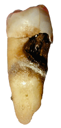
Cervical decay on a premolar
Image via Wikipedia

[amazon_link asins=’B000LT0SK2,B06XPFKHQX,B01GSV4JLU,B01JRGOAAI,B00124YDMY,B07196MR1Y,B000Y4S38I,B0052EBR3I,B000COIT8Y’ template=’ProductCarousel’ store=’finmeacur-20′ marketplace=’US’ link_id=’00b8a5d6-4c13-11e7-ab51-f1b1f69f4aa3′]

Modern lifestyle habits help prevent tooth decay, according to a new study.
……………..…CLICK & SEE.

According to a review of the scientific evidence over the past 150 years, the effects of fluoride toothpaste, good oral hygiene and health education, might override the effects of food alone on tooth decay.

Professor Monty Duggal, an author of the review, said that it’s not enough to just look at what we eat when talking about tooth decay, as other factors seem to be as important.

Fluoride toothpaste changes the effect that some foods have on the teeth, as do other good oral hygiene practices’.

“Future research should investigate a number of lifestyle factors together with different foods that might affect tooth decay. Times have changed and with that, the foods we eat, and how we care for our teeth,” Duggal said.

The overall aim of the review was to look at the evidence for the claim that sugar was the main cause of dental caries (tooth decay).

The researchers concluded that out of 31 studies carefully reviewed, the majority did not find a relationship between the amount of sugar consumed and dental caries, but the frequency of consumption may be important.

Most people now know the best way to prevent tooth decay is to brush with fluoride toothpaste twice a day, especially before going to bed, courtesy dental health education.

The research has been published online in a Supplement to the journal Obesity Reviews.

Sources:The Times Of India

Reblog this post [with Zemanta]
css.php