Categories
News on Health & Science

Broccoli Sprouts Curb Bladder Cancer

Broccoli has always been known to fight cancer. Now, a new study has found that eating concentrated extract of freeze dried broccoli sprouts may curb the development of bladder tumours.

.click to see the picture
4-5 days. These healthy sprouts contain a high percentage of sulforaphane, a compound proven to inhibit the development of cancer. Though not bitter, these sprouts do possess “a little zing” that adds flavor to salads.

.Click to see

Dr. Yuesheng Zhang, PhD, lead researcher and professor of oncology at Roswell Park Cancer Institute, said the study carried out in an animal model provides strong evidence that eating vegetables is beneficial in bladder cancer prevention.

The team led by Zhang tested the ability of the concentrate to prevent bladder tumours in five groups of rats.

The first was a control group, while the second group was given only the broccoli extract to test for safety. The remaining three groups were given a chemical, N-butyl-N-(4-hydroxybutyl) nitrosamine (BBN) in drinking water, which induces bladder cancer.

Out of the three two were given the broccoli extract in diet, two weeks before the carcinogenic chemical was delivered.

The findings revealed that the control group and the group given only the extract developed no tumours, and there was no toxicity from the extract in the rats.

They found that nearly 96 per cent of animals given only BBN developed an average of almost two tumours each of varying sizes.

And about 74 per cent of animals given a low dose of the extract developed cancer, and the number of tumours per rat was 1.39. The group given the high dose of extract had even fewer tumours.

Though animals that had the most protection against development of bladder cancer were given high doses of the extract, Zhang said humans at increased risk for this cancer likely do not need to eat huge amounts of broccoli sprouts in order to derive protective benefits.

The report will appear in the March 1 issue of Cancer Research , a journal of the American Association for Cancer Research.

Click to leanr more aboutBROCCOLI: THE CROWN JEWEL OF NUTRITION”.

Sources: The Times Of India

Enhanced by Zemanta
Categories
Ailmemts & Remedies

Brain Tumor

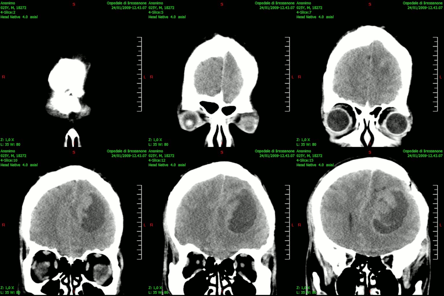
English: TAC_Brain_tumor_glioblastoma-Coronal_...
English: TAC_Brain_tumor_glioblastoma-Coronal_plane Italiano: Immagine TAC della zona cerebrale, identificando un tumore di tipo glioblastoma (Photo credit: Wikipedia)

A brain tumor is any intracranial tumor created by abnormal and uncontrolled cell division, normally either in the brain itself (neurons, glial cells (astrocytes, oligodendrocytes, ependymal cells), lymphatic tissue, blood vessels), in the cranial nerves (myelin-producing Schwann cells), in the brain envelopes (meninges), skull, pituitary and pineal gland, or spread from cancers primarily located in other organs (metastatic tumors). Primary (true) brain tumors are commonly located in the posterior cranial fossa in children and in the anterior two-thirds of the cerebral hemispheres in adults, although they can affect any part of the brain. In the United States in the year 2005, it was estimated that there were 43,800 new cases of brain tumors (Central Brain Tumor Registry of the United States, Primary Brain Tumors in the United States, Statistical Report, 2005 – 2006), which accounted for 1.4 percent of all cancers, 2.4 percent of all cancer deaths, and 20–25 percent of pediatric cancers. Ultimately, it is estimated that there are 13,000 deaths/year as a result of brain tumors……….click & see

Classification

Primary tumors
Tumors occurring in the brain include: astrocytoma, pilocytic astrocytoma, dysembryoplastic neuroepithelial tumor, oligodendrogliomas, ependymoma, glioblastoma multiforme, mixed gliomas, oligoastrocytomas, medulloblastoma, retinoblastoma, neuroblastoma, germinoma and teratoma.

Most primary brain tumors originate from glia (gliomas) such as astrocytes (astrocytomas), oligodendrocytes (oligodendrogliomas), or ependymal cells (ependymoma). There are also mixed forms, with both an astrocytic and an oligodendroglial cell component. These are called mixed gliomas or oligoastrocytomas. Plus, mixed glio-neuronal tumors (tumors displaying a neuronal, as well as a glial component, e.g. gangliogliomas, disembryoplastic neuroepithelial tumors) and tumors originating from neuronal cells (e.g. gangliocytoma, central gangliocytoma) can also be encountered.

Other varieties of primary brain tumors include: primitive neuroectodermal tumors (PNET, e.g. medulloblastoma, medulloepithelioma, neuroblastoma, retinoblastoma, ependymoblastoma), tumors of the pineal parenchyma (e.g. pineocytoma, pineoblastoma), ependymal cell tumors, choroid plexus tumors, neuroepithelial tumors of uncertain origin (e.g. gliomatosis cerebri, astroblastoma), etc.

From a histological perspective, astrocytomas, oligondedrogliomas, oligoastrocytomas, and teratomas may be benign or malignant. Glioblastoma multiforme represents the most aggressive variety of malignant glioma. At the opposite end of the spectrum, there are so-called pilocytic astrocytomas, a distinct variety of astrocytic tumors. The majority of them are located in the posterior cranial fossa, affect mainly children and young adults, and have a clinically favorable course and prognosis. Teratomas and other germ cell tumors also may have a favorable prognosis, although they have the capacity to grow very large.

Another type of primary intracranial tumor is primary cerebral lymphoma, also known as primary CNS lymphoma, which is a type of non-Hodgkin’s lymphoma that is much more prevalent in those with severe immunosuppression, e.g. AIDS.

In contrast to other types of cancer, primary brain tumors rarely metastasize, and in this rare event, the tumor cells spread within the skull and spinal canal through the cerebrospinal fluid, rather than via bloodstream to other organs.

There are various classification systems currently in use for primary brain tumors, the most common being the World Health Organization (WHO) brain tumor classification, introduced in 1993.

Secondary tumors and non-tumor lesions
Secondary or metastatic brain tumors originate from malignant tumors (cancers) located primarily in other organs. Their incidence is higher than that of primary brain tumors. The most frequent types of metastatic brain tumors originate in the lung, skin (malignant melanoma), kidney (hypernephroma), breast (breast carcinoma), and colon (colon carcinoma). These tumor cells reach the brain via the blood-stream.

Some non-tumoral masses and lesions can mimic tumors of the central nervous system. These include tuberculosis of the brain, cerebral abscess (commonly in toxoplasmosis), and hamartomas (for example, in tuberous sclerosis and von Recklinghausen neurofibromatosis).

Symptoms of brain tumors may depend on two factors: tumor size (volume) and tumor location. The time point of symptom onset in the course of disease correlates in many cases with the nature of the tumor (“benign”, i.e. slow-growing/late symptom onset, or malignant (fast growing/early symptom onset).

Many low-grade (benign) tumors can remain asymptomatic (symptom-free) for years and they may accidentally be discovered by imaging exams for unrelated reasons (such as a minor trauma).

New onset of epilepsy is a frequent reason for seeking medical attention in brain tumor cases.

Large tumors or tumors with extensive perifocal swelling edema inevitably lead to elevated intracranial pressure (intracranial hypertension), which translates clinically into headaches, vomiting (sometimes without nausea), altered state of consciousness (somnolence, coma), dilatation of the pupil on the side of the lesion (anisocoria), papilledema (prominent optic disc at the funduscopic examination). However, even small tumors obstructing the passage of cerebrospinal fluid (CSF) may cause early signs of increased intracranial pressure. Increased intracranial pressure may result in herniation (i.e. displacement) of certain parts of the brain, such as the cerebellar tonsils or the temporal uncus, resulting in lethal brainstem compression. In young children, elevated intracranial pressure may cause an increase in the diameter of the skull and bulging of the fontanelles.

Depending on the tumor location and the damage it may have caused to surrounding brain structures, either through compression or infiltration, any type of focal neurologic symptoms may occur, such as cognitive and behavioral impairment, personality changes, hemiparesis, (hemi) hypesthesia, aphasia, ataxia, visual field impairment, facial paralysis, double vision, tremor etc. These symptoms are not specific for brain tumors – they may be caused by a large variety of neurologic conditions (e.g. stroke, traumatic brain injury). What counts, however, is the location of the lesion and the functional systems (e.g. motor, sensory, visual, etc.) it affects.

A bilateral temporal visual field defect (bitemporal hemianopia—due to compression of the optic chiasm), often associated with endocrine disfunction—either hypopituitarism or hyperproduction of pituitary hormones and hyperprolactinemia is suggestive of a pituitary tumor.

Brain tumors in infants and children
In 2000 approximately 2.76 children per 100,000 will be affected by a CNS tumor in the United States each year. This rate has been increasing and by 2005 was 3.0 children per 100,000. This is approximately 2,500-3,000 pediatric brain tumors occurring each year in the US. The tumor incidence is increasing by about 2.7% per year. The CNS Cancer survival rate in children is approximately 60%. However, this rate varies with the age of onset (younger has higher mortality) and cancer type.

In children under 2, about 70% of brain tumors are medulloblastoma, ependymoma, and low-grade glioma. Less commonly, and seen usually in infants, are teratoma and atypical teratoid rhabdoid tumor

Diagnosis
Although there is no specific clinical symptom or sign for brain tumors, slowly progressive focal neurologic signs and signs of elevated intracranial pressure, as well as epilepsy in a patient with a negative history for epilepsy should raise red flags. However, a sudden onset of symptoms, such as an epileptic seizure in a patient with no prior history of epilepsy, sudden intracranial hypertension (this may be due to bleeding within the tumor, brain swelling or obstruction of cerebrospinal fluid’s passage) is also possible.

Symptoms include phantom odors and tastes. Often, in the case of metastatic tumors, the smell of vulcanized rubber is prevalent.

Imaging plays a central role in the diagnosis of brain tumors. Early imaging methods—invasive and sometimes dangerous—such as pneumoencephalography and cerebral angiography, have been abandoned in recent times in favor of non-invasive, high-resolution modalities, such as computed tomography (CT) and especially magnetic resonance imaging (MRI). Benign brain tumors often show up as hypodense (darker than brain tissue) mass lesions on cranial CT-scans. On MRI, they appear either hypo- (darker than brain tissue) or isointense (same intensity as brain tissue) on T1-weighted scans, or hyperintense (brighter than brain tissue) on T2-weighted MRI. Perifocal edema also appears hyperintense on T2-weighted MRI. Contrast agent uptake, sometimes in characteristic patterns, can be demonstrated on either CT or MRI-scans in most malignant primary and metastatic brain tumors. This is due to the fact that these tumors disrupt the normal functioning of the blood-brain barrier and lead to an increase in its permeability.

Electrophysiological exams, such as electroencephalography (EEG) play a marginal role in the diagnosis of brain tumors.

The definitive diagnosis of brain tumor can only be confirmed by histological examination of tumor tissue samples obtained either by means of brain biopsy or open surgery. The histologic examination is essential for determining the appropriate treatment and the correct prognosis.

Treatment and prognosis
Meningiomas, with the exception of some tumors located at the skull base, can be successfully removed surgically, but the chances are less than 50%. In more difficult cases, stereotactic radiosurgery, such as Gamma Knife radiosurgery, remains a viable option.

Most pituitary adenomas can be removed surgically, often using a minimally invasive approach through the nasal cavity and skull base (trans-nasal, trans-sphenoidal approach). Large pituitary adenomas require a craniotomy (opening of the skull) for their removal. Radiotherapy, including stereotactic approaches, is reserved for the inoperable cases.

Although there is no generally accepted therapeutic management for primary brain tumors, a surgical attempt at tumor removal or at least cytoreduction (that is, removal of as much tumor as possible, in order to reduce the number of tumor cells available for proliferation) is considered in most cases. However, due to the infiltrative nature of these lesions, tumor recurrence, even following an apparently complete surgical removal, is not uncommon. Postoperative radiotherapy and chemotherapy are integral parts of the therapeutic standard for malignant tumors. Radiotherapy may also be administered in cases of “low-grade” gliomas, when a significant tumor burden reduction could not be achieved surgically.

Survival rates in primary brain tumors depend on the type of tumor, age, functional status of the patient, the extent of surgical tumor removal, to mention just a few factors.

Patients with benign gliomas may survive for many years while survival in most cases of glioblastoma multiforme is limited to a few months after diagnosis.

The main treatment option for single metastatic tumors is surgical removal, followed by radiotherapy and/or chemotherapy. Multiple metastatic tumors are generally treated with radiotherapy and chemotherapy. Stereotactic radiosurgery, such as Gamma Knife radiosurgery, remains a viable option. However, the prognosis in such cases is determined by the primary tumor, and it is generally poor.

A shunt operation is used not as a cure but to relieve the symptoms. The hydrocephalus caused by the blocking drainage of the cerebrospinal fluid can be removed with this operation.

Click for more knowledge on
Brain Tumor Causes, Symptoms, Diagnosis, Treatment

Click for Homeopathic Treatment….…………(1)………(2).…..…(3).………(4)

Natural Brain Tumor Cure

Disclaimer: This information is not meant to be a substitute for professional medical advise or help. It is always best to consult with a Physician about serious health concerns. This information is in no way intended to diagnose or prescribe remedies.This is purely for educational purpose.

Resources:http://en.wikipedia.org/wiki/Brain_tumor

Enhanced by Zemanta
Categories
Ailmemts & Remedies

Brain Tumor

[amazon_link asins=’0992941806,B0084K0FBM,B01FUR1100,0989974006,B01LY2L9QW,B0731BS5TL,0805079688,069265626X,0199897794′ template=’ProductCarousel’ store=’finmeacur-20′ marketplace=’US’ link_id=’27b61790-a740-11e7-b012-a726291336ef’]

A brain tumor is any intracranial tumor created by abnormal and uncontrolled cell division, normally either in the brain itself (neurons, glial cells (astrocytes, oligodendrocytes, ependymal cells), lymphatic tissue, blood vessels), in the cranial nerves (myelin-producing Schwann cells), in the brain envelopes (meninges), skull, pituitary and pineal gland, or spread from cancers primarily located in other organs (metastatic tumors). Primary (true) brain tumors are commonly located in the posterior cranial fossa in children and in the anterior two-thirds of the cerebral hemispheres in adults, although they can affect any part of the brain. In the United States in the year 2005, it was estimated that there were 43,800 new cases of brain tumors (Central Brain Tumor Registry of the United States, Primary Brain Tumors in the United States, Statistical Report, 2005 – 2006) , which accounted for 1.4 percent of all cancers, 2.4 percent of all cancer deaths. it is estimated that there are 13,000 deaths/year as a result of brain tumors.

CLICK & SEE 

Brain tumors may be cancerous or non-cancerous. however, unlike most tumors elsewhere in the body, cancerous and non-cancerous brain tumors may be equally serious. The seriousness of a brain tumor depends on its location, size and rate of growth. Both types of tumors can compress nearby tissue, causing pressure to build up inside the skull.

Tumors that arise from brain tissue or from the meninges, the membranes that cover the brain, are called primary tumors and may be cancerous or non-cancerous. most primary tumors arise from nerve cells. less commonly, tumors may develop from the cells that support nerve cells or from the cells of the meninges. Primary brain tumors that develop in the pituitary gland at the base of the brain are discussed in the article on pituitary tumors.

Secondary brain tumors are more common than primary tumors. They are always cancerous, having developed from cells that have spread to the brain from cancers in other areas, such as the breast. several secondary tumors may develop simultaneously.

Brain tumors are slightly more common in males and usually develop between age 60 and 70. certain types of tumors, such as neuroblastomas, affect only children.

Primary tumors
Tumors occurring in the brain include: astrocytoma, pilocytic astrocytoma, dysembryoplastic neuroepithelial tumor, oligodendrogliomas, ependymoma, glioblastoma multiforme, mixed gliomas, oligoastrocytomas, medulloblastoma, retinoblastoma, neuroblastoma, germinoma and teratoma.

Most primary brain tumors originate from glia (gliomas) such as astrocytes (astrocytomas), oligodendrocytes (oligodendrogliomas), or ependymal cells (ependymoma). There are also mixed forms, with both an astrocytic and an oligodendroglial cell component. These are called mixed gliomas or oligoastrocytomas. Plus, mixed glio-neuronal tumors (tumors displaying a neuronal, as well as a glial component, e.g. gangliogliomas, disembryoplastic neuroepithelial tumors) and tumors originating from neuronal cells (e.g. gangliocytoma, central gangliocytoma) can also be encountered.

Other varieties of primary brain tumors include: primitive neuroectodermal tumors (PNET, e.g. medulloblastoma, medulloepithelioma, neuroblastoma, retinoblastoma, ependymoblastoma), tumors of the pineal parenchyma (e.g. pineocytoma, pineoblastoma), ependymal cell tumors, choroid plexus tumors, neuroepithelial tumors of uncertain origin (e.g. gliomatosis cerebri, astroblastoma), etc.

From a histological perspective, astrocytomas, oligondedrogliomas, oligoastrocytomas, and teratomas may be benign or malignant. Glioblastoma multiforme represents the most aggressive variety of malignant glioma. At the opposite end of the spectrum, there are so-called pilocytic astrocytomas, a distinct variety of astrocytic tumors. The majority of them are located in the posterior cranial fossa, affect mainly children and young adults, and have a clinically favorable course and prognosis. Teratomas and other germ cell tumors also may have a favorable prognosis, although they have the capacity to grow very large.

Another type of primary intracranial tumor is primary cerebral lymphoma, also known as primary CNS lymphoma, which is a type of non-Hodgkin’s lymphoma that is much more prevalent in those with severe immunosuppression, e.g. AIDS.

In contrast to other types of cancer, primary brain tumors rarely metastasize, and in this rare event, the tumor cells spread within the skull and spinal canal through the cerebrospinal fluid, rather than via bloodstream to other organs.

There are various classification systems currently in use for primary brain tumors, the most common being the World Health Organization (WHO) brain tumor classification, introduced in 1993.

Secondary tumors and non-tumor lesions:
Secondary or metastatic brain tumors originate from malignant tumors (cancers) located primarily in other organs. Their incidence is higher than that of primary brain tumors. The most frequent types of metastatic brain tumors originate in the lung, skin (malignant melanoma), kidney (hypernephroma), breast (breast carcinoma), and colon (colon carcinoma). These tumor cells reach the brain via the blood-stream.

Some non-tumoral masses and lesions can mimic tumors of the central nervous system. These include tuberculosis of the brain, cerebral abscess (commonly in toxoplasmosis), and hamartomas (for example, in tuberous sclerosis and von Recklinghausen neurofibromatosis).

Symptoms of brain tumors may depend on two factors: tumor size (volume) and tumor location. The time point of symptom onset in the course of disease correlates in many cases with the nature of the tumor (“benign”, i.e. slow-growing/late symptom onset, or malignant (fast growing/early symptom onset).

Many low-grade (benign) tumors can remain asymptomatic (symptom-free) for years and they may accidentally be discovered by imaging exams for unrelated reasons (such as a minor trauma).

New onset of epilepsy is a frequent reason for seeking medical attention in brain tumor cases.

Large tumors or tumors with extensive perifocal swelling edema inevitably lead to elevated intracranial pressure (intracranial hypertension), which translates clinically into headaches, vomiting (sometimes without nausea), altered state of consciousness (somnolence, coma), dilatation of the pupil on the side of the lesion (anisocoria), papilledema (prominent optic disc at the funduscopic examination). However, even small tumors obstructing the passage of cerebrospinal fluid (CSF) may cause early signs of increased intracranial pressure. Increased intracranial pressure may result in herniation (i.e. displacement) of certain parts of the brain, such as the cerebellar tonsils or the temporal uncus, resulting in lethal brainstem compression. In young children, elevated intracranial pressure may cause an increase in the diameter of the skull and bulging of the fontanelles.

Depending on the tumor location and the damage it may have caused to surrounding brain structures, either through compression or infiltration, any type of focal neurologic symptoms may occur, such as cognitive and behavioral impairment, personality changes, hemiparesis, (hemi) hypesthesia, aphasia, ataxia, visual field impairment, facial paralysis, double vision, tremor etc. These symptoms are not specific for brain tumors – they may be caused by a large variety of neurologic conditions (e.g. stroke, traumatic brain injury). What counts, however, is the location of the lesion and the functional systems (e.g. motor, sensory, visual, etc.) it affects.

A bilateral temporal visual field defect (bitemporal hemianopia—due to compression of the optic chiasm), often associated with endocrine disfunction—either hypopituitarism or hyperproduction of pituitary hormones and hyperprolactinemia is suggestive of a pituitary tumor.

Brain tumors in infants and children:

Approximately 2,500-3,000 pediatric brain tumors occurring each year in the US. The tumor incidence is increasing by about 2.7% per year. The CNS Cancer survival rate in children is approximately 60%. However, this rate varies with the age of onset (younger has higher mortality) and cancer type.

In children under 2, about 70% of brain tumors are medulloblastoma, ependymoma, and low-grade glioma. Less commonly, and seen usually in infants, are teratoma and atypical teratoid rhabdoid tumor

Symptoms:
Symptoms usually occur when a primary tumor or a metastasis compresses part of the brain or raises the pressure inside the skull. they may include:

· Headache that is usually more severe in the morning and is worsened by coughing or bending over.
· Nausea and vomiting.
· Blurry vision.

Other symptoms tend to be related to whichever area of the brain is affected by the tumor and may include:

· Slurred speech.
· Strabismus due to partial paralysis of the eye muscles.
· Difficulty reading and writing.
· Change of personality.
· Numbness and weakness of the limbs on one side of the body.

A tumor may also cause seizures. Sometimes, a tumor blocks the flow of the cerebrospinal fluid that circulates in and around the brain and spinal cord. As a result, the pressure inside the ventricles increases and leads to further compression of brain tissue. Left untreated, drowsiness can develop, which may eventually progress to coma and death.

Diagnosis:
Although there is no specific clinical symptom or sign for brain tumors, slowly progressive focal neurologic signs and signs of elevated intracranial pressure, as well as epilepsy in a patient with a negative history for epilepsy should raise red flags. However, a sudden onset of symptoms, such as an epileptic seizure in a patient with no prior history of epilepsy, sudden intracranial hypertension (this may be due to bleeding within the tumor, brain swelling or obstruction of cerebrospinal fluid’s passage) is also possible.

Symptoms include phantom odors and tastes. Often, in the case of metastatic tumors, the smell of galvanised vulcan rubber is prevalent.

Imaging plays a central role in the diagnosis of brain tumors. Early imaging methods  invasive and sometimes dangerous  such as pneumoencephalography and cerebral angiography, have been abandoned in recent times in favor of non-invasive, high-resolution modalities, such as computed tomography (CT) and especially magnetic resonance imaging (MRI). Benign brain tumors often show up as hypodense (darker than brain tissue) mass lesions on cranial CT-scans. On MRI, they appear either hypo- (darker than brain tissue) or isointense (same intensity as brain tissue) on T1-weighted scans, or hyperintense (brighter than brain tissue) on T2-weighted MRI. Perifocal edema also appears hyperintense on T2-weighted MRI. Contrast agent uptake, sometimes in characteristic patterns, can be demonstrated on either CT or MRI-scans in most malignant primary and metastatic brain tumors. This is due to the fact that these tumors disrupt the normal functioning of the blood-brain barrier and lead to an increase in its permeability.
CT scan of brain showing brain cancer to left parietal lobe in the peri-ventricular

Electrophysiological exams, such as electroencephalography (EEG) play a marginal role in the diagnosis of brain tumors.

The definitive diagnosis of brain tumor can only be confirmed by histological examination of tumor tissue samples obtained either by means of brain biopsy or open surgery. The histologic examination is essential for determining the appropriate treatment and the correct prognosis.

Treatment and prognosis:
Meningiomas, with the exception of some tumors located at the skull base, can be successfully removed surgically, but the chances are less than 50%. In more difficult cases, stereotactic radiosurgery, such as Gamma Knife radiosurgery, remains a viable option.

Most pituitary adenomas can be removed surgically, often using a minimally invasive approach through the nasal cavity and skull base (trans-nasal, trans-sphenoidal approach). Large pituitary adenomas require a craniotomy (opening of the skull) for their removal. Radiotherapy, including stereotactic approaches, is reserved for the inoperable cases.

Although there is no generally accepted therapeutic management for primary brain tumors, a surgical attempt at tumor removal or at least cytoreduction (that is, removal of as much tumor as possible, in order to reduce the number of tumor cells available for proliferation) is considered in most cases. However, due to the infiltrative nature of these lesions, tumor recurrence, even following an apparently complete surgical removal, is not uncommon. Postoperative radiotherapy and chemotherapy are integral parts of the therapeutic standard for malignant tumors. Radiotherapy may also be administered in cases of “low-grade” gliomas, when a significant tumor burden reduction could not be achieved surgically.

Survival rates in primary brain tumors depend on the type of tumor, age, functional status of the patient, the extent of surgical tumor removal, to mention just a few factors.

Patients with benign gliomas may survive for many years while survival in most cases of glioblastoma multiforme is limited to a few months after diagnosis.

The main treatment option for single metastatic tumors is surgical removal, followed by radiotherapy and/or chemotherapy. Multiple metastatic tumors are generally treated with radiotherapy and chemotherapy. Stereotactic radiosurgery, such as Gamma Knife radiosurgery, remains a viable option. However, the prognosis in such cases is determined by the primary tumor, and it is generally poor.

A shunt operation is used not as a cure but to relieve the symptoms. The hydrocephalus caused by the blocking drainage of the cerebrospinal fluid can be removed with this operation.

The prognosis is usually better for slow growing noncancerous tumors, and many people with such a tumor can be completely cured. For other tumors, the prognosis depends on the type of cell affected and whether the tumor can be surgically removed. About 1 in 4 people is alive 2 years after the initial diagnosis of a primary cancerous brain tumor, but few people live longer than 5 years. Most people with brain metastases do not live longer than 6 months, although in rare cases, a person with a single metastatic tumor may be cured. All types of brain tumor carry a risk of permanently damaging nearby brain tissue.

Recomended Ayurvedic Therapy: Nasya , Rakthmokshan

Ayurvedic and Herbal Treatment of Brain Tumor

Brain Tumor as related to Herbal Medicine

Homeopathy: Treatment of Brain Tumor

Disclaimer: This information is not meant to be a substitute for professional medical advise or help. It is always best to consult with a Physician about serious health concerns. This information is in no way intended to diagnose or prescribe remedies.

Resources:
http://en.wikipedia.org/wiki/Brain_tumor
http://www.charak.com/DiseasePage.asp?thx=1&id=12

Reblog this post [with Zemanta]
Categories
Ailmemts & Remedies

Amenorrhea

[amazon_link asins=’099723668X,B00W80QG76,B01LYWY2AI,B00DNN6HB2,B06XP2Q44N,B01M2C0D3E,B00G6BHQWW,160327863X’ template=’ProductCarousel’ store=’finmeacur-20′ marketplace=’US’ link_id=’9eec3a79-24c8-11e7-b9ed-432f231982a0′]

There are two types of amenorrhea, primary and secondary. Primary amenorrhea is the term used to describe menstruation that as not started at all by age 16. once menstruation has started at puberty, it is normal for it to stop during pregnancy, for a few months following childbirth, while breast-feeding, after ceasing to take oral contraceptive pills, and permanently at menopause. If menstrual periods stop at any other time for at least 3 continuous months, the condition is known as secondary amenorrhea.

CLICK & SEE

What are the causes?
Amenorrhea is often caused by disturbance in the female sex hormones, which may be brought on by factors such as stress or depression. Excessive exercise and extreme or sudden weight loss may also lead to such hormonal disturbances and are common causes of amenorrhea in athletes, gymnasts, and ballot dancers. Hormonal changes may lead to primary or secondary amenorrhea, depending on when they occur.

Primary amenorrhea is a characteristic feature of delayed puberty, and may be caused by a chromosomal abnormality. The failure of menstrual periods to start at puberty may also be due to a condition in which the hymen (the thin membrane over the vagina) has no opening, and menstrual blood cannot leave the body. in rare cases, the uterus is absent from birth, and therefore no menstruation can occur.

Secondary amenorrhea may be due to a pituitary gland disorder, such as pituitary tumor, or it may be due to a premature menopause, in which menstrual periods cease before age 35. Other possible causes are disorders of the ovaries, such as polycystic ovary syndrome, and treatments such as chemotherapy and radiation therapy, that can result in damage to the ovaries.

What might be done?
Treatment is not needed if amenorrhea occurs for a few months after stopping oral contraceptives or during pregnancy or breast-feeding. Enstruation usually resumes within a few months of giving birth if you are not breast-feeding or within a month of stopping breast-feeding. If you are near menopause, amenorrhea will be permanent.

Amenorrhea that occurs at any other time should be investigated. Your doctor will examine you and may do a pregnancy test. You may also need to have blood tests to measure hormone levels, ultrasound scanning of the ovaries and uterus, and ct scanning of the pituitary gland.

treatment of the underlying disorder induces menstruation in most cases. if the cause cannot be treated, hormonal treatment may be used to start menstruation. amenorrhea due to weight loss, stress, or excessive exercise should clear up once the problem is overcome.

Recommended Ayurvedic Therapy: Virechan , Basti

Click to learn more about Amenorrhea……(1).……(2).……(3)…….(4)

Homeopathic treatment of Amenorrhea ………………………………..(1).(2)…..(3)

Herbal Home Remedy of Amenorrhea ………………………….(1).……..(2)

Parsley is the most beneficial herb for the treatment of Amenorrhea

Disclaimer: This information is not meant to be a substitute for professional medical advise or help. It is always best to consult with a Physician about serious health concerns. This information is in no way intended to diagnose or prescribe remedies.This is purely for educational purpose.

Sources: http://www.charak.com/DiseasePage.asp?thx=1&id=278

Categories
News on Health & Science

Sunshine Vitamin Repairs Your Skin

[amazon_link asins=’B00IDWP4IA,B01G6SAJO6′ template=’ProductCarousel’ store=’finmeacur-20′ marketplace=’US’ link_id=’c967d22e-1a11-11e7-9af0-1d71a3cf7a31′]

The Sunshine Vitamin Repairs Your Skin and Boosts Your Immune System
Sunshine may be able to help fight skin diseases and cancer by attracting immune cells to the skin surface……....click & see

Sunshine causes the skin to produce vitamin D3, and immune cells in the skin, known as dendritic cells, can convert vitamin D3 into its active form. The active vitamin D3 then causes T-cells to make surface changes that allow them to migrate to the uppermost layer of the skin.

T-cells destroy damaged and infected cells, and also regulate other immune cells.

The skin disorder psoriasis is sometimes treated with vitamin D3 creams; these creams may work by moving T-cells into the skin.

Add skin repair to the growing legion of benefits associated with getting your body’s optimal dose of vitamin D from the sun. According to scientists, these new findings may explain how T-cells get routed to the surface of your skin via the sun when it has been exposed to damage.

Of course, the real trick about getting the optimal amount of vitamin D is getting the appropriate and safe amount of sun exposure , a big challenge right now for people living in the Northern Hemisphere.

Ideally, you should have exposure to the sun, however, safe tanning beds are another option. You can see the list of safe U.S. tanning beds.

Just a reminder, if you choose to take a high-quality cod liver oil to increase your vitamin D levels while also getting valuable omega-3 fats, It is strongly urged that you to have your blood levels checked regularly, as it is potentially dangerous not to do so.

Source:www.mercola.com

css.php