Categories
Diagnonistic Test

ERCP (Endoscopic Retrograde Cholangiopancreatography)

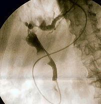
Fluoroscopic image of :en:common bile duct sto...
Image via Wikipedia

[amazon_link asins=’1455723673,1118769414,1493923196,1556641958′ template=’ProductCarousel’ store=’finmeacur-20′ marketplace=’US’ link_id=’84171de6-4815-11e7-874d-29b026a2b537′]

click to see
Endoscopic retrograde cholangiopancreatography (en-doh-SKAH-pik REH-troh-grayd koh-LAN-jee-oh-PANG-kree-uh-TAH-gruh-fee) (ERCP) enables the physician to diagnose problems in the liver, gallbladder, bile ducts, and pancreas. The liver is a large organ that, among other things, makes a liquid called bile that helps with digestion. The gallbladder is a small, pear-shaped organ that stores bile until it is needed for digestion. The bile ducts are tubes that carry bile from the liver to the gallbladder and small intestine. These ducts are sometimes called the biliary tree. The pancreas is a large gland that produces chemicals that help with digestion and hormones such as insulin.
.click to see
ERCP is used primarily to diagnose and treat conditions of the bile ducts, including gallstones, inflammatory strictures (scars), leaks (from trauma and surgery), and cancer. ERCP combines the use of x rays and an endoscope, which is a long, flexible, lighted tube. Through the endoscope, the physician can see the inside of the stomach and duodenum, and inject dyes into the ducts in the biliary tree and pancreas so they can be seen on x rays.

For the procedure, you will lie on your left side on an examining table in an x-ray room. You will be given medication to help numb the back of your throat and a sedative to help you relax during the exam. You will swallow the endoscope, and the physician will then guide the scope through your esophagus, stomach, and duodenum until it reaches the spot where the ducts of the biliary tree and pancreas open into the duodenum. At this time, you will be turned to lie flat on your stomach, and the physician will pass a small plastic tube through the scope. Through the tube, the physician will inject a dye into the ducts to make them show up clearly on x rays. X rays are taken as soon as the dye is injected.

If the exam shows a gallstone or narrowing of the ducts, the physician can insert instruments into the scope to remove or relieve the obstruction. Also, tissue samples (biopsy) can be taken for further testing.

Possible complications of ERCP include pancreatitis (inflammation of the pancreas), infection, bleeding, and perforation of the duodenum. Except for pancreatitis, such problems are uncommon. You may have tenderness or a lump where the sedative was injected, but that should go away in a few days.

ERCP takes 30 minutes to 2 hours. You may have some discomfort when the physician blows air into the duodenum and injects the dye into the ducts. However, the pain medicine and sedative should keep you from feeling too much discomfort. After the procedure, you will need to stay at the hospital for 1 to 2 hours until the sedative wears off. The physician will make sure you do not have signs of complications before you leave. If any kind of treatment is done during ERCP, such as removing a gallstone, you may need to stay in the hospital overnight.

Preparation:-
Your stomach and duodenum must be empty for the procedure to be accurate and safe. You will not be able to eat or drink anything after midnight the night before the procedure, or for 6 to 8 hours beforehand, depending on the time of your procedure. Also, the physician will need to know whether you have any allergies, especially to iodine, which is in the dye. You must also arrange for someone to take you home—you will not be allowed to drive because of the sedatives. The physician may give you other special instructions.

For More Information:-
American Gastroenterological Association (AGA)
National Office
4930 Del Ray Avenue
Bethesda, MD 20814
Phone: 301–654–2055
Fax: 301–654–5920
Email: info@gastro.org
Internet: www.gastro.org

National Digestive Diseases Information Clearinghouse
2 Information Way
Bethesda, MD 20892–3570
Phone: 1–800–891–5389
TTY: 1–866–569–1162
Fax: 703–738–4929
Email: nddic@info.niddk.nih.gov
Internet: www.digestive.niddk.nih.gov

Sources:http://digestive.niddk.nih.gov/ddiseases/pubs/ercp/index.htm

Enhanced by Zemanta
Categories
Healthy Tips

How Mind Can Suppress Hunger Pangs

You might not be keeping a check on the amount of calories you’re consuming during a party, but your brain will, say Yale University researchers, who have identified a molecule that tells brain that the stomach is full – and signals it’s time to say no to a second piece of Delicious food and push back from the dining table.
....click to see the picture
.…………
In a study on rodents, the researchers have discovered that one type of lipid produced in the gut, called N-acylphosphatidylethanolamines or NAPEs, rises after eating fatty foods.

The NAPEs enter the bloodstream and go straight to the brain, where they concentrate in a brain region that controls food intake and energy expenditure.

Led by Gerald I. Shulman, Yale professor of medicine and cellular & molecular physiology and a Howard Hughes Medical Institute investigator, the researchers suggested that the molecule may help regulate how much animals and people eat.

NAPEs are synthesized and secreted into the blood by the small intestine after fatty foods are eaten. The researchers found that mice and rats injected regularly with NAPEs ate less food and lost weight. In addition, treatment with NAPEs appeared to reduce the activity of “hunger” neurons in the brain while stimulating activity in neurons that are believed to play a role in reducing appetite.

In the last two decades, scientists have made great inroads toward understanding how the body communicates with the brain to control food intake. Till date, hormones such as leptin that act as regulators of this complex system have proved disappointing when tested as potential weight-loss treatments in humans.

The researchers are now planning to investigate how the new findings apply to humans.The team will first study non-human primates to determine if NAPE on centrations increase in a similar fashion after fat ingestion.

Then, Shulman said: “If chronic NAPE treatment is well tolerated and can cause weight loss by a reduction of food intake, we would have strong impetus to move forward with human NAPE trials.” The findings are published in the latest issue of the journal Cell.

Sources: The Times Of India

Enhanced by Zemanta
Categories
Featured

The Digestive System and How It Works

[amazon_link asins=’B009DQO8LY,B0189AHZWI,B06ZZ5D55N,0071441964,B00DGBMGPW,B0189AFZ4I,B00ICXAH2I,B00ICXAH14,1571457895′ template=’ProductCarousel’ store=’finmeacur-20′ marketplace=’US’ link_id=’d8f2fa7d-7b28-11e7-adde-d52a37586dde’]

The digestive system is a series of hollow organs joined in a long, twisting tube from the mouth to the anus(see the figure below) . Inside this tube is a lining called the mucosa. In the mouth, stomach, and small intestine, the mucosa contains tiny glands that produce juices to help digest food…..click & see

Two solid organs, the liver and the pancreas, produce digestive juices that reach the intestine through small tubes. In addition, parts of other organ systems (for instance, nerves and blood) play a major role in the digestive system.

Why is digestion important?

When we eat such things as bread, meat, and vegetables, they are not in a form that the body can use as nourishment. Our food and drink must be changed into smaller molecules of nutrients before they can be absorbed into the blood and carried to cells throughout the body. Digestion is the process by which food and drink are broken down into their smallest parts so that the body can use them to build and nourish cells and to provide energy.

How is food digested?

Digestion involves the mixing of food, its movement through the digestive tract, and the chemical breakdown of the large molecules of food into smaller molecules. Digestion begins in the mouth, when we chew and swallow, and is completed in the small intestine. The chemical process varies somewhat for different kinds of food.

Movement of Food Through the System

The large, hollow organs of the digestive system contain muscle that enables their walls to move. The movement of organ walls can propel food and liquid and also can mix the contents within each organ. Typical movement of the esophagus, stomach, and intestine is called peristalsis. The action of peristalsis looks like an ocean wave moving through the muscle. The muscle of the organ produces a narrowing and then propels the narrowed portion slowly down the length of the organ. These waves of narrowing push the food and fluid in front of them through each hollow organ.

click & see

The first major muscle movement occurs when food or liquid is swallowed. Although we are able to start swallowing by choice, once the swallow begins, it becomes involuntary and proceeds under the control of the nerves.

The esophagus is the organ into which the swallowed food is pushed. It connects the throat above with the stomach below. At the junction of the esophagus and stomach, there is a ringlike valve closing the passage between the two organs. However, as the food approaches the closed ring, the surrounding muscles relax and allow the food to pass.

The food then enters the stomach, which has three mechanical tasks to do. First, the stomach must store the swallowed food and liquid. This requires the muscle of the upper part of the stomach to relax and accept large volumes of swallowed material. The second job is to mix up the food, liquid, and digestive juice produced by the stomach. The lower part of the stomach mixes these materials by its muscle action. The third task of the stomach is to empty its contents slowly into the small intestine.

Several factors affect emptying of the stomach, including the nature of the food (mainly its fat and protein content) and the degree of muscle action of the emptying stomach and the next organ to receive the contents (the small intestine). As the food is digested in the small intestine and dissolved into the juices from the pancreas, liver, and intestine, the contents of the intestine are mixed and pushed forward to allow further digestion.

Finally, all of the digested nutrients are absorbed through the intestinal walls. The waste products of this process include undigested parts of the food, known as fiber, and older cells that have been shed from the mucosa. These materials are propelled into the colon, where they remain, usually for a day or two, until the feces are expelled by a bowel movement.

Production of Digestive Juices

The glands that act first are in the mouth—the salivary glands. Saliva produced by these glands contains an enzyme that begins to digest the starch from food into smaller molecules.

The next set of digestive glands is in the stomach lining. They produce stomach acid and an enzyme that digests protein. One of the unsolved puzzles of the digestive system is why the acid juice of the stomach does not dissolve the tissue of the stomach itself. In most people, the stomach mucosa is able to resist the juice, although food and other tissues of the body cannot.

After the stomach empties the food and juice mixture into the small intestine, the juices of two other digestive organs mix with the food to continue the process of digestion. One of these organs is the pancreas. It produces a juice that contains a wide array of enzymes to break down the carbohydrate, fat, and protein in food. Other enzymes that are active in the process come from glands in the wall of the intestine or even a part of that wall.

The liver produces yet another digestive juice—bile. The bile is stored between meals in the gallbladder. At mealtime, it is squeezed out of the gallbladder into the bile ducts to reach the intestine and mix with the fat in our food. The bile acids dissolve the fat into the watery contents of the intestine, much like detergents that dissolve grease from a frying pan. After the fat is dissolved, it is digested by enzymes from the pancreas and the lining of the intestine.

Absorption and Transport of Nutrients

Digested molecules of food, as well as water and minerals from the diet, are absorbed from the cavity of the upper small intestine. Most absorbed materials cross the mucosa into the blood and are carried off in the bloodstream to other parts of the body for storage or further chemical change. As already noted, this part of the process varies with different types of nutrients.

Carbohydrates. It is recommended that about 55 to 60 percent of total daily calories be from carbohydrates. Some of our most common foods contain mostly carbohydrates. Examples are bread, potatoes, legumes, rice, spaghetti, fruits, and vegetables. Many of these foods contain both starch and fiber.

The digestible carbohydrates are broken into simpler molecules by enzymes in the saliva, in juice produced by the pancreas, and in the lining of the small intestine. Starch is digested in two steps: First, an enzyme in the saliva and pancreatic juice breaks the starch into molecules called maltose; then an enzyme in the lining of the small intestine (maltase) splits the maltose into glucose molecules that can be absorbed into the blood. Glucose is carried through the bloodstream to the liver, where it is stored or used to provide energy for the work of the body.

Table sugar is another carbohydrate that must be digested to be useful. An enzyme in the lining of the small intestine digests table sugar into glucose and fructose, each of which can be absorbed from the intestinal cavity into the blood. Milk contains yet another type of sugar, lactose, which is changed into absorbable molecules by an enzyme called lactase, also found in the intestinal lining.

Protein. Foods such as meat, eggs, and beans consist of giant molecules of protein that must be digested by enzymes before they can be used to build and repair body tissues. An enzyme in the juice of the stomach starts the digestion of swallowed protein. Further digestion of the protein is completed in the small intestine. Here, several enzymes from the pancreatic juice and the lining of the intestine carry out the breakdown of huge protein molecules into small molecules called amino acids. These small molecules can be absorbed from the hollow of the small intestine into the blood and then be carried to all parts of the body to build the walls and other parts of cells.

Fats. Fat molecules are a rich source of energy for the body. The first step in digestion of a fat such as butter is to dissolve it into the watery content of the intestinal cavity. The bile acids produced by the liver act as natural detergents to dissolve fat in water and allow the enzymes to break the large fat molecules into smaller molecules, some of which are fatty acids and cholesterol. The bile acids combine with the fatty acids and cholesterol and help these molecules to move into the cells of the mucosa. In these cells the small molecules are formed back into large molecules, most of which pass into vessels (called lymphatics) near the intestine. These small vessels carry the reformed fat to the veins of the chest, and the blood carries the fat to storage depots in different parts of the body.

Vitamins. Another vital part of our food that is absorbed from the small intestine is the class of chemicals we call vitamins. The two different types of vitamins are classified by the fluid in which they can be dissolved: water-soluble vitamins (all the B vitamins and vitamin C) and fat-soluble vitamins (vitamins A, D, and K).

Water and salt. Most of the material absorbed from the cavity of the small intestine is water in which salt is dissolved. The salt and water come from the food and liquid we swallow and the juices secreted by the many digestive glands.

How is the digestive process controlled?

Hormone Regulators

A fascinating feature of the digestive system is that it contains its own regulators. The major hormones that control the functions of the digestive system are produced and released by cells in the mucosa of the stomach and small intestine. These hormones are released into the blood of the digestive tract, travel back to the heart and through the arteries, and return to the digestive system, where they stimulate digestive juices and cause organ movement.

The hormones that control digestion are gastrin, secretin, and cholecystokinin (CCK):

  • Gastrin causes the stomach to produce an acid for dissolving and digesting some foods. It is also necessary for the normal growth of the lining of the stomach, small intestine, and colon.
  • Secretin causes the pancreas to send out a digestive juice that is rich in bicarbonate. It stimulates the stomach to produce pepsin, an enzyme that digests protein, and it also stimulates the liver to produce bile.
  • CCK causes the pancreas to grow and to produce the enzymes of pancreatic juice, and it causes the gallbladder to empty.

Additional hormones in the digestive system regulate appetite:

  • Ghrelin is produced in the stomach and upper intestine in the absence of food in the digestive system and stimulates appetite.
  • Peptide YY is produced in the GI tract in response to a meal in the system and inhibits appetite.

Both of these hormones work on the brain to help regulate the intake of food for energy.

Nerve Regulators

Two types of nerves help to control the action of the digestive system. Extrinsic (outside) nerves come to the digestive organs from the unconscious part of the brain or from the spinal cord. They release a chemical called acetylcholine and another called adrenaline. Acetylcholine causes the muscle of the digestive organs to squeeze with more force and increase the “push” of food and juice through the digestive tract. Acetylcholine also causes the stomach and pancreas to produce more digestive juice. Adrenaline relaxes the muscle of the stomach and intestine and decreases the flow of blood to these organs.

Even more important, though, are the intrinsic (inside) nerves, which make up a very dense network embedded in the walls of the esophagus, stomach, small intestine, and colon. The intrinsic nerves are triggered to act when the walls of the hollow organs are stretched by food. They release many different substances that speed up or delay the movement of food and the production of juices by the digestive organs.

National Digestive Diseases Information Clearinghouse

2 Information Way
Bethesda, MD 20892–3570
Phone: 1–800–891–5389
Fax: 703–738–4929
Email: nddic@info.niddk.nih.gov
Internet: www.digestive.niddk.nih.gov

Sources:http://digestive.niddk.nih.gov/ddiseases/pubs/yrdd/index.htm#fig
Categories
Ailmemts & Remedies

Gastroparesis

[amazon_link asins=’162315698X,B006ZEFMCQ,1612436455,1461168643,B072MP4P4Q,0998676101,B007KVY0M6,B01N47LYUG,B0060A2SBI’ template=’ProductCarousel’ store=’finmeacur-20′ marketplace=’US’ link_id=’4aae8911-7b28-11e7-8deb-59225e18f247′]

What is gastroparesis?

Gastroparesis, also called delayed gastric emptying, is a disorder in which the stomach takes too long to empty its contents. Normally, the stomach contracts to move food down into the small intestine for digestion. The vagus nerve controls the movement of food from the stomach through the digestive tract. Gastroparesis occurs when the vagus nerve is damaged and the muscles of the stomach and intestines do not work normally. Food then moves slowly or stops moving through the digestive tract.

…………..CLICK & SEE

What causes gastroparesis?

The most common cause of gastroparesis is diabetes. People with diabetes have high blood glucose, also called blood sugar, which in turn causes chemical changes in nerves and damages the blood vessels that carry oxygen and nutrients to the nerves. Over time, high blood glucose can damage the vagus nerve.

Some other causes of gastroparesis are

  • surgery on the stomach or vagus nerve
  • viral infections
  • anorexia nervosa or bulimia
  • medications—anticholinergics and narcotics—that slow contractions in the intestine
  • gastroesophageal reflux disease
  • smooth muscle disorders, such as amyloidosis and scleroderma
  • nervous system diseases, including abdominal migraine and Parkinson’s disease
  • metabolic disorders, including hypothyroidism

Many people have what is called idiopathic gastroparesis, meaning the cause is unknown and cannot be found even after medical tests.

What are the complications of gastroparesis?

If food lingers too long in the stomach, it can cause bacterial overgrowth from the fermentation of food. Also, the food can harden into solid masses called bezoars that may cause nausea, vomiting, and obstruction in the stomach. Bezoars can be dangerous if they block the passage of food into the small intestine.

Gastroparesis can make diabetes worse by making blood glucose control more difficult. When food that has been delayed in the stomach finally enters the small intestine and is absorbed, blood glucose levels rise. Since gastroparesis makes stomach emptying unpredictable, a person’s blood glucose levels can be erratic and difficult to control.

How is gastroparesis diagnosed?

After performing a full physical exam and taking your medical history, your doctor may order several blood tests to check blood counts and chemical and electrolyte levels. To rule out an obstruction or other conditions, the doctor may perform the following tests:

  • Upper endoscopy. After giving you a sedative to help you become drowsy, the doctor passes a long, thin tube called an endoscope through your mouth and gently guides it down the throat, also called the esophagus, into the stomach. Through the endoscope, the doctor can look at the lining of the stomach to check for any abnormalities.
  • Ultrasound. To rule out gallbladder disease and pancreatitis as sources of the problem, you may have an ultrasound test, which uses harmless sound waves to outline and define the shape of the gallbladder and pancreas.
  • Barium x ray. After fasting for 12 hours, you will drink a thick liquid called barium, which coats the stomach, making it show up on the x ray. If you have diabetes, your doctor may have special instructions about fasting. Normally, the stomach will be empty of all food after 12 hours of fasting. Gastroparesis is likely if the x ray shows food in the stomach. Because a person with gastroparesis can sometimes have normal emptying, the doctor may repeat the test another day if gastroparesis is suspected.

Once other causes have been ruled out, the doctor will perform one of the following gastric emptying tests to confirm a diagnosis of gastroparesis.

  • Gastric emptying scintigraphy. This test involves eating a bland meal, such as eggs or egg substitute, that contains a small amount of a radioactive substance, called radioisotope, that shows up on scans. The dose of radiation from the radioisotope is not dangerous. The scan measures the rate of gastric emptying at 1, 2, 3, and 4 hours. When more than 10 percent of the meal is still in the stomach at 4 hours, the diagnosis of gastroparesis is confirmed.
  • Breath test. After ingestion of a meal containing a small amount of isotope, breath samples are taken to measure the presence of the isotope in carbon dioxide, which is expelled when a person exhales. The results reveal how fast the stomach is emptying.
  • SmartPill. Approved by the U.S. Food and Drug Administration (FDA) in 2006, the SmartPill is a small device in capsule form that can be swallowed.The device then moves through the digestive tract and collects information about its progress that is sent to a cell phone-sized receiver worn around your waist or neck. When the capsule is passed from the body with the stool in a couple of days, you take the receiver back to the doctor, who enters the information into a computer.

How is gastroparesis treated?

Treatment of gastroparesis depends on the severity of the symptoms. In most cases, treatment does not cure gastroparesis—it is usually a chronic condition. Treatment helps you manage the condition so you can be as healthy and comfortable as possible.

Medication

Several medications are used to treat gastroparesis. Your doctor may try different medications or combinations to find the most effective treatment. Discussing the risk of side effects of any medication with your doctor is important.

  • Metoclopramide (Reglan). This drug stimulates stomach muscle contractions to help emptying. Metoclopramide also helps reduce nausea and vomiting. Metoclopramide is taken 20 to 30 minutes before meals and at bedtime. Side effects of this drug include fatigue, sleepiness, depression, anxiety, and problems with physical movement.
  • Erythromycin. This antibiotic also improves stomach emptying. It works by increasing the contractions that move food through the stomach. Side effects include nausea, vomiting, and abdominal cramps.
  • Domperidone. This drug works like metoclopramide to improve stomach emptying and decrease nausea and vomiting. The FDA is reviewing domperidone, which has been used elsewhere in the world to treat gastroparesis. Use of the drug is restricted in the United States.
  • Other medications. Other medications may be used to treat symptoms and problems related to gastroparesis. For example, an antiemetic can help with nausea and vomiting. Antibiotics will clear up a bacterial infection. If you have a bezoar in the stomach, the doctor may use an endoscope to inject medication into it to dissolve it.

Dietary Changes

Changing your eating habits can help control gastroparesis. Your doctor or dietitian may prescribe six small meals a day instead of three large ones. If less food enters the stomach each time you eat, it may not become overly full. In more severe cases, a liquid or pureed diet may be prescribed.

The doctor may recommend that you avoid high-fat and high-fiber foods. Fat naturally slows digestion—a problem you do not need if you have gastroparesis—and fiber is difficult to digest. Some high-fiber foods like oranges and broccoli contain material that cannot be digested. Avoid these foods because the indigestible part will remain in the stomach too long and possibly form bezoars.

Feeding Tube

If a liquid or pureed diet does not work, you may need surgery to insert a feeding tube. The tube, called a jejunostomy, is inserted through the skin on your abdomen into the small intestine. The feeding tube bypasses the stomach and places nutrients and medication directly into the small intestine. These products are then digested and delivered to your bloodstream quickly. You will receive special liquid food to use with the tube. The jejunostomy is used only when gastroparesis is severe or the tube is necessary to stabilize blood glucose levels in people with diabetes.

Parenteral Nutrition

Parenteral nutrition refers to delivering nutrients directly into the bloodstream, bypassing the digestive system. The doctor places a thin tube called a catheter in a chest vein, leaving an opening to it outside the skin. For feeding, you attach a bag containing liquid nutrients or medication to the catheter. The fluid enters your bloodstream through the vein. Your doctor will tell you what type of liquid nutrition to use.

This approach is an alternative to the jejunostomy tube and is usually a temporary method to get you through a difficult period with gastroparesis. Parenteral nutrition is used only when gastroparesis is severe and is not helped by other methods.

Gastric Electrical Stimulation

A gastric neurostimulator is a surgically implanted battery-operated device that releases mild electrical pulses to help control nausea and vomiting associated with gastroparesis. This option is available to people whose nausea and vomiting do not improve with medications. Further studies will help determine who will benefit most from this procedure, which is available in a few centers across the United States.

Botulinum Toxin

The use of botulinum toxin has been associated with improvement in symptoms of gastroparesis in some patients; however, further research on this form of therapy is needed.

What if I have diabetes and gastroparesis?

The primary treatment goals for gastroparesis related to diabetes are to improve stomach emptying and regain control of blood glucose levels. Treatment includes dietary changes, insulin, oral medications, and, in severe cases, a feeding tube and parenteral nutrition.

Dietary Changes

The doctor will suggest dietary changes such as six smaller meals to help restore your blood glucose to more normal levels before testing you for gastroparesis. In some cases, the doctor or dietitian may suggest you try eating several liquid or pureed meals a day until your blood glucose levels are stable and the symptoms improve. Liquid meals provide all the nutrients found in solid foods, but can pass through the stomach more easily and quickly.

Insulin for Blood Glucose Control

If you have gastroparesis, food is being absorbed more slowly and at unpredictable times. To control blood glucose, you may need to

  • take insulin more often or change the type of insulin you take
  • take your insulin after you eat instead of before
  • check your blood glucose levels frequently after you eat and administer insulin whenever necessary

Your doctor will give you specific instructions for taking insulin based on your particular needs.

Hope Through Research

The National Institute of Diabetes and Digestive and Kidney Diseases’ Division of Digestive Diseases and Nutrition supports basic and clinical research into gastrointestinal motility disorders, including gastroparesis. Among other areas, researchers are studying whether experimental medications can relieve or reduce symptoms of gastroparesis, such as bloating, abdominal pain, nausea, and vomiting, or shorten the time the stomach needs to empty its contents following a meal.

Points to Remember

  • Gastroparesis is the result of damage to the vagus nerve, which controls the movement of food through the digestive system. Instead of moving through the digestive tract normally, the food is retained in the stomach.
  • Gastroparesis may occur in people with type 1 diabetes or type 2 diabetes. The vagus nerve becomes damaged after years of high blood glucose, resulting in gastroparesis. In turn, gastroparesis contributes to poor blood glucose control.
  • Symptoms of gastroparesis include early fullness, abdominal pain, stomach spasms, heartburn, nausea, vomiting, bloating, gastroesophageal reflux, lack of appetite, and weight loss.
  • Gastroparesis is diagnosed with tests such as x rays, manometry, and gastric emptying scans.
  • Treatment includes dietary changes, oral medications, adjustments in insulin injections for people with diabetes, a jejunostomy tube, parenteral nutrition, gastric neurostimulators, or botulinum toxin.

For More Information

American College of Gastroenterology
P.O. Box 342260
Bethesda, MD 20827–2260
Phone: 301–263–9000
Internet: www.acg.gi.org

American Diabetes Association
1701 North Beauregard Street
Alexandria, VA 22311
Phone: 1–800–342–2383
Email: AskADA@diabetes.org
Internet: www.diabetes.org

International Foundation for Functional Gastrointestinal Disorders
P.O. Box 170864
Milwaukee, WI 53217
Phone: 1–888–964–2001 or 414–964–1799
Fax: 414–964–7176
Email: iffgd@iffgd.org
Internet: www.iffgd.org

Sources:http://digestive.niddk.nih.gov/ddiseases/pubs/gastroparesis/index.htm

Categories
News on Health & Science

Gas And Flatulence After Meals

[amazon_link asins=’B000FGXLQE,B01N7SXM9Y,B0010EBEQE,B015Y91HY4,B015Y8HCYE,B00WIRFT0E,B01LMOHGCW,B00EPLL9HS,B00065KDVU’ template=’ProductCarousel’ store=’finmeacur-20′ marketplace=’US’ link_id=’08d10700-74d2-11e7-bbba-7d3694a3086a’]

Avoid high-fat meals :
Eating a high-fat meal can generate a large amount of carbon dioxide, some of which is released as gas. That’s because carbon dioxide is produced in the small intestine when bicarbonate is released to neutralise stomach acid and fat during meals.
Eat smaller, more frequent meals instead of three large meals

1. Eat smaller, more frequent meals instead of three large meals.
2. Avoid high-fat meals.
3. Consult your doctor to rule out the possibility of fat malabsorption. Signs of fat malabsorption include loose and light-coloured stools.

Odorous Flatulence and Gas :
Gas that has a strong odour usually results from the metabolism of sulfur-containing proteins and amino acids in the intestines.
1. Chew meat and other protein foods carefully. Avoid excessive protein in your diet.
2. Taking activated charcoal tablets can help to remove the odour.

Eating Foods that Produce Gas:
Certain foods are inherently gas-producing. Gas-producing foods include beans, cabbage, onions, brussels sprouts, cauliflower, broccoli, fluffy wheat products such as bread, apples, peaches, pears, prunes, corn, oats, potatoes, milk, ice cream, and soft cheese.

Foods that produce minimal gas include rice, bananas, citrus, grapes, hard cheese, meat, eggs, peanut butter, non-carbonated beverages, and yogurt made with live bacteria.

When someone has persisting bloating and flatulence, lab tests and x-rays are first conducted to exclude the presence of medical disease. Colorectal cancer often presents with the symptoms of abdomen discomfort and bloating. Celiac disease and inflammatory bowel disease may have similar symptoms.
It’s important to remember that gas and bloating are vague symptoms that can be associated with many medical diseases, so consultation with your primary care provider should always be the first step.

Source: The Times Of India

css.php